Advertisement

Main menu

  • Home
  • Content
    • Current Issue
    • Accepted Manuscripts
    • Article Preview
    • Past Issue Archive
    • Video Articles
    • AJNR Case Collection
    • Case of the Week Archive
    • Case of the Month Archive
    • Classic Case Archive
  • Special Collections
    • AJNR Awards
    • Low-Field MRI
    • Alzheimer Disease
    • ASNR Foundation Special Collection
    • Photon-Counting CT
    • View All
  • Multimedia
    • AJNR Podcasts
    • AJNR SCANtastic
    • Trainee Corner
    • MRI Safety Corner
    • Imaging Protocols
  • For Authors
    • Submit a Manuscript
    • Submit a Video Article
    • Submit an eLetter to the Editor/Response
    • Manuscript Submission Guidelines
    • Statistical Tips
    • Fast Publishing of Accepted Manuscripts
    • Graphical Abstract Preparation
    • Imaging Protocol Submission
    • Author Policies
  • About Us
    • About AJNR
    • Editorial Board
    • Editorial Board Alumni
  • More
    • Become a Reviewer/Academy of Reviewers
    • Subscribers
    • Permissions
    • Alerts
    • Feedback
    • Advertisers
    • ASNR Home

User menu

  • Alerts
  • Log in

Search

  • Advanced search
American Journal of Neuroradiology

American Journal of Neuroradiology

ASHNR American Society of Functional Neuroradiology ASHNR American Society of Pediatric Neuroradiology ASSR
  • Alerts
  • Log in

Advanced Search

  • Home
  • Content
    • Current Issue
    • Accepted Manuscripts
    • Article Preview
    • Past Issue Archive
    • Video Articles
    • AJNR Case Collection
    • Case of the Week Archive
    • Case of the Month Archive
    • Classic Case Archive
  • Special Collections
    • AJNR Awards
    • Low-Field MRI
    • Alzheimer Disease
    • ASNR Foundation Special Collection
    • Photon-Counting CT
    • View All
  • Multimedia
    • AJNR Podcasts
    • AJNR SCANtastic
    • Trainee Corner
    • MRI Safety Corner
    • Imaging Protocols
  • For Authors
    • Submit a Manuscript
    • Submit a Video Article
    • Submit an eLetter to the Editor/Response
    • Manuscript Submission Guidelines
    • Statistical Tips
    • Fast Publishing of Accepted Manuscripts
    • Graphical Abstract Preparation
    • Imaging Protocol Submission
    • Author Policies
  • About Us
    • About AJNR
    • Editorial Board
    • Editorial Board Alumni
  • More
    • Become a Reviewer/Academy of Reviewers
    • Subscribers
    • Permissions
    • Alerts
    • Feedback
    • Advertisers
    • ASNR Home
  • Follow AJNR on Twitter
  • Visit AJNR on Facebook
  • Follow AJNR on Instagram
  • Join AJNR on LinkedIn
  • RSS Feeds

AJNR Awards, New Junior Editors, and more. Read the latest AJNR updates

OtherBRAIN

Acute Korsakoff Syndrome Following Mammillothalamic Tract Infarction

Yuichiro Yoneoka, Norio Takeda, Akira Inoue, Yasuo Ibuchi, Takashi Kumagai, Tsutomu Sugai, Ken-ichiro Takeda and Kaoru Ueda
American Journal of Neuroradiology June 2004, 25 (6) 964-968;
Yuichiro Yoneoka
  • Find this author on Google Scholar
  • Find this author on PubMed
  • Search for this author on this site
Norio Takeda
  • Find this author on Google Scholar
  • Find this author on PubMed
  • Search for this author on this site
Akira Inoue
  • Find this author on Google Scholar
  • Find this author on PubMed
  • Search for this author on this site
Yasuo Ibuchi
  • Find this author on Google Scholar
  • Find this author on PubMed
  • Search for this author on this site
Takashi Kumagai
  • Find this author on Google Scholar
  • Find this author on PubMed
  • Search for this author on this site
Tsutomu Sugai
  • Find this author on Google Scholar
  • Find this author on PubMed
  • Search for this author on this site
Ken-ichiro Takeda
  • Find this author on Google Scholar
  • Find this author on PubMed
  • Search for this author on this site
Kaoru Ueda
  • Find this author on Google Scholar
  • Find this author on PubMed
  • Search for this author on this site
  • Article
  • Figures & Data
  • Info & Metrics
  • Responses
  • References
  • PDF
Loading

Abstract

Summary: There are limited case reports of structural lesions causing Korsakoff syndrome. This report describes acute Korsakoff syndrome following localized, bilateral infarction of the mammillothalamic tracts (MTTs). Axial T2-weighted imaging revealed the lesions at the lateral wall level of the third ventricle and diffusion-weighted imaging confirmed that the left lesion was new and the right old. Korsakoff syndrome persisted 6 months after the onset. This case suggests that bilateral MTT dysfunction can lead to Korsakoff syndrome.

Since Korsakoff syndrome was first described in 1887, the syndrome has been associated with thiamine deficiency. Almost all of the cases described in the literature that were unrelated to alcoholism were related to malnutrition or malabsorption, including persistent emesis, intravenous feeding, gastrointestinal carcinoma, dialysis, and AIDS (1). There are limited case reports of structural lesions causing Korsakoff syndrome, although some causative tumors of the central nervous system or mass lesions have been described (2–5). Despite agreement on the neuropathologic process of this syndrome, the critical lesion sites for memory disorder have been debated. We present a case of Korsakoff syndrome following small infarctions involving the mammillothalamic tracts (MTTs) and consider the pathogenesis and structural involvement of Korsakoff syndrome by using conventional MR imaging.

Case Report

A 56-year-old right-handed diabetic man was brought to the emergency room because of persistent memory disturbance over the course of the previous 3 days. The patient was an executive of a nationwide agricultural cooperative who had no problems with cognition until this abrupt onset of the memory disturbance. At presentation, he could not remember recent conversations, events of the day, or where he had placed objects in his house. His conversation repeatedly contained confabulations. In addition to anterograde amnesia, he also had retrograde amnesia and could not recall events of the previous 4 years. He did not believe that his father had died, which occurred 2 years before this interview. Mental status testing revealed that he was alert and attentive but disoriented about month and year. The level of general intelligence, previously learned skills, immediate recall, and ability to calculate in short formulae were retained. Serial-sevens could be performed up to 86, at which point he lost concentration and stopped. Naming items, item recognition, repetitions, comprehension, reading, and writing were normal. The patient did not exhibit hemiparesis, hemisensory loss, ataxia, and nystagmus. CT scans and conventional MR images obtained 3 days after symptom onset showed small infarctions in the corpus callosum on the right, in the right caudate head, and on the right and left MTTs. The left MTT lesion was depicted by diffusion-weighted (DW) imaging and confirmed that it was new (Figs 1 and 2). The medial temporal regions were shown to be intact at CT and MR imaging (Fig 1). He was treated conservatively for cerebral infarctions associated with Korsakoff syndrome, and peroral aspirin was substituted for an intravenous antithrombin agent, which was the initial therapy. His diabetes mellitus was also treated intensively by a diabetiologist, but the patient’s amnesia persisted.

Fig 1.
  • Download figure
  • Open in new tab
  • Download powerpoint
Fig 1.

T2-weighted images examined 3 days after onset (A and B) show multiple ischemic lesions in the midpons, the head of the right caudate nucleus, and the left anterior thalamus. A DW image confirms the left anterior thalamic lesion as a new ischemic lesion (D). Medial temporal lobes appear intact both on T2-weighted and DW images (A and C).

Fig 2.
  • Download figure
  • Open in new tab
  • Download powerpoint
Fig 2.

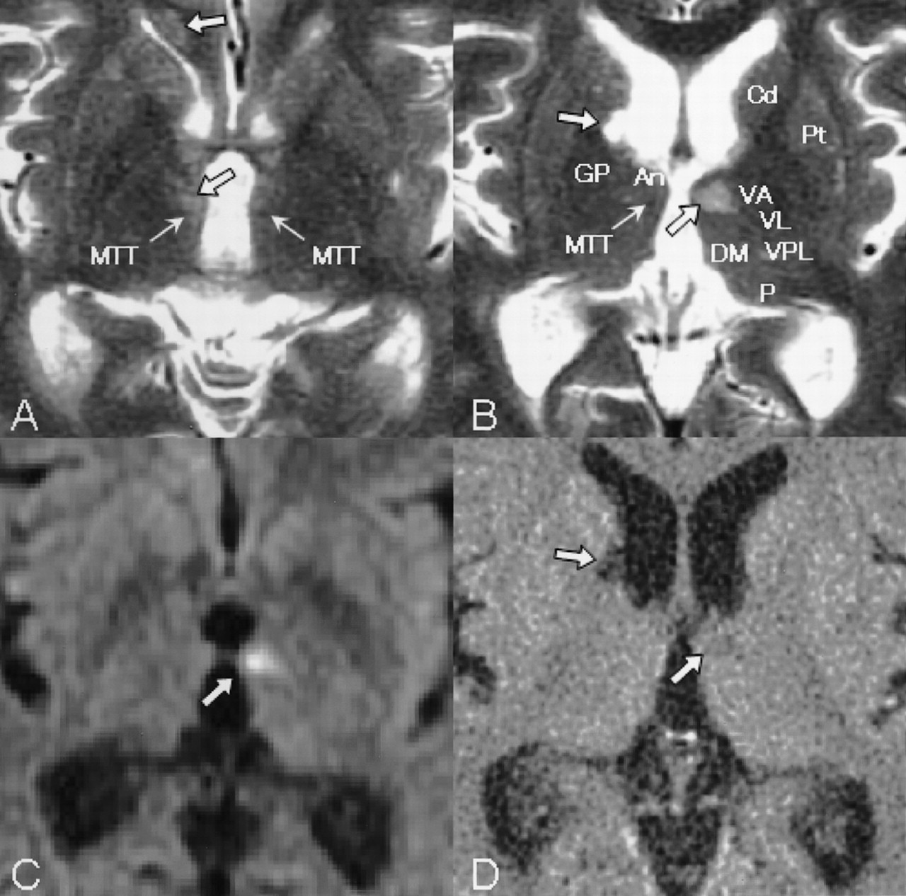
Area of detail on T2-weighted images examined 3 days after onset (A and B) demonstrate anatomic structures, such as the anterior nucleus (An), caudate nucleus (Cd), dorsomedial nucleus (DM), globus pallidus (GP), MTT, pulvinar (P), putamen (Pt), ventral anterior nucleus (VA), ventral lateral nucleus (VL), and ventral posterolateral nucleus (VPL). An infarction on the left MTT (C, upswept thick arrow) demonstrated on a DW image is identical to that on the T2-weighted image (B, upswept thick arrow) and on the fluid-attenuated inversion recovery image, which was seen on a follow-up CT scan 4 weeks after onset (D, upswept thick arrow), which was the same as on MR images at admission. Older ischemic lesions were also seen in the corpus callosum on the right (A, upper descending thick arrow), on the right MTT (A, lower descending thick arrow), and in the head of the right caudate nucleus (B and D, descending thick arrow). Thick arrows show ischemic lesions, and thin arrows, MTTs.

Mental status testing 4 weeks after onset revealed no improvement of his memory. Serial-sevens could be performed to 79, although he gave up because of frustration. He still did not accept the fact that his father had died, although he said that he had noticed something odd about his memory. A follow-up CT scan showed that a small infarction of the left MTT was the same as that on previous images examined 4 weeks earlier.

Eight weeks after onset, his amnesia persisted. Mental status testing results were almost the same as those obtained 4 weeks after onset. Follow-up MR imaging 8 weeks after onset demonstrated small infarctions of both MTTs (Figs 3–5). The infarction over the right MTT was thought to be much older than the left, because the right MTT lesion was hypointense at DW imaging performed at admission and 8 weeks after onset. Six months after onset, his memory showed no improvement during a regular follow-up evaluation at the outpatient clinic.

Fig 3.
  • Download figure
  • Open in new tab
  • Download powerpoint
Fig 3.

T2-weighted images obtained 8 weeks after onset show ischemic lesions in the corpus callosum on the right and the right anterior thalamus (C and D). The infarction of the left MTT (D) is identical to that on MR images obtained at admission (Fig 1). DW imaging shows no acute ischemic lesions (E and F). The medial temporal lobes (A and B) are intact, which is the same finding as that on the MR images obtained at admission (Fig 1).

Fig 4.
  • Download figure
  • Open in new tab
  • Download powerpoint
Fig 4.

Magnified T2-weighted images examined 8 weeks after onset demonstrate that both MTTs are affected (A and B), which are not visible on DW images (C). They are not acute ischemic lesions as shown on DW images (C) and on fluid attenuation inversion recovery images (D).

Fig 5.
  • Download figure
  • Open in new tab
  • Download powerpoint
Fig 5.

Schematic figure of the limbic system and its connections. The hippocampus projects to the septal nuclei and the mammillary body via the fornix. The mammillary body projects to the anterior nucleus of the thalamus via the MTT. The anterior nucleus of the thalamus projects to the cingulated cortex via the internal capsule. The cingulated cortex projects back to the hippocampus via the cingulum bundle. Both MTT lesions can interrupt the input from the hippocampus, resulting in dysfunction of the neurocircuit in the limbic system.

Discussion

The clinical diagnosis of Korsakoff syndrome is based on the identification of an amnesic syndrome with a history of alcohol abuse or malnutrition. The diagnosis can be made when memory deficits are more prominent than other cognitive disorders in the domain of language, visuoperceptual functioning, problem solving, and judgment (2). The patient described in this report presented with a syndrome that mirrored Korsakoff syndrome: memory dysfunction with retrograde amnesia, anterograde amnesia, limited insight into dysfunction, and confabulation despite no history of chronic alcoholism or malnutrition.

The two above-mentioned findings—acute memory disorder identical to Korsakoff syndrome and MR imaging findings of bilateral MTT infarctions (the left lesion fresh and the right old)—indicate that the sites of involvement in acute Korsakoff syndrome are both MTTs, leading to bilateral MTT dysfunction.

Although there is agreement on the neuropathologic mechanisms of Korsakoff syndrome, the critical lesion sites for memory disorder have been debated. From the viewpoint of the critical lesion(s) for amnesic syndrome, the thalamus and the mammillary bodies are most commonly implicated (6). Victor et al (7) pointed out that all 24 patients in whom the medial-dorsal nucleus of the thalamus was affected had a clinical history of persistent memory impairment (Korsakoff syndrome). Mair et al (8) provided a careful pathologic and neuropsychological description of two patients with Korsakoff syndrome in whom autopsies showed lesions in the mammillary bodies and the midline and anterior portion of the thalamus but not in the medial-dorsal nuclei, which was closely replicated by Mayes et al (9). On the other hand, Heilman et al (10) reported Korsakoff syndrome resulting from bilateral fornix lesions.

Recent evidence suggests that the circuit involving the mammillary bodies, the MTT and the anterior thalamus rather than the medial dorsal nucleus of the thalamus, is particularly critical in the formation of new memories (6). Visser et al (11) showed anterograde amnesia in alcoholic Korsakoff syndrome is associated with atrophy of the nuclei in the midline of the thalamus, but not with atrophy of the mamillary bodies, the hippocampus, or the parahippocampal gyrus. In addition, Harding et al (12) concluded that neuronal loss in the anterior thalamic nuclei was found consistently only in alcoholic Korsakoff psychosis, although neurodegeneration of the hypothalamic mammillary nuclei and the mediodorsal thalamic nuclei was substantial in those with nonamnesic alcoholism and those with amnesic alcoholism with Wernicke encephalopathy.

From animal experiments, Mishkin (13, 14) argued that there are two limbic circuits in which combined lesions are required to produce severe amnesia: hippocampal (medial limbic) and amygdaloid (basolateral limbic) circuits. The anterior and dorsomedial thalamus and the medial temporal lobes are the anatomic locations (“nodal points”) wherein these two circuits converge and are, therefore, particularly vulnerable to the effect of discrete structural lesions (15–19). Consequently, lesions of both MTTs projecting to the anterior nucleus of the thalamus can affect the nodal points, resulting in a severe amnesic state (Fig 5).

Determining the contribution of an individual region in memory is difficult, because it is unusual to find anatomically circumscribed damage to only one structural region in a clinical setting. In our case, however, we encountered infarctions in both MTTs depicted by DW imaging. The left lesion was new, the right was old, and the medial temporal lobes (Fig 3) were normal. It is interesting to note that, from the viewpoint of the clinical course, the patient had no memory or cognitive disorder when he developed the right MTT lesion. We speculate that the right MTT lesion did not cause clinical symptoms. Figure 4 shows small lesions in the MTTs and demonstrates intact ventral anterior nucleus, ventral lateral nucleus, and ventral posterolateral nucleus on both sides. Thus, in this case, the pathogenetic candidate of acute Korsakoff syndrome was thought to be both the affected MTTs and the left MTT ischemia acted as a trigger of Korsakoff syndrome. Both MTT lesions may have cut off hippocampal input through the mammillary bodies, into the anterior nucleus, thereby producing a severe amnesic state.

References

  1. ↵
    Shear PK, Sullivan EV, Lane B, Pfefferbaum A. Mammillary body and cerebellar shrinkage in chronic alcoholics with and without amnesia. Alcohol Clin Exp Res 1996;20:1489–1495
    CrossRefPubMed
  2. ↵
    Toth C, Voll C, Macaulay R. Primary CNS lymphoma as a cause of Korsakoff syndrome. Surg Neurol 2002;57:41–45
    PubMed
  3. Konovalov AN, Dobrokhotova TA, Voronina IA, Urakov SV. Case of Korsakoff syndrome and colloid cyst of the 3rd ventricle [in Russian] Zh Nevrol Psikhiatr Im S S Korsakova 1998;98:49–51
  4. Yarde WL, Kepes JJ, O’Boynick P. Craniopharyngioma presenting as Korsakoff psychosis. Kans Med 1995;96:22–23, 34
    PubMed
  5. ↵
    Neciga EG, Peralta AG, Polaina M, et al. Cyst of the septum pellucidum and Korsakoff’s psychosis. Eur Neurol 1989;29:99–101
    PubMed
  6. ↵
    Kopelman MD. The Korsakoff syndrome. Br J Psychiatry 1995;166:154–173
    Abstract/FREE Full Text
  7. ↵
    Victor M, Adams RD, Collins GH. The Wernicke-Korsakoff syndrome: a clinical and pathological study of 245 patients, 82 with post-mortem examinations. Contemp Neurol Ser 1971;7:1–206
    PubMed
  8. ↵
    Mair WG, Warrington EK, Weiskrantz L. Memory disorder in Korsakoff’s psychosis: a neuropathological and neuropsychological investigation of two cases. Brain 1979;102:749–783
    FREE Full Text
  9. ↵
    Mayes AR, Meudell PR, Mann D, Pickering A. Location of lesions in Korsakoff’s syndrome: neuropsychological and neuropathological data on two patients. Cortex 1988;24:367–388
    PubMed
  10. ↵
    Heilman KM, Sypert GW. Korsakoff’s syndrome resulting from bilateral fornix lesions. Neurology 1977;27:490–493
    Abstract/FREE Full Text
  11. ↵
    Visser PJ, Krabbendam L, Verhey FR, et al. Brain correlates of memory dysfunction in alcoholic Korsakoff’s syndrome. J Neurol Neurosurg Psychiatry 1999;67:774–778
    Abstract/FREE Full Text
  12. ↵
    Harding A, Halliday G, Caine D, Kril J. Degeneration of anterior thalamic nuclei differentiates alcoholics with amnesia. Brain 2000;123:141–154
    Abstract/FREE Full Text
  13. ↵
    Mishkin M. Memory in monkeys severely impaired by combined but not by separate removal of amygdala and hippocampus. Nature 1978;273:297–298
    CrossRefPubMed
  14. ↵
    Mishkin M. A memory system in the monkey. Philos Trans R Soc Lond B Biol Sci298:85–95
  15. ↵
    Speedie LJ, Heilman KM. Amnestic disturbance following infarction of the left dorsomedial nucleus of the thalamus. Neuropsychologia 1982;20:597–604
    CrossRefPubMed
  16. Markowitsch HJ. Can amnesia be caused by damage of a single brain structure? Cortex 1984;20:27–45
    PubMed
  17. Valenstein E, Bowers D, Verfaellie M, et al. Retrosplenial amnesia. Brain 1987;110:1631–1646
    Abstract/FREE Full Text
  18. von Cramon DY, Hebel N, Schuri U. A contribution to the anatomical basis of thalamic amnesia. Brain 1985;108:993–1008
    Abstract/FREE Full Text
  19. ↵
    Graff-Radford NR, Tranel D, Van Hoesen GW, Brandt JP. Diencephalic amnesia. Brain 1990;113:1–25
    Abstract/FREE Full Text
  • Received September 7, 2003.
  • Accepted after revision October 20, 2003.
  • Copyright © American Society of Neuroradiology
View Abstract
PreviousNext
Back to top

In this issue

American Journal of Neuroradiology: 25 (6)
American Journal of Neuroradiology
Vol. 25, Issue 6
1 Jun 2004
  • Table of Contents
  • Index by author
Advertisement
Print
Download PDF
Email Article

Thank you for your interest in spreading the word on American Journal of Neuroradiology.

NOTE: We only request your email address so that the person you are recommending the page to knows that you wanted them to see it, and that it is not junk mail. We do not capture any email address.

Enter multiple addresses on separate lines or separate them with commas.
Acute Korsakoff Syndrome Following Mammillothalamic Tract Infarction
(Your Name) has sent you a message from American Journal of Neuroradiology
(Your Name) thought you would like to see the American Journal of Neuroradiology web site.
CAPTCHA
This question is for testing whether or not you are a human visitor and to prevent automated spam submissions.
Cite this article
Yuichiro Yoneoka, Norio Takeda, Akira Inoue, Yasuo Ibuchi, Takashi Kumagai, Tsutomu Sugai, Ken-ichiro Takeda, Kaoru Ueda
Acute Korsakoff Syndrome Following Mammillothalamic Tract Infarction
American Journal of Neuroradiology Jun 2004, 25 (6) 964-968;

Citation Manager Formats

  • BibTeX
  • Bookends
  • EasyBib
  • EndNote (tagged)
  • EndNote 8 (xml)
  • Medlars
  • Mendeley
  • Papers
  • RefWorks Tagged
  • Ref Manager
  • RIS
  • Zotero
0 Responses
Respond to this article
Share
Bookmark this article
Acute Korsakoff Syndrome Following Mammillothalamic Tract Infarction
Yuichiro Yoneoka, Norio Takeda, Akira Inoue, Yasuo Ibuchi, Takashi Kumagai, Tsutomu Sugai, Ken-ichiro Takeda, Kaoru Ueda
American Journal of Neuroradiology Jun 2004, 25 (6) 964-968;
del.icio.us logo Twitter logo Facebook logo Mendeley logo
  • Tweet Widget
  • Facebook Like
  • Google Plus One

Jump to section

  • Article
    • Abstract
    • Case Report
    • Discussion
    • References
  • Figures & Data
  • Info & Metrics
  • Responses
  • References
  • PDF

Related Articles

  • No related articles found.
  • PubMed
  • Google Scholar

Cited By...

  • Mammillothalamic Disconnection Alters Hippocampocortical Oscillatory Activity and Microstructure: Implications for Diencephalic Amnesia
  • Mammillothalamic disconnection alters hippocampo-cortical oscillatory activity and microstructure: Implications for diencephalic amnesia
  • Crossref
  • Google Scholar

This article has not yet been cited by articles in journals that are participating in Crossref Cited-by Linking.

More in this TOC Section

  • Evaluating the Effects of White Matter Multiple Sclerosis Lesions on the Volume Estimation of 6 Brain Tissue Segmentation Methods
  • Quiet PROPELLER MRI Techniques Match the Quality of Conventional PROPELLER Brain Imaging Techniques
  • Predictors of Reperfusion in Patients with Acute Ischemic Stroke
Show more BRAIN

Similar Articles

Advertisement

Indexed Content

  • Current Issue
  • Accepted Manuscripts
  • Article Preview
  • Past Issues
  • Editorials
  • Editor's Choice
  • Fellows' Journal Club
  • Letters to the Editor
  • Video Articles

Cases

  • Case Collection
  • Archive - Case of the Week
  • Archive - Case of the Month
  • Archive - Classic Case

More from AJNR

  • Trainee Corner
  • Imaging Protocols
  • MRI Safety Corner

Multimedia

  • AJNR Podcasts
  • AJNR Scantastics

Resources

  • Turnaround Time
  • Submit a Manuscript
  • Submit a Video Article
  • Submit an eLetter to the Editor/Response
  • Manuscript Submission Guidelines
  • Statistical Tips
  • Fast Publishing of Accepted Manuscripts
  • Graphical Abstract Preparation
  • Imaging Protocol Submission
  • Evidence-Based Medicine Level Guide
  • Publishing Checklists
  • Author Policies
  • Become a Reviewer/Academy of Reviewers
  • News and Updates

About Us

  • About AJNR
  • Editorial Board
  • Editorial Board Alumni
  • Alerts
  • Permissions
  • Not an AJNR Subscriber? Join Now
  • Advertise with Us
  • Librarian Resources
  • Feedback
  • Terms and Conditions
  • AJNR Editorial Board Alumni

American Society of Neuroradiology

  • Not an ASNR Member? Join Now

© 2025 by the American Society of Neuroradiology All rights, including for text and data mining, AI training, and similar technologies, are reserved.
Print ISSN: 0195-6108 Online ISSN: 1936-959X

Powered by HighWire