
Article Figures & Data
Figures
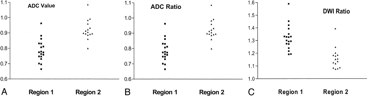
- Fig 1.
Images obtained in three patients in group B who had hyperintense regions at initial DWI that appeared normal at follow-up. Region 1 is DWI-hyperintense tissue that was abnormal at follow-up imaging. Region 2 is DWI-hyperintense tissue that was normal at follow-up imaging.
A, ADC values in square millimeters per second.
B, ADC ratios.
C, DWI ratios.
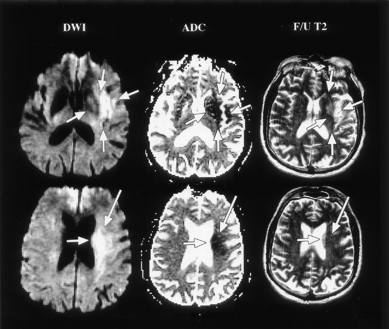
- Fig 2.
Reversal of DWI-hyperintense lesion after thrombolysis in a 68-year-old man with right-sided weakness and dysarthria. The patient had a left MCA occlusion that was successfully treated with IA tissue plasminogen activator at 6 hours. Initial DWI (row 1) and ADC (row 2) images demonstrate abnormality involving the left basal ganglia, insula, and subinsular region (short arrows) and in the left corona radiata (long arrow). At 3 days, follow-up (F/U) T2-weighted images demonstrate no abnormality in the left corona radiata. Lesions in the basal ganglia, insula, and subinsular region are unchanged. At discharge, the right-sided weakness had resolved.


Tables
Data All Patients (n = 16) Group A (n = 13) Group B (n = 3) Sex (M:F) 8:8 5:8 3:0 Age, y 68.3 (57–88) 69.0 (57–88) 65.3 (57–71) Time to treatment, h 5.1 (2.8–6.5) 5.2 (4.2–6.5) 4.5 (2.8–6.3) Initial NIHSS Score 17.2 (11–21) 17.2 (11–21) 17 (14–21) Follow-up mRS score 4.6 (2–6) 4.8 (2–6) 3.7 (2–5) Clot location 8 MCA stem 8 ICA T 5 MCA stem 8 ICA T 3 MCA stem Recanalization 2 TIMI 3, 9 TIMI 2, 5 TIMI 1 9 TIMI 2, 4 TIMI 1 2 TIMI 3, 1 TIMI 1 Follow-up study, d 3.2 (1–7) 3.1 (1–7) 3.7 (3–5) Note.—Data are the mean (range) unless otherwise specified. ICA indicates internal carotid artery.
Patient/Age, y/Sex Clot Location Initial NIHSS Score DWI Volume, cm3 Follow-Up Volume, cm3 Time to Treatment, h Angiographic Result after Thrombolysis TIMI Grade mRS Score at Discharge 1/57/M* L M1/M2 21 16.81 16.40 3.75 M1, ID and SD open 3 2 2/68/M* L M1 16 37.31 24.00 6 M1, ID and SD open 3 4 3/71/M* L M1/M2 14 42.40 80.45 4.5 M1 clot persistent with some penetration, moderate collaterals 1 5 4/75/F R ICA T 17 122.09 276.75 5.33 R ICA T clot persistent with some penetration 1 6 5/57/M R ICA T 19 118.12 147.5 5 ICA, A1, M1 and SD patent, ID clot persistent 2 5 6/71/F LM1/M2 11 3.07 4.39 5.25 M1 and SD open, ID open except for angular artery 2 2 7/76/M R M1 18 35.81 112.79 5.25 M1 and SD open, ID clot persistent 2 4 8/75/F L ICA T 18 25.51 28.16 5.33 ICA, M1 and ID open, partial filling of SD, A1 clot persistent 2 5 9/88/M L ICA T 21 120.56 217.26 4.67 ICA, M1 and ID open, partial filling of SD, A1 clot persistent 2 5 10/82/M R ICA T 20 216.44 420.45 4.75 R ICA T clot persistent with some penetration 1 6 11/78/F L ICA T 15 80.68 672.45 5 L ICA T clot persistent with some penetration 1 6 12/72/F L M1 21 30.56 199.31 4.75 M1 partially open with minimal filling of SD 2 6 13/20/F R ICA T 18 35.86 157.47 6 RICA and M1 open, partial filling of SD, ID occluded 2 5 14/73/F R M1 21 39.08 132.76 6 M1 open, partial filling of SD and ID 2 5 15/69/F R ICA T 14 12.9 212.7 4.25 R ICA T clot persistent with some penetration 1 5 16/61/M L M1 11 14.08 46.37 5.25 M1 and SD open, ID occluded 2 3 Note.—ICA indicates internal carotid artery; ID, inferior division of the MCA; M1, M1 segment of the MCA; NS, not significant; and SD, superior division of the MCA.
* Patients in group B.
Group Initial Volume, cm3 Follow-up Volume, cm3 Change, % All (n = 16) 59.45 (3.07–216.4) 171.8 (4.39–672.4) 200.0 (−35.7 to 1548) Group A (n = 13) 65.75 (3.07–216.4) 202.2 (4.39–672.4) 207.5 (10.3–1548) Group B (n = 3) 32.17 (16.81–42.40) 40.28 (16.4–80.45) 25.2 (−35.7 to 89.8) Note.—Data are the mean (range).
In this issue


Jump to section
Related Articles
Cited By...
- Quantitative Analysis of Hypoperfusion in Acute Stroke: Arterial Spin Labeling Versus Dynamic Susceptibility Contrast
- Imaging-based selection for intra-arterial stroke therapies
- Periprocedural Arterial Spin Labeling and Dynamic Susceptibility Contrast Perfusion in Detection of Cerebral Blood Flow in Patients With Acute Ischemic Syndrome
- Diffusion weighted imaging reversibility in the brainstem following successful recanalization of acute basilar artery occlusion
- Does Diffusion-Weighted Imaging Represent the Ischemic Core? An Evidence-Based Systematic Review
- Reduction of Diffusion-Weighted MRI Lesion Volume After Early Moderate Hypothermia in Ischemic Stroke