
Article Figures & Data
Figures
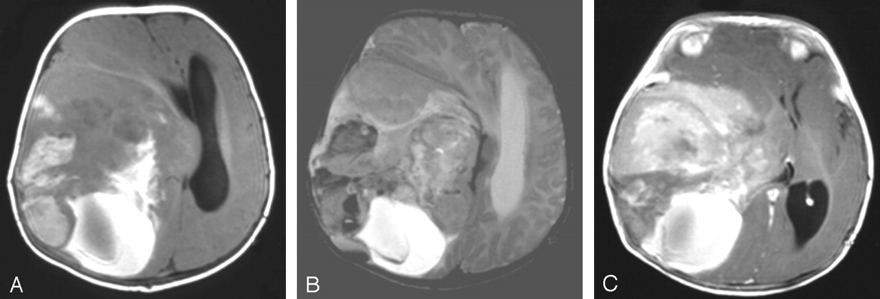
- Fig 1.
Case 1. Axial T1- (A), axial T2- (B), and axial postgadolinium T1-weighted (C) MR images show a large supratentorial and heterogeneous tumor involving the frontotemporoparietal lobes with a major mass effect. The solid part of the tumor has two components. The peripheral part is heterogeneous and moderately hyperintense on T1-weighted images and shows low signal intensity on T2-weighted images suggestive of intratumoral bleeding; the medial part is enhanced after gadolinium injection. The posterior cystic part of the lesion is of high signal intensity on T1- and T2-weighted images, which is compatible with a recent bleeding episode. Radiologic appearance is suggestive of a malignant lesion.
- Fig 2.
Case 3. Axial postgadolinium T1- (A), coronal postgadolinium T1- (B), and axial T2-weighted (C) MR images show a typically large supratentorial mass with a peripheral solid and a deep cystic component. The peripheral solid component is enhanced after gadolinium injection (A and B) and is associated with a meningeal thickening and enhancement. Note the major mass effect on the midline and the ventricles.
- Fig 3.
Standard hematoxylin coloration (magnification ×250) shows the fusiform astrocytic cells forming a dense fascicle pattern.
- Fig 4.
DIG with marked neurofilament immunostain (magnification ×400) due to the presence of neural cells (black arrow).


Tables
Clinical findings
Finding Case (No.) 1 2 3 4 5 6 Sex/age (mo) M/5 M/39 M/5 F/2.5 F/48 M/5 Symptom duration 3 wk 1 mo 2 mo 2.5 mo 2 mo 0 Increasing head circumference Y N N Y N N Seizures N Y Y N N N Increased ICP Y N N N Y N Other symptoms Skull deformation . . . . . . . . . Visual field . . . Treatment Total excision Total excision Subtotal excision Total excision Partial excision Total excision Follow-up 18 mo, norm dvpt, upper limb paralysis 14 mo, norm dvpt 3 y 6 mo, norm dvpt, dysphasia 24 mo norm dvpt 13 mo death 7 y, norm dvpt Note.—Yes (Y), no (N), normal development (norm dvpt).
In this issue


Jump to section
Related Articles
Cited By...
- No citing articles found.