
Article Figures & Data
Figures
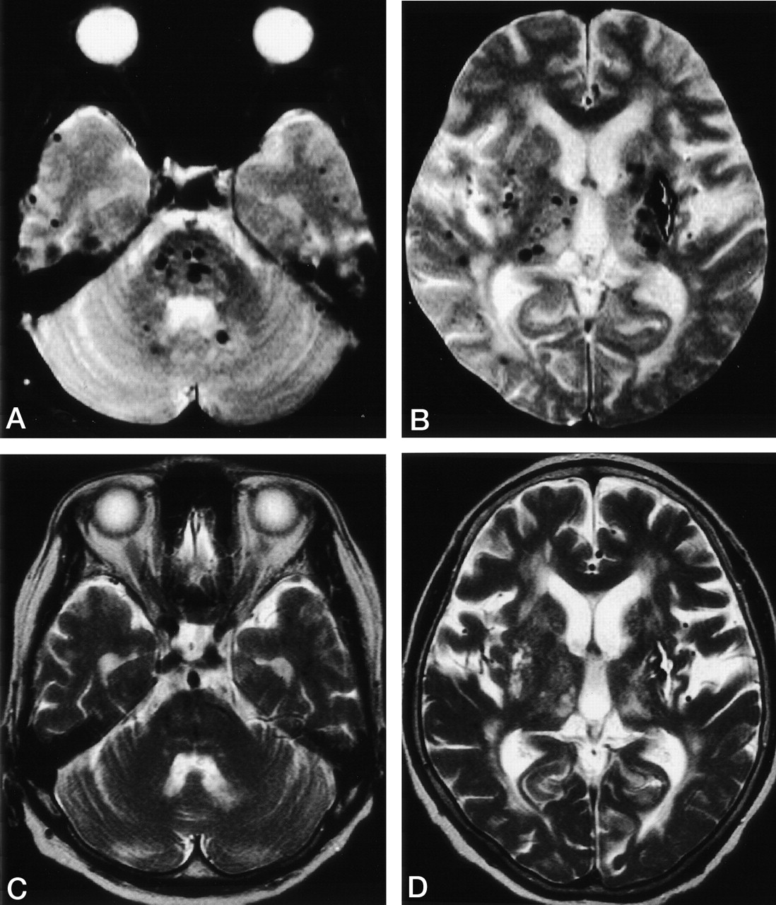
- Fig 1.
MR images of a 75-year-old patient with intracerebral hemorrhage in the right parietal lobe 8 years after the occurrence of intracerebral hemorrhage in the left putamen.
A and B, T2*-weighted gradient-echo images (800/26; flip angle, 20 degrees) reveal multiple foci of signal intensity loss (microbleeds) in the brain stem, cerebellum, basal ganglia, and cerebral hemispheres. In addition, old intracerebral hemorrhage is evident in the left putamen.
C and D, T2-weighted spin-echo images (4500/112) show the site of old intracerebral hemorrhage in the left putamen, but microbleeds are not evident.

Tables
Primary Stroke Recurrent Stroke P Patients, n (M/F) 102 (57/45) 54 (38/16) .0777 Age, yr (SD) 69.0 (12.7) 68.6 (11.4) .8377 Hypertension, n (%) 65 (63.7) 38 (70.4) .4045 Atrial fibrillation, n (%) 14 (13.7) 5 (9.3) .4171 Diabetes mellitus, n (%) 23 (22.5) 9 (16.7) .3867 Hyperlipidemia, n (%) 20 (19.6) 10 (18.5) .8695 Intracranial large-artery disease, n (%) 38 (37.3) 16 (29.6) .3409 Leukoaraiosis, grade (SD) 1.04 (0.91) 1.74 (0.99) <.0001 Note.—M indicates male; F, female.
Microbleeds n (%) Grade of Microbleeds Absent (grade 0), n Mild (grade 1), n Moderate (grade 2), n Severe (grade 3), n Stroke subtype Atherothrombotic (n = 22) 5 (22.7) 17 3 2 0 Cardioembolic (n = 13) 0 (0) 13 0 0 0 Lacunar (n = 31) 7 (22.6) 24 4 0 3 Intracerebral hemorrhage (n = 36) 17 (47.2) 19 5 10 2 Total (n = 102) 29 (28.4) 73 12 12 5 Antiplatelet or Anticoagulation Therapy after Previous Stroke Patients, n Microbleeds, n Combination of stroke subtype* Intracerebral hemorrhage/intracerebral hemorrhage 9 8 Lacunar/lacunar + 6 2 Intracerebral hemorrhage/lacunar 9 8 Intracerebral hemorrhage (three times) 4 4 Lacunar/intracerebral hemorrhage − 3 2 Atherothrombotic/lacunar + 3 1 Intracerebral hemorrhage/atherothrombotic 2 1 Atherothrombotic/atherothrombotic − 2 1 Lacunar/atherothrombotic − 2 2 Lacunar/lacunar − 2 1 Lacunar/atherothrombotic + 2 1 Lacunar/intracerebral hemorrhage + 2 2 Cardioembolic/cardioembolic + 2 1 Atherothrombotic/atherothrombotic + 2 2 Intracerebral hemorrhage/cardioembolic 1 0 Lacunar (three times) + 1 1 Atherothrombotic/lacunar − 1 0 Cardioembolic/cardioembolic − 1 0 Total 54 37 * Combination of stroke subtype expressed as previous stroke subtype/latest stroke subtype; Present is indicated by + and absent by −.
- TABLE 4:
Prevalence and grade of microbleeds in patients with each combination of subtypes of recurrent stroke
Microbleeds n (%) Grade of Microbleeds Absent (grade 0), n Mild (grade 1), n Moderate (grade 2), n Severe (grade 3), n Combination of stroke subtype Intracerebral hemorrhage alone (n = 13) 12 (92.3) 1 1 4 7 Intracerebral hemorrhage and ischemic stroke (n = 17) 13 (76.5) 4 1 5 7 Ischemic stroke alone (n = 24) 12 (50.0) 12 4 5 3 Total (n = 54) 37 (68.5) 17 6 14 17 Variable Odds Ratio 95% CI P Age 0.992 0.957–1.029 .6771 Sex 1.764 0.754–4.125 .1905 Hypertension 1.986 0.823–4.794 .1271 Atrial fibrillation 0.209 0.047–0.934 .0404 Diabetes mellitus 1.257 0.495–3.192 .6306 Hyperlipidemia 0.566 0.205–1.564 .2721 Intracranial large-artery diseases 0.981 0.425–2.264 .9642 Leukoaraiosis 5.079 2.125–12.143 .0003 Recurrent stroke 4.487 1.989–10.120 .0003 Note.—CI indicates confidence interval.


