Advertisement

Main menu

  • Home
  • Content
    • Current Issue
    • Accepted Manuscripts
    • Article Preview
    • Past Issue Archive
    • Video Articles
    • AJNR Case Collection
    • Case of the Week Archive
    • Case of the Month Archive
    • Classic Case Archive
  • Special Collections
    • AJNR Awards
    • Low-Field MRI
    • Alzheimer Disease
    • ASNR Foundation Special Collection
    • Photon-Counting CT
    • View All
  • Multimedia
    • AJNR Podcasts
    • AJNR SCANtastic
    • Trainee Corner
    • MRI Safety Corner
    • Imaging Protocols
  • For Authors
    • Submit a Manuscript
    • Submit a Video Article
    • Submit an eLetter to the Editor/Response
    • Manuscript Submission Guidelines
    • Statistical Tips
    • Fast Publishing of Accepted Manuscripts
    • Graphical Abstract Preparation
    • Imaging Protocol Submission
    • Author Policies
  • About Us
    • About AJNR
    • Editorial Board
    • Editorial Board Alumni
  • More
    • Become a Reviewer/Academy of Reviewers
    • Subscribers
    • Permissions
    • Alerts
    • Feedback
    • Advertisers
    • ASNR Home

User menu

  • Alerts
  • Log in

Search

  • Advanced search
American Journal of Neuroradiology

American Journal of Neuroradiology

ASHNR American Society of Functional Neuroradiology ASHNR American Society of Pediatric Neuroradiology ASSR
  • Alerts
  • Log in

Advanced Search

  • Home
  • Content
    • Current Issue
    • Accepted Manuscripts
    • Article Preview
    • Past Issue Archive
    • Video Articles
    • AJNR Case Collection
    • Case of the Week Archive
    • Case of the Month Archive
    • Classic Case Archive
  • Special Collections
    • AJNR Awards
    • Low-Field MRI
    • Alzheimer Disease
    • ASNR Foundation Special Collection
    • Photon-Counting CT
    • View All
  • Multimedia
    • AJNR Podcasts
    • AJNR SCANtastic
    • Trainee Corner
    • MRI Safety Corner
    • Imaging Protocols
  • For Authors
    • Submit a Manuscript
    • Submit a Video Article
    • Submit an eLetter to the Editor/Response
    • Manuscript Submission Guidelines
    • Statistical Tips
    • Fast Publishing of Accepted Manuscripts
    • Graphical Abstract Preparation
    • Imaging Protocol Submission
    • Author Policies
  • About Us
    • About AJNR
    • Editorial Board
    • Editorial Board Alumni
  • More
    • Become a Reviewer/Academy of Reviewers
    • Subscribers
    • Permissions
    • Alerts
    • Feedback
    • Advertisers
    • ASNR Home
  • Follow AJNR on Twitter
  • Visit AJNR on Facebook
  • Follow AJNR on Instagram
  • Join AJNR on LinkedIn
  • RSS Feeds

AJNR Awards, New Junior Editors, and more. Read the latest AJNR updates

Research ArticleNeurointervention

A Novel Two-Stage Technique for Construction of Experimental Aneurysms

James V. Byrne and Neil Hubbard
American Journal of Neuroradiology February 2004, 25 (2) 319-321;
James V. Byrne
  • Find this author on Google Scholar
  • Find this author on PubMed
  • Search for this author on this site
Neil Hubbard
  • Find this author on Google Scholar
  • Find this author on PubMed
  • Search for this author on this site
  • Article
  • Figures & Data
  • Info & Metrics
  • Responses
  • References
  • PDF
Loading

Abstract

Summary: A two-staged technique for construction of experimental aneurysms in swine is described. The jugular vein is initially anastomosed to the carotid artery. Spontaneous hypertrophy of the vein is modeled by placing restraining sutures to create a fusiform aneurysm. Subsequent endovascular closure of this aneurysm leaves a sidewall aneurysm pouch. Staging allows maturation of the anastomosis, and the technique provides additional targets for endovascular training. Trial has been successful in three animals.

Experimental saccular aneurysms have for many years been constructed by researchers from vein homografts in a variety of animal types (1). There is no consensus on the optimum species for aneurysm construction, but swine are widely used because they are suitable in size for testing endovascular devices, are ethically acceptable, and have a hematology profile similar to that of humans (2). However, in swine, the free graft lateral wall vein pouch experimental aneurysm is liable to spontaneously rupture during the short term (3) (Y. Murayama, personal communication). This report describes a two-stage procedure to create fusiform or saccular experimental aneurysms that overcomes this problem.

Description of the Technique

The procedure involves anastomosis of the external jugular vein to the carotid artery to establish an arteriovenous fistula (AVF) and delayed endovascular closure of the fistula to create a saccular aneurysm.

Surgical Technique: Stage 1

To establish an AVF, the external jugular vein is exposed and cleaned of surrounding tissues for a length of 5 cm. The cranial end is ligated and the distal exposed portion mobilized to allow it to be swung medially under the sternocleidomastoid muscle. The cervical carotid artery is then exposed and a 4.5-mm arteriotomy made on its anterolateral aspect after the application of temporary vascular clamps. A venotomy is made in the mobilized vein and the two vessels anastomosed side-to-side with a running 60 nylon suture. To assist the anastomosis, temporary stay sutures are placed at the ligated end of the vein and back bleeding is controlled by placing a vascular sling around the caudal part of the vein.

After removal of the arterial clamps and vascular sling, blood flow in the vein and artery distal to the fistula is confirmed visually. Loops of 30 silk sutures are then placed 1.5 cm apart around the draining vein to narrow the vessel above and below the section to subsequently be occluded. The wound is closed in layers (Fig 1).

Fig 1.
  • Download figure
  • Open in new tab
  • Download powerpoint
Fig 1.

Line drawing shows the arteriovenous anastomosis between the carotid artery and mobilized external jugular vein. A side-to-side anastomosis is performed 1–2 cm below the cranial end of the ligated vein. Restraining sutures are then applied to induce a pouch in the draining vein.

Endovascular Technique: Stage 2

The second stage procedure is performed 1 to 2 weeks later. The vein inevitably enlarges during the interval so that fusiform dilations develop immediately proximal to each suture. To convert the fistula into an aneurysm, an endovascular balloon (1815 DSB; Target Therapeutics, Inc., Fremont, CA) is placed via a percutaneous femoral puncture between the sutures under fluoroscopic control. Once the fistula is closed, a proximal vein pouch aneurysm remains for testing embolization devices or other studies. Both stages are performed after the induction of general anesthesia. The surgical techniques were developed and performed according to a protocol approved by the University Ethics Panel and the United Kingdom Government Home Office.

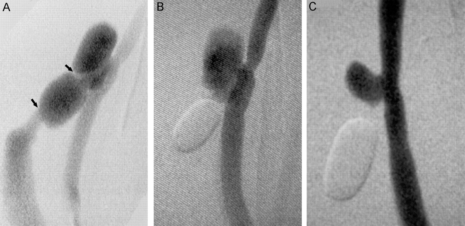
Unilateral AVF were created in three pigs weighing 25 to 30 kg each. Follow-up intra-arterial transfemoral angiography was performed 7, 12, and 18 days later, respectively. All the AVFs were patent with enlargement of the draining vein (Fig 2A). Pouch aneurysms were successfully created after closure of AVF between the restraining sutures in all animals (Fig 2B). One animal was followed without embolization, and repeat angiography after an additional 14 days showed a patent but smaller residual aneurysm pouch (Fig 2C).

Fig 2.
  • Download figure
  • Open in new tab
  • Download powerpoint
Fig 2.

Stage 1 of surgical technique.

A, Intra-arterial angiogram obtained 7 days after creation of a fistula by anastomosis of the external jugular vein (side-to-side) with the cervical carotid artery. Two fusiform pouches are present in the draining vein because restraining sutures were placed to restrict enlargement of the vein at the site of the arrows.

B, A balloon has been deployed in the distal vein pouch to close the fistula and convert the upper pouch into a sidewall aneurysm.

C, Angiogram obtained 14 days after closure of the fistula. The sidewall aneurysm has remodeled.

Discussion

The conventional vein graft pouch continues to be used for the construction of in vivo sidewall experimental aneurysms by researchers because the surgical technique is relatively simple. The described modification is intended to improve the reliability of aneurysm production without adding to the surgical complexity. Descriptions of techniques to createexperimental aneurysms that involve arteriovenous anastomosis have been presented. Black (4), in amodification of his original operation, established an AVF and then, after a short period (approximately 60 min) of observation, ligated the draining vein to create a sidewall aneurysm. This was performed so that the pouch could be more accurately sized, based on the principle that matching the sac to neck size made spontaneous thrombosis less likely. Others have exploited the technique to create terminal and bifurcation aneurysms (5). The use of a restraining suture to modify the anticipated enlargement of the draining vein has not previously been described.

The sidewall experimental aneurysm, particularly when created in swine, is liable to spontaneously thrombose as well as rupture (2, 3). Black (4) found that restricting the length of the aneurysm sac (ie, reducing the ratio of sac-to-neck dimensions) reduced the incidence of spontaneous thrombosis, presumably by altering intrasaccular blood flow patterns. Thus, species and aneurysm geometry are both factors. Our modification may also be liable to thrombosis for the same reasons, and more work is needed to assess this possibility.

Increasing the complexity of aneurysm preparation, as proposed, has to be justified. The AVF allows the anastomosis between artery and vein to mature and seems to avoid the danger of spontaneous rupture. The phenomena, which have also been described as occurring in rats (6) but not dogs (7), occur during the first week after surgery (3). As a result, embolization has to be performed early, usually at the time or within 48 hr of surgery. This is not optimum because recently manipulated vessels are more likely to develop spasm during endovascular catheterization. The delay between the two stages is therefore an advantage, and if the model is to be used for training, balloon or coil embolization of the distal vein pouch can be incorporated in a teaching program. For experimentation, the model allows the creation of fusiform aneurysms and has potential for further modification. For example, surgical modifications, such as ligating the caudal rather than cranial end of the vein, would reduce the angle between parent artery and pouch so that a stent could be placed more easily.

The response observed in vessels after surgical construction of AVF has been studied in patients and animals. Involved arteries and veins enlarge in response to increased blood flow. Hypertrophy of vein wall media has been observed as early as 1 week after arteriovenous anastomosis, and by 8 weeks, the media muscle layers appear similar to that of arteries, although no elastic lamina develops (8). The hemodynamic changes associated with establishing and then closing an AVF warrant further study because arteries enlarge in response to increased shear stress but intimal thickening and remodeling occur if blood flow (and therefore shear stress) is then reduced (9). Such vascular changes mean that previously described aneurysm flow dynamics (7) cannot be assumed in this model.

References

  1. ↵
    German WJ, Black SP. Experimental production of carotid aneurysms. New Engl J Med 1954;250:104–106
  2. ↵
    Massoud TF, Guglielmi G, Ji C, Viñuela F, Duckweiler GR. Experimental saccular aneurysms. Neuroradiology 1994;36:537–546
    CrossRefPubMed
  3. ↵
    Byrne JV, Hubbard N, Morris JH. Endovascular coil occlusion of experimental aneurysms: partial treatment does not prevent subsequent rupture. Neurol Res 1994;16:425–427
    PubMed
  4. ↵
    Black SP. Experimental saccular aneurysm by an arterio-venous fistula method. Mo Med 1963;4:340–343
  5. ↵
    Massoud TF, Ji C, Guglielmi G, Viñuela F, Robert J. Experimental models of bifurcation and terminal aneurysms: construction technique in swine. AJNR Am J Neuroradiol 1994;15:938–944
    Abstract/FREE Full Text
  6. ↵
    Nishikawa M, Smith RD, Yonekawa Y. Experimental intracranial aneurysms. Surg Neurol 1977;7:241–244
    PubMed
  7. ↵
    Strother CM, Graves VB, Rappe A. Aneurysm hemodynamics: an experimental study. AJNR Am J Neuroradiol 1992;13:1089–1095
    Abstract/FREE Full Text
  8. ↵
    Ozek C, Zhang F, Lineaweaver WC, Chin BT, Newlin L, Eiman T, Buncke HJ. Arterialization of the venous system in a rat lower limb model. Br J Plast Surg 1997;50:402–407
    PubMed
  9. ↵
    Zhuang Y-J, Singh TM, Zarins CK, Masuda H. Sequential increases and decreases in blood flow stimulates progressive intimal thickening. Eur J Vasc Endovasc Surg 1998;16:301–310
    CrossRefPubMed
  • Received June 12, 2003.
  • Accepted after revision July 29, 2003.
  • Copyright © American Society of Neuroradiology
PreviousNext
Back to top

In this issue

American Journal of Neuroradiology: 25 (2)
American Journal of Neuroradiology
Vol. 25, Issue 2
1 Feb 2004
  • Table of Contents
  • Index by author
Advertisement
Print
Download PDF
Email Article

Thank you for your interest in spreading the word on American Journal of Neuroradiology.

NOTE: We only request your email address so that the person you are recommending the page to knows that you wanted them to see it, and that it is not junk mail. We do not capture any email address.

Enter multiple addresses on separate lines or separate them with commas.
A Novel Two-Stage Technique for Construction of Experimental Aneurysms
(Your Name) has sent you a message from American Journal of Neuroradiology
(Your Name) thought you would like to see the American Journal of Neuroradiology web site.
CAPTCHA
This question is for testing whether or not you are a human visitor and to prevent automated spam submissions.
Cite this article
James V. Byrne, Neil Hubbard
A Novel Two-Stage Technique for Construction of Experimental Aneurysms
American Journal of Neuroradiology Feb 2004, 25 (2) 319-321;

Citation Manager Formats

  • BibTeX
  • Bookends
  • EasyBib
  • EndNote (tagged)
  • EndNote 8 (xml)
  • Medlars
  • Mendeley
  • Papers
  • RefWorks Tagged
  • Ref Manager
  • RIS
  • Zotero
0 Responses
Respond to this article
Share
Bookmark this article
A Novel Two-Stage Technique for Construction of Experimental Aneurysms
James V. Byrne, Neil Hubbard
American Journal of Neuroradiology Feb 2004, 25 (2) 319-321;
del.icio.us logo Twitter logo Facebook logo Mendeley logo
  • Tweet Widget
  • Facebook Like
  • Google Plus One

Jump to section

  • Article
    • Abstract
    • Description of the Technique
    • Discussion
    • References
  • Figures & Data
  • Info & Metrics
  • Responses
  • References
  • PDF

Related Articles

  • No related articles found.
  • PubMed
  • Google Scholar

Cited By...

  • No citing articles found.
  • Crossref
  • Google Scholar

This article has not yet been cited by articles in journals that are participating in Crossref Cited-by Linking.

More in this TOC Section

  • Factors Associated with Major Re-recanalization
  • A Key Factor Shapes LS-DAVFs EVT Outcome
  • Optimizing Voxel Size in 3D Rotational Angiography
Show more Neurointervention

Similar Articles

Advertisement

Indexed Content

  • Current Issue
  • Accepted Manuscripts
  • Article Preview
  • Past Issues
  • Editorials
  • Editor's Choice
  • Fellows' Journal Club
  • Letters to the Editor
  • Video Articles

Cases

  • Case Collection
  • Archive - Case of the Week
  • Archive - Case of the Month
  • Archive - Classic Case

More from AJNR

  • Trainee Corner
  • Imaging Protocols
  • MRI Safety Corner
  • Book Reviews

Multimedia

  • AJNR Podcasts
  • AJNR Scantastics

Resources

  • Turnaround Time
  • Submit a Manuscript
  • Submit a Video Article
  • Submit an eLetter to the Editor/Response
  • Manuscript Submission Guidelines
  • Statistical Tips
  • Fast Publishing of Accepted Manuscripts
  • Graphical Abstract Preparation
  • Imaging Protocol Submission
  • Evidence-Based Medicine Level Guide
  • Publishing Checklists
  • Author Policies
  • Become a Reviewer/Academy of Reviewers
  • News and Updates

About Us

  • About AJNR
  • Editorial Board
  • Editorial Board Alumni
  • Alerts
  • Permissions
  • Not an AJNR Subscriber? Join Now
  • Advertise with Us
  • Librarian Resources
  • Feedback
  • Terms and Conditions
  • AJNR Editorial Board Alumni

American Society of Neuroradiology

  • Not an ASNR Member? Join Now

© 2025 by the American Society of Neuroradiology All rights, including for text and data mining, AI training, and similar technologies, are reserved.
Print ISSN: 0195-6108 Online ISSN: 1936-959X

Powered by HighWire