
Article Figures & Data
Figures
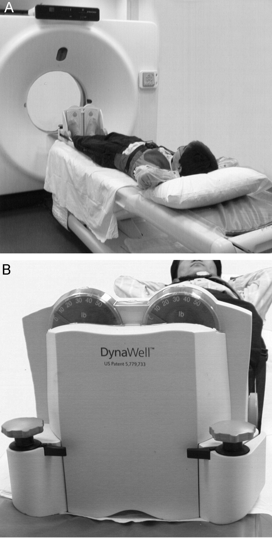
- Fig 1.
Patient in position during axial compression.
A and B, Device consists of nonmagnetic harness/jacket with straps connected to a footplate. By tightening or loosening the adjustment knobs on the compression part, the load can be regulated and equally distributed to both legs. The applied load can be measured by using scales on the footplate.
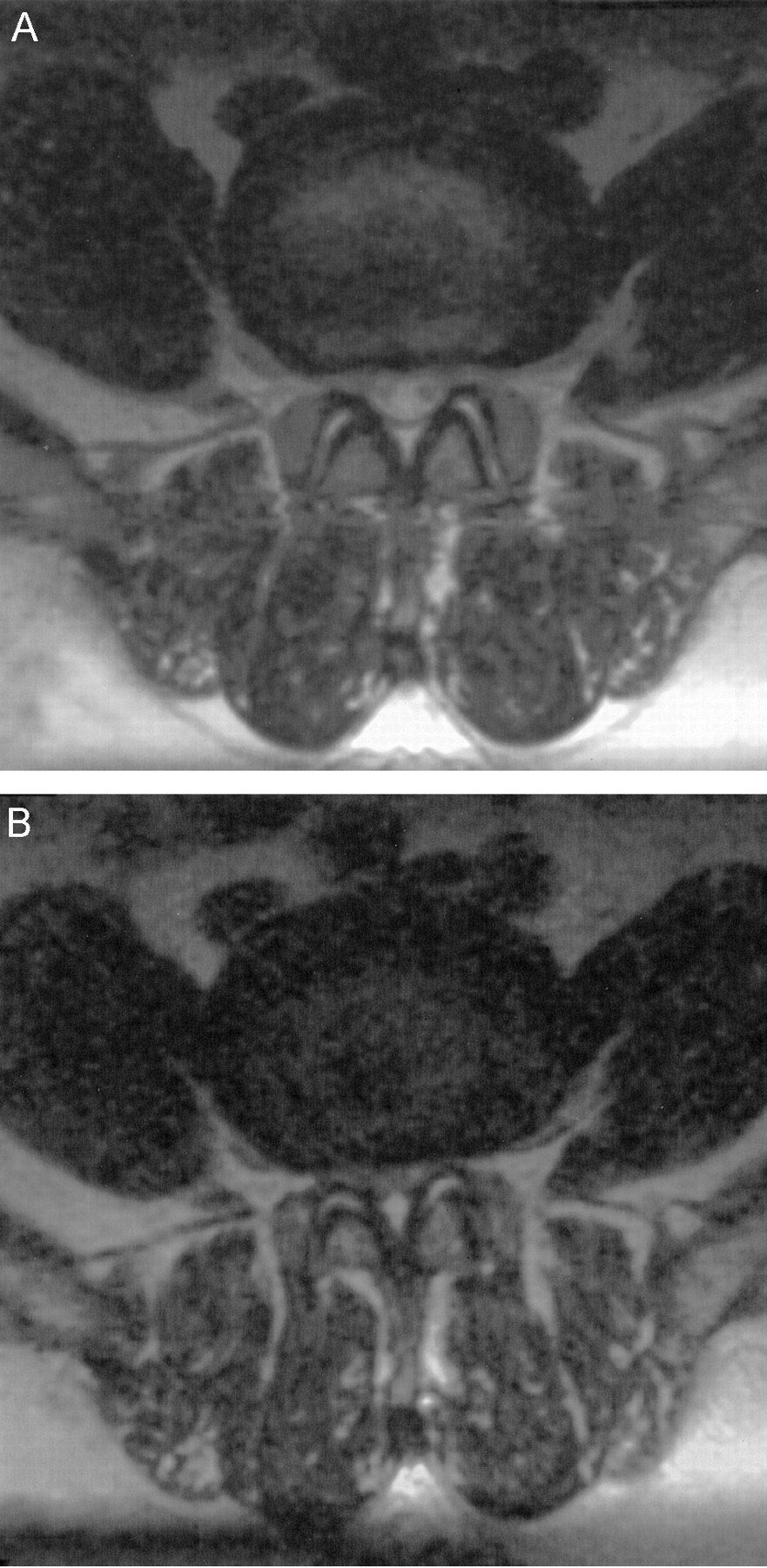
- Fig 2.
Routine and axially loaded MR images of a 56-year-old man with bilateral sciatica and claudication. All three neurosurgeons changed the treatment decision from conservative therapy to decompression surgery for this patient based on the additional information provided by the axially loaded MR images.
A, Routine T2-weighted image obtained at L4–L5 shows mild spinal stenosis.
B, Axially loaded T2-weighted image obtained at L4–L5 shows severe spinal stenosis, deformation of dural sac and bilateral lateral recesses, and prominence of the dorsal fat pad.
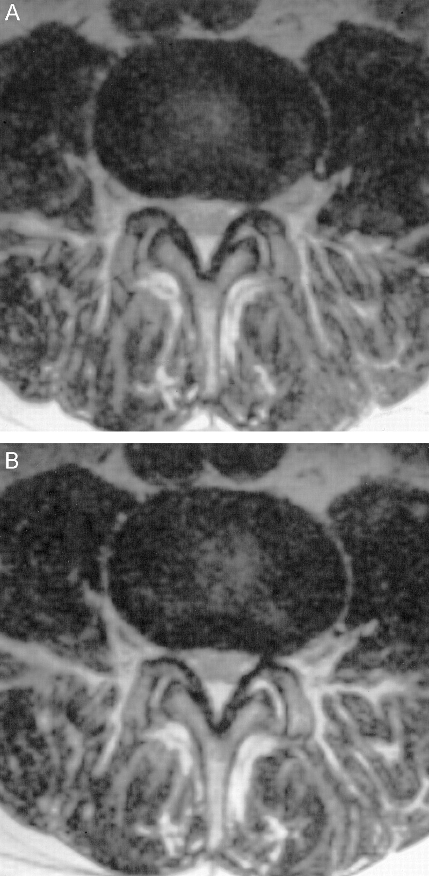
- Fig 3.
Routine and axially loaded MR images of a 75-year-old man with right sciatica. Decompression surgery had been performed 2 years before this study. All three surgeons changed the treatment decision from conservative therapy to decompression surgery based on the additional information provided by the axially loaded images.
A, Routine T2-weighted image obtained at L4–L5 shows deformation of dural sac and thickening of ligamentum flavum, especially on the left side.
B, Axially loaded T2-weighted image obtained at L4–L5 shows a right synovial cyst (arrow) that was not shown by the routine MR image. Prominent deformation of the dural sac and thickening of the ligamentum flavum can be seen.
- Fig 4.
Routine and axially loaded MR images of a 54-year-old man with right sciatica and claudication. None of the three neurosurgeons changed the treatment decision based on the information provided by the axially loaded images.
A, Routine T2-weighted image obtained at L4–L5 shows mild spinal stenosis and bilateral foraminal stenosis.
B, Axially loaded T2-weighted image obtained at L4–L5 shows minimal accentuation of spinal stenosis.


