Advertisement

Main menu

  • Home
  • Content
    • Current Issue
    • Accepted Manuscripts
    • Article Preview
    • Past Issue Archive
    • Video Articles
    • AJNR Case Collection
    • Case of the Week Archive
    • Case of the Month Archive
    • Classic Case Archive
  • Special Collections
    • Advancing NeuroMRI with High-Relaxivity Contrast Agents
    • AJNR Awards
    • Low-Field MRI
    • Alzheimer Disease
    • ASNR Foundation Special Collection
    • Photon-Counting CT
    • View All
  • Multimedia
    • AJNR Podcasts
    • AJNR SCANtastic
    • Trainee Corner
    • MRI Safety Corner
    • Imaging Protocols
  • For Authors
    • Submit a Manuscript
    • Submit a Video Article
    • Submit an eLetter to the Editor/Response
    • Manuscript Submission Guidelines
    • Statistical Tips
    • Fast Publishing of Accepted Manuscripts
    • Graphical Abstract Preparation
    • Imaging Protocol Submission
    • Author Policies
  • About Us
    • About AJNR
    • Editorial Board
    • Editorial Board Alumni
  • More
    • Become a Reviewer/Academy of Reviewers
    • Subscribers
    • Permissions
    • Alerts
    • Feedback
    • Advertisers
    • ASNR Home

User menu

  • Alerts
  • Log in

Search

  • Advanced search
American Journal of Neuroradiology

American Journal of Neuroradiology

ASHNR American Society of Functional Neuroradiology ASHNR American Society of Pediatric Neuroradiology ASSR
  • Alerts
  • Log in

Advanced Search

  • Home
  • Content
    • Current Issue
    • Accepted Manuscripts
    • Article Preview
    • Past Issue Archive
    • Video Articles
    • AJNR Case Collection
    • Case of the Week Archive
    • Case of the Month Archive
    • Classic Case Archive
  • Special Collections
    • Advancing NeuroMRI with High-Relaxivity Contrast Agents
    • AJNR Awards
    • Low-Field MRI
    • Alzheimer Disease
    • ASNR Foundation Special Collection
    • Photon-Counting CT
    • View All
  • Multimedia
    • AJNR Podcasts
    • AJNR SCANtastic
    • Trainee Corner
    • MRI Safety Corner
    • Imaging Protocols
  • For Authors
    • Submit a Manuscript
    • Submit a Video Article
    • Submit an eLetter to the Editor/Response
    • Manuscript Submission Guidelines
    • Statistical Tips
    • Fast Publishing of Accepted Manuscripts
    • Graphical Abstract Preparation
    • Imaging Protocol Submission
    • Author Policies
  • About Us
    • About AJNR
    • Editorial Board
    • Editorial Board Alumni
  • More
    • Become a Reviewer/Academy of Reviewers
    • Subscribers
    • Permissions
    • Alerts
    • Feedback
    • Advertisers
    • ASNR Home
  • Follow AJNR on Twitter
  • Visit AJNR on Facebook
  • Follow AJNR on Instagram
  • Join AJNR on LinkedIn
  • RSS Feeds

AJNR Awards, New Junior Editors, and more. Read the latest AJNR updates


Improved Turnaround Times | Median time to first decision: 12 days

Research ArticleBrain

Acute Methotrexate Neurotoxicity: Findings on Diffusion-Weighted Imaging and Correlation with Clinical Outcome

Nancy Rollins, Naomi Winick, Robert Bash and Timothy Booth
American Journal of Neuroradiology November 2004, 25 (10) 1688-1695;
Nancy Rollins
  • Find this author on Google Scholar
  • Find this author on PubMed
  • Search for this author on this site
Naomi Winick
  • Find this author on Google Scholar
  • Find this author on PubMed
  • Search for this author on this site
Robert Bash
  • Find this author on Google Scholar
  • Find this author on PubMed
  • Search for this author on this site
Timothy Booth
  • Find this author on Google Scholar
  • Find this author on PubMed
  • Search for this author on this site
  • Article
  • Figures & Data
  • Info & Metrics
  • Responses
  • References
  • PDF
Loading

Article Figures & Data

Figures

  • Tables
  • Fig 1.
    • Download figure
    • Open in new tab
    • Download powerpoint
    Fig 1.

    Patient 1 presented with nonfluent aphasia and bilateral upper-extremity weakness.

    A, Axial DW image is limited by motion artifact. Areas of restricted diffusion in the centrum semiovale account for the arm weakness. Nonfluent aphasia may be subcortical.

    B, Axial FLAIR image obtained 39 months later shows minimal abnormal T2 signal intensity consistent with demyelination. There are no residual neurologic deficits.
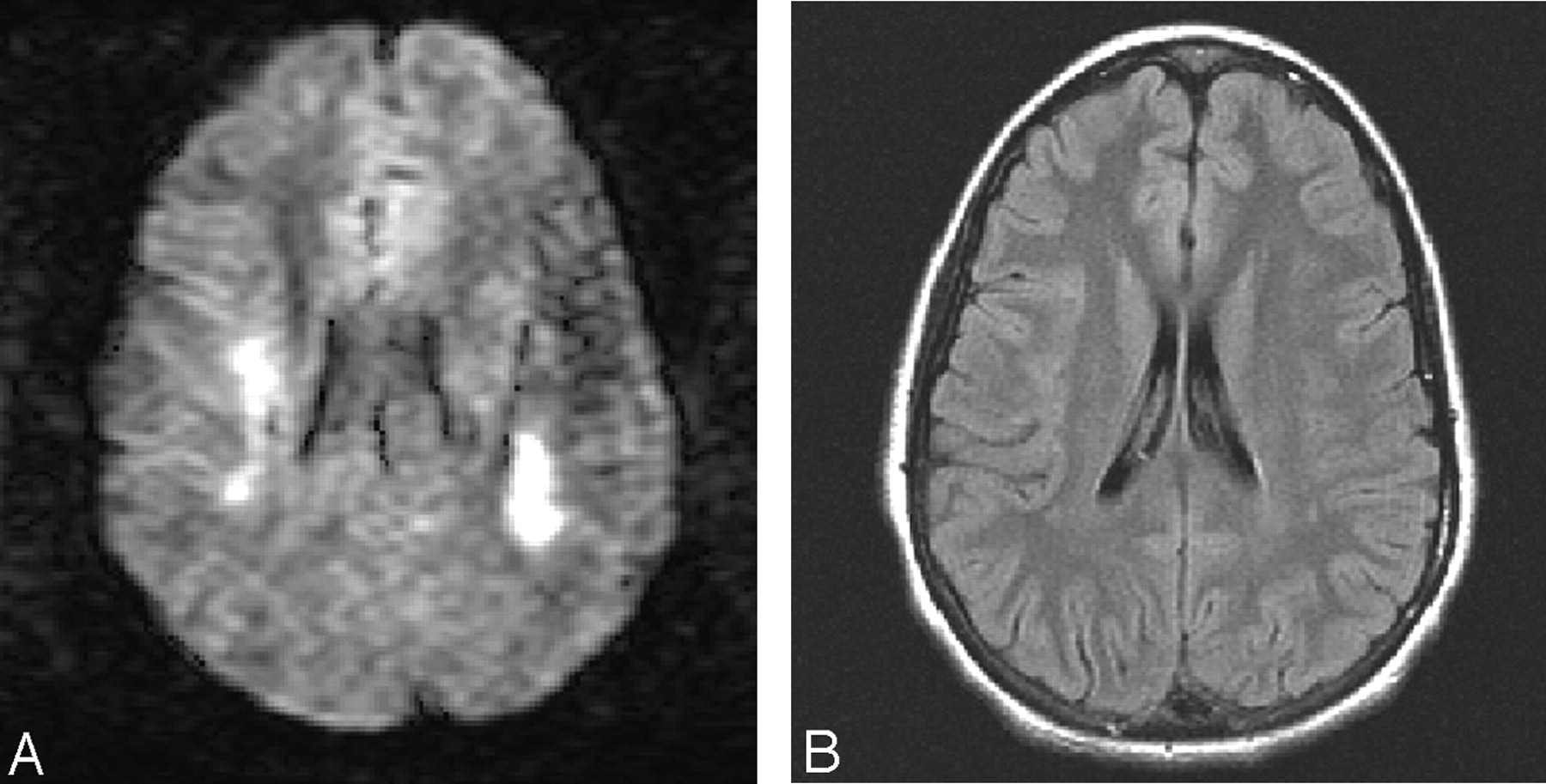
  • Fig 2.
    • Download figure
    • Open in new tab
    • Download powerpoint
    Fig 2.

    Patient 2 presented with nonfluent aphasia and left-sided hemiparesis and hemisensory loss.

    A, Axial ADC map shows asymmetric areas of restricted diffusion in the centrum semiovale. Image distortion is due to orthodontia. Right-sided lesion is correlated with left-sided hemiparesis, whereas the left-sided white matter lesion was not accompanied by a motor deficit.

    B, Axial FLAIR image obtained 8 weeks later shows large, confluent areas of presumed demyelination in the right centrum semiovale similar in size to the DW abnormality. Left cerebral white matter lesion that was clinically unapparent at presentation is smaller than the white matter lesion associated with neurologic deficit at presentation. Neurologic deficits had resolved.

  • Fig 3.
    • Download figure
    • Open in new tab
    • Download powerpoint
    Fig 3.

    Patient 3 had steroid-induced IDDM and presented during induction. Initial strokelike event was associated with aphasia and right hemiparesis and hemisensory loss, which resolved. Second event occurred 8 weeks later and was associated with aphasia, left hemiparesis, and focal seizure, all of which resolved.

    A and B, DW image (A) and ADC map (B) at initial presentation show large areas of restricted diffusion in the centrum semiovale (arrows). Bilateral diffusion abnormalities were associated with unilateral hemimotor-sensory deficit.

    C and D, Axial FLAIR images obtained 8 weeks later shows extensive presumed demyelination in deep white matter. Note absence of abnormal signal intensity in the right parietal region.

    E and F, Axial DW image (E) and ADC map (F) show restricted diffusion in the right parietal cortex and subcortical white matter. Diffusion abnormalities were correlated with left hemiparesis and left focal seizure. Hyperintensity in the left anterior centrum semiovale (arrow in E) is due to T2 shine-through; ADC map shows a corresponding area of hyperintensity (arrow in F).

    G, Axial FLAIR image obatined 28 months later when the patient was asymptomatic shows more-extensive white matter abnormalities. Note absence of gliosis in the right parietal region.
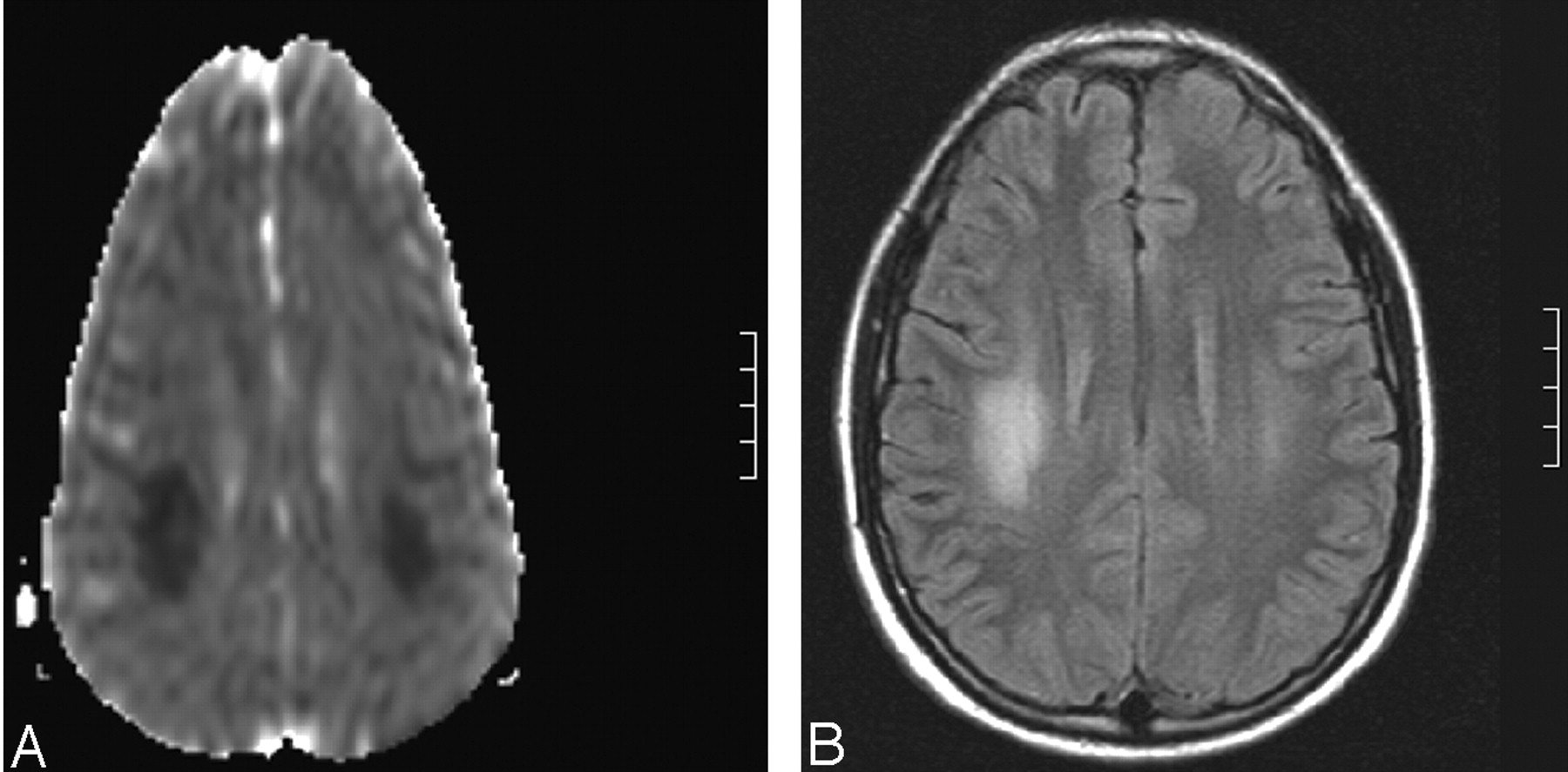
  • Fig 4.
    • Download figure
    • Open in new tab
    • Download powerpoint
    Fig 4.

    Patient 4 presented with nonfluent aphasia and alternating fluctuating hemiparesis and left facial droop.

    A and B, Axial DW images show asymmetric regions of restricted diffusion in the anterior centrum semiovale that do not explain the motor neurologic deficits.

    C, Follow-up MR image obtained 39 months later shows minimal scattered demyelination. The patient was neurologically intact.

  • Fig 5.
    • Download figure
    • Open in new tab
    • Download powerpoint
    Fig 5.

    Patient 5 presented with nonfluent aphasia, mild right facial weakness, right hemiparesis, and left-arm sensory deficit.

    A, DW image shows a small area of restricted diffusion with the left precentral region (arrow) that does not fully explain the neurologic deficits.

    B, Axial FLAIR image obtained 13 months later shows no signal-intensity abnormality in the left precentral region.

    C, Image shows small areas of presumed demyelination in the centrum semiovale. The patient was neurologically intact.

Tables

  • Figures
  • TABLE 1:
    • View popup
    TABLE 2:

    MR findings in acute MTX neurotoxicity

    Patient/AgeSymptomsSite of Restricted DiffusionInterval to Next MR StudyT2 Abnormalities on Next MR StudyClinical Follow-Up (mo)
    1/15/FAphasia, bilateral arm weakness R < LBilateral posterior centrum semiovale13 moMinimal involvement of R cenetrum semiovale25
    2/14/MAphasia, L upper-extremity weaknessBilateral posterior centrum semiovale6 wkAsymmetric involvement of centrum semiovale R > L9
    3/15/F
        InitialAphasia, R-sided weaknessBilateral posterior centrum semiovale8 wkBilateral centrum semiovale42
        RecurrenceRecurrence of aphasia, L hemiparesis, and focal seizure 8 wk laterR parietal cortex and white matter28 moProgressive involvement of centrum semiovale, normal cortex
    4/14/MAphasia, fluctuating bilateral hemiparesis, L facial droopBilateral asymmetric anterior centrum semiovale39 moMinimal scattered involvement of bilateral centrum semiovale49
    5/12/FAphasia, R-sided weakness, mild R facial droopL precentral subcortical white matter13 moMinimal scattered involvement of bilateral centrum semiovale23
PreviousNext
Back to top

In this issue

American Journal of Neuroradiology: 25 (10)
American Journal of Neuroradiology
Vol. 25, Issue 10
1 Nov 2004
  • Table of Contents
  • Index by author
Advertisement
Print
Download PDF
Email Article

Thank you for your interest in spreading the word on American Journal of Neuroradiology.

NOTE: We only request your email address so that the person you are recommending the page to knows that you wanted them to see it, and that it is not junk mail. We do not capture any email address.

Enter multiple addresses on separate lines or separate them with commas.
Acute Methotrexate Neurotoxicity: Findings on Diffusion-Weighted Imaging and Correlation with Clinical Outcome
(Your Name) has sent you a message from American Journal of Neuroradiology
(Your Name) thought you would like to see the American Journal of Neuroradiology web site.
CAPTCHA
This question is for testing whether or not you are a human visitor and to prevent automated spam submissions.
Cite this article
Nancy Rollins, Naomi Winick, Robert Bash, Timothy Booth
Acute Methotrexate Neurotoxicity: Findings on Diffusion-Weighted Imaging and Correlation with Clinical Outcome
American Journal of Neuroradiology Nov 2004, 25 (10) 1688-1695;

Citation Manager Formats

  • BibTeX
  • Bookends
  • EasyBib
  • EndNote (tagged)
  • EndNote 8 (xml)
  • Medlars
  • Mendeley
  • Papers
  • RefWorks Tagged
  • Ref Manager
  • RIS
  • Zotero
0 Responses
Respond to this article
Share
Bookmark this article
Acute Methotrexate Neurotoxicity: Findings on Diffusion-Weighted Imaging and Correlation with Clinical Outcome
Nancy Rollins, Naomi Winick, Robert Bash, Timothy Booth
American Journal of Neuroradiology Nov 2004, 25 (10) 1688-1695;
del.icio.us logo Twitter logo Facebook logo Mendeley logo
  • Tweet Widget
  • Facebook Like
  • Google Plus One

Jump to section

  • Article
    • Abstract
    • Methods
    • Results
    • Discussion
    • Conclusion
    • Acknowledgments
    • References
  • Figures & Data
  • Info & Metrics
  • Responses
  • References
  • PDF

Related Articles

  • No related articles found.
  • PubMed
  • Google Scholar

Cited By...

  • Alternating hemiparesis and orolingual apraxia as manifestations of methotrexate neurotoxicity in a paediatric case of acute lymphoblastic leukaemia
  • Methotrexate-Induced Neurotoxicity and Leukoencephalopathy in Childhood Acute Lymphoblastic Leukemia
  • "Dazed and diffused": making sense of diffusion abnormalities in neurologic pathologies
  • Ms Hassanzadeh, et al reply
  • Nonconvulsive Status Epilepticus and Leucoencephalopathy After High-Dose Methotrexate
  • Delayed leukoencephalopathy with stroke-like presentation in chemotherapy recipients
  • Seizures and epilepsy in oncological practice: causes, course, mechanisms and treatment
  • Crossref
  • Google Scholar

This article has not yet been cited by articles in journals that are participating in Crossref Cited-by Linking.

More in this TOC Section

  • Multimodal CT Provides Improved Performance for Lacunar Infarct Detection
  • Optimal MRI Sequence for Identifying Occlusion Location in Acute Stroke: Which Value of Time-Resolved Contrast-Enhanced MRA?
  • Evaluating the Effects of White Matter Multiple Sclerosis Lesions on the Volume Estimation of 6 Brain Tissue Segmentation Methods
Show more Brain

Similar Articles

Advertisement

Indexed Content

  • Current Issue
  • Accepted Manuscripts
  • Article Preview
  • Past Issues
  • Editorials
  • Editor's Choice
  • Fellows' Journal Club
  • Letters to the Editor
  • Video Articles

Cases

  • Case Collection
  • Archive - Case of the Week
  • Archive - Case of the Month
  • Archive - Classic Case

More from AJNR

  • Trainee Corner
  • Imaging Protocols
  • MRI Safety Corner
  • Book Reviews

Multimedia

  • AJNR Podcasts
  • AJNR Scantastics

Resources

  • Turnaround Time
  • Submit a Manuscript
  • Submit a Video Article
  • Submit an eLetter to the Editor/Response
  • Manuscript Submission Guidelines
  • Statistical Tips
  • Fast Publishing of Accepted Manuscripts
  • Graphical Abstract Preparation
  • Imaging Protocol Submission
  • Evidence-Based Medicine Level Guide
  • Publishing Checklists
  • Author Policies
  • Become a Reviewer/Academy of Reviewers
  • News and Updates

About Us

  • About AJNR
  • Editorial Board
  • Editorial Board Alumni
  • Alerts
  • Permissions
  • Not an AJNR Subscriber? Join Now
  • Advertise with Us
  • Librarian Resources
  • Feedback
  • Terms and Conditions
  • AJNR Editorial Board Alumni

American Society of Neuroradiology

  • Not an ASNR Member? Join Now

© 2025 by the American Society of Neuroradiology All rights, including for text and data mining, AI training, and similar technologies, are reserved.
Print ISSN: 0195-6108 Online ISSN: 1936-959X

Powered by HighWire