
Article Figures & Data
Figures
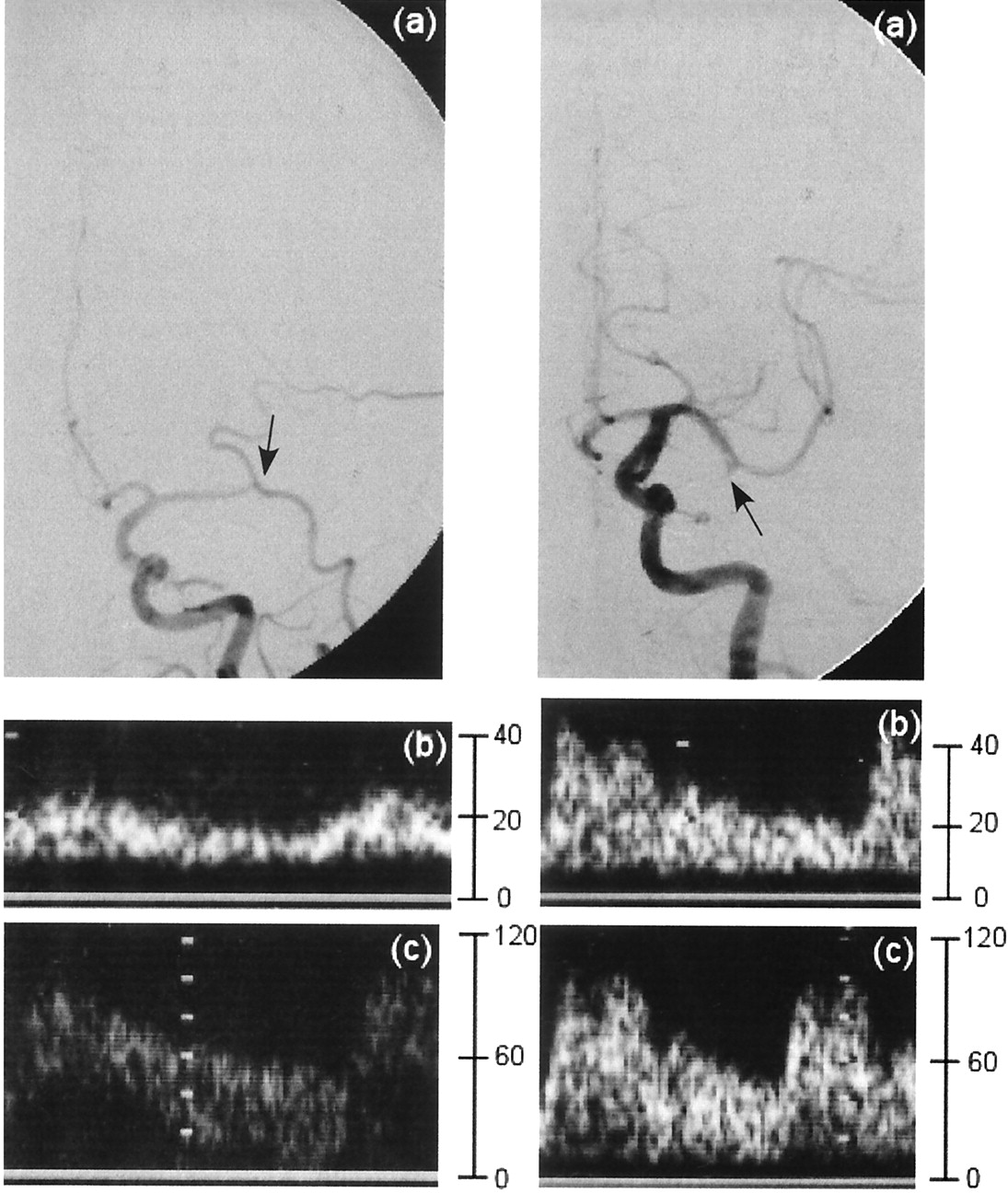
- Fig 1.
Left anteroposterior carotid angiograms in representative cases evaluated with cerebral angiography and TCCS. Y axis is represented blood flow velocity (cm/s). Left image shows occlusion of the horizontal portion of the left MCA (a). Occlusion site overlies the external carotid artery branch. Doppler waveforms of the left (b) and right (c) MCAs show EDVs of 14.5 and 49.9 cm/s, respectively. Right image shows occlusion of the branch in the left MCA (a). Doppler waveforms of the left (b) and right (c) MCAs show EDVs of 18.2 and 44.6 cm/s, respectively.
- Fig 2.
Scattergrams. Top, Mean EDVs (±2 SDs) for the control, MO, and MB groups are 40.5 ± 11.5, 12.2 ± 3.6, and 19.6 ± 4.8, respectively (P < .001, Scheffé test). Bottom, Mean end-diastolic ratios (±2 SDs) for the control, MO, and MB groups are 1.2 ± 0.1, 4.2 ± 1.5, and 1.8 ± 0.5, respectively (P < .001, Scheffé test).
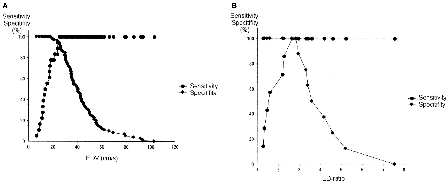
- Fig 3.
Sensitivity-specificity curves.
A, Predicting MO or MB by EDV. Optimal threshold value for EDV is 25 cm/s.
B, Differentiating MO from MB by the end-diastolic ratio. Optimal threshold value for the ratio is 2.7.
- Fig 4.
Algorithm for diagnosing MO and MB by using the EDV and end-diastolic ratio on TCCS.


