
Article Figures & Data
Figures
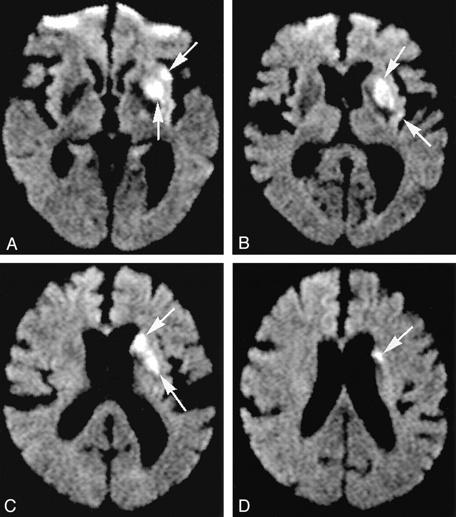
- Fig 1.
54-year-old man with right hemiparesis.
A–D, Diffusion-weighted images (b = 1000 s/mm2) illustrate volume of abnormality. Three-dimensional volume of abnormality was found by summing the areas of abnormality on each section (arrows) and multiplying by section thickness. The same approach was used to compute volumes of MR perfusion abnormality.
- Fig 2.
Scatterplot compares extents of CBF abnormality on single-slice CT perfusion scans with their corresponding extent of CBF abnormality on multislice MR perfusion images. Kendall τ b correlation coefficient for comparison of extent of CT-CBF with extent of MR-CBF was 0.60 (P = .003).
- Fig 3.
90-year-old woman with acute right hemiparesis and aphasia 2 hours before imaging.
A, CT perfusion CBV map shows well-circumscribed area of very low CBV (arrows).
B, MR perfusion CBV map. Correlation of volume of MR-CBV abnormality with area of CT-CBV abnormality approached statistical significance in our study.
C, CT perfusion CBF map shows a larger abnormality than seen on the CBV map. CBF in the range of 0–10 mL/100 g/min is displayed as blue (arrows).
D, MR perfusion CBF map with areas of low relative CBF (< 50%) displayed as blue (arrows). Good correlation was found in our study between extents of 2D CT-CBF abnormality and 3D MR-CBF abnormality.
E, CT perfusion MTT map shows the largest extent of abnormality. Values of MTT greater than 6 seconds are displayed as red (arrows).
F, MR MTT map. Good correlation was found in our study between 2D CT-MTT abnormalities and 3D MR-MTT abnormalities (arrows).
G, Diffusion-weighted MR image (b = 1000 s/mm2) obtained 4 hours after symptom onset through the basal ganglia shows abnormality in the left frontal lobe (arrows). Good correlation was found in our study between extent of CT-CBV abnormality and extent of diffusion-weighted abnormality. Good correlation was also found with extents of MR-CBV and MR-MTT abnormalities.


Tables
Statistical correlation of extents of CT and MR perfusion abnormalities with extent of initial abnormality on diffusion-weighted images
Perfusion Parameter Correlation Coefficient* P Value CT-CBV 0.47 .02 CT-CBF 0.38 .05 CT-MTT 0.32 .11 MR-CBV 0.69 .001 MR-CBF 0.41 .04 MR-MTT 0.67 .002 * Kendall τ b correlation coefficients.
In this issue


Jump to section
Related Articles
Cited By...
- Rescan Time Delays in Ischemic Stroke Imaging: A Retrospective Observation and Analysis of Causes and Clinical Impact
- Early Basal Ganglia Hyperperfusion on CT Perfusion in Acute Ischemic Stroke: A Marker of Irreversible Damage?
- Comparison of Computed Tomographic and Magnetic Resonance Perfusion Measurements in Acute Ischemic Stroke: Back-to-Back Quantitative Analysis
- Diagnostic value of CT perfusion imaging for parotid neoplasms
- CT perfusion-guided patient selection for endovascular recanalization in acute ischemic stroke: a multicenter study
- Perfusion Deficits Detected by Arterial Spin-Labeling in Patients with TIA with Negative Diffusion and Vascular Imaging
- Dramatically Reducing Imaging-to-Recanalization Time in Acute Ischemic Stroke: Making Choices
- Cerebral Blood Flow Is the Optimal CT Perfusion Parameter for Assessing Infarct Core
- Perfusion CT in Acute Ischemic Stroke: A Qualitative and Quantitative Comparison of Deconvolution and Maximum Slope Approach
- Evaluation of CT Perfusion in the Setting of Cerebral Ischemia: Patterns and Pitfalls
- A Comparison of Computed Tomography Perfusion-Guided and Time-Guided Endovascular Treatments for Patients With Acute Ischemic Stroke
- Identification of Infarct Core and Penumbra in Acute Stroke Using CT Perfusion Source Images
- Recommendations for Imaging of Acute Ischemic Stroke: A Scientific Statement From the American Heart Association
- Perfusion CT in Patients with Acute Ischemic Stroke Treated with Intra-Arterial Thrombolysis: Predictive Value of Infarct Core Size on Clinical Outcome
- Management of Stroke in Infants and Children: A Scientific Statement From a Special Writing Group of the American Heart Association Stroke Council and the Council on Cardiovascular Disease in the Young
- Reproducibility of Quantitative CT Brain Perfusion Measurements in Patients with Symptomatic Unilateral Carotid Artery Stenosis
- Identification of Penumbra and Infarct in Acute Ischemic Stroke Using Computed Tomography Perfusion-Derived Blood Flow and Blood Volume Measurements
- Perfusion-CT Assessment of Infarct Core and Penumbra: Receiver Operating Characteristic Curve Analysis in 130 Patients Suspected of Acute Hemispheric Stroke
- Visual evaluation of perfusion computed tomography in acute stroke accurately estimates infarct volume and tissue viability
- Comparative Overview of Brain Perfusion Imaging Techniques
- Comparison of Perfusion Computed Tomography and Computed Tomography Angiography Source Images With Perfusion-Weighted Imaging and Diffusion-Weighted Imaging in Patients With Acute Stroke of Less Than 6 Hours' Duration