
Article Figures & Data
Figures
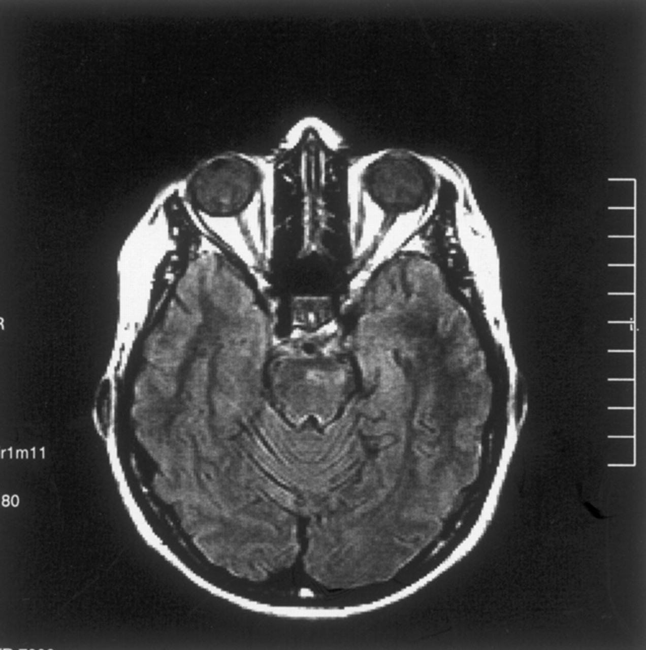
- Fig 1.
Images obtained during the acute stage.
A, Fluid-attenuated inversion recovery image reveals a left-sided hyperintense lesion in the pons.
B, T1-weighted image reveals a focal high signal intensity change, suggesting hemorrhage or presence of myelin breakdown products.
C, Contrast-enhanced T1-weighted image shows enhancement in the corresponding region.
D, Diffusion-weighted (b = 1000 s/mm2) image reveals high signal intensity in the lesion, suggesting restricted diffusion (see E).
E, ADC map (same section as that shown in D) reveals high signal intensity and a high ADC value (1.22 × 10⁻³ mm2/s), compared with the normal side of the pons (0.86 × 10⁻³ mm2/s) and compared with the temporal white matter (0.80 × 10⁻³ mm2/s). This is consistent with presence of increased diffusion, hence vasogenic edema.
F, Proton MR spectroscopy (1500/40) reveals that there is no lactic acid peak, excluding acute infarct. Major peaks are normal.
- Fig 2.
Images obtained at the 16-month follow-up examination.
A, T2-weighted image reveals a small remaining focus of gliosis.
B, Diffusion-weighted (b = 1000 s/mm2) image is normal appearing.
C, ADC map (same section as that shown in B) reveals high signal intensity and a high ADC value (1.20 × 10⁻³ mm2/s), consistent with tissue disintegration due to gliosis.
- Fig 3.
Fluid-attenuated inversion recovery image obtained at the 2-year follow-up examination reveals focal high signal intensity secondary to presumed gliosis.


