
Article Figures & Data
Figures
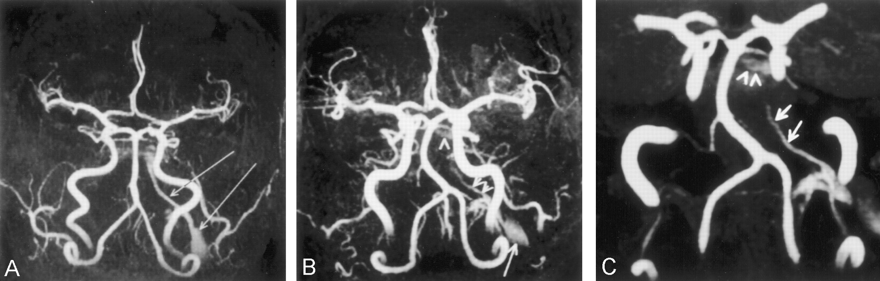
- Fig 1.
Abnormal vascular signal intensity was seen in the left jugular bulb, inferior petrosal sinus, and cavernous sinus.
A, 3D time-of-flight cranial arterial MR angiogram of the first patient shows high signal intensity in the left cavernous sinus together with the left inferior petrosal sinus and internal jugular vein at the level of the jugular bulb (arrows).
B, MR angiogram of the second patient shows abnormal flow-related enhancement in the left inferior petrosal sinus because of a fistula (arrowhead, cavernous sinus; double arrowheads, inferior petrosal sinus; arrow, jugular bulb).
C, Subvolumetric reconstructed maximum intensity projection image of the third patient shows more details of the inferior petrosal and cavernous sinuses (arrowheads, cavernous sinus; arrows, inferior petrosal sinus).
- Fig 2.
When the flow signal intensity in the inferior petrosal sinus was saturated proximally, the flow signal intensity in the left inferior petrosal sinus was saturated whereas the flow signal intensity in the right inferior petrosal sinus was not.
A, Section (lines) and saturation band (rectangle) position.
B, Axial view fluid-attenuated fast low angle shot 2D image. Right transverse sinus and right jugular bulb (arrowheads) and right inferior petrosal sinus (arrow) show bright signal intensity. No vascular signal intensity can be seen in the left inferior petrosal sinus and internal carotid arteries.
- Fig 3.
When the flow signal intensity in the distal inferior petrosal sinus was saturated, the flow signal intensity in the right inferior petrosal sinus was saturated whereas the flow signal intensity in the left inferior petrosal sinus was not.
A, Section (lines) and saturation band (rectangle) position.
B, Axial view fast low angle shot 2D image. Bilateral internal carotid arteries (arrowheads) and left inferior petrosal sinus (arrow) show bright signal intensity.
- Fig 4.
Axial T2-weighted MR image shows normal flow void in the right internal jugular vein (arrows), whereas flow-related enhancement can be seen in the left internal jugular (arrow) vein due to sluggish flow.
- Fig 5.
Contrast-enhanced MR angiograms show left brachiocephalic vein stenosis (arrow).
A, Before subtraction of the arterial signals (in the third patient).
B, After subtraction of the arterial signals (in the first patient).


In this issue


Jump to section
Related Articles
Cited By...
- Normal Flow Signal of the Pterygoid Plexus on 3T MRA in Patients without DAVF of the Cavernous Sinus
- Evaluation of Dural Arteriovenous Fistulas with 4D Contrast-Enhanced MR Angiography at 3T
- Sonographic Findings of Physiologic Left Brachiocephalic Vein Compression in a Case Initially Misdiagnosed as a Left Internal Jugular Vein Thrombus
- Detection of intracranial venous reflux in patients of transient global amnesia