Advertisement

Main menu

  • Home
  • Content
    • Current Issue
    • Accepted Manuscripts
    • Article Preview
    • Past Issue Archive
    • Video Articles
    • AJNR Case Collection
    • Case of the Week Archive
    • Case of the Month Archive
    • Classic Case Archive
  • Special Collections
    • AJNR Awards
    • Low-Field MRI
    • Alzheimer Disease
    • ASNR Foundation Special Collection
    • Photon-Counting CT
    • View All
  • Multimedia
    • AJNR Podcasts
    • AJNR SCANtastic
    • Trainee Corner
    • MRI Safety Corner
    • Imaging Protocols
  • For Authors
    • Submit a Manuscript
    • Submit a Video Article
    • Submit an eLetter to the Editor/Response
    • Manuscript Submission Guidelines
    • Statistical Tips
    • Fast Publishing of Accepted Manuscripts
    • Graphical Abstract Preparation
    • Imaging Protocol Submission
    • Author Policies
  • About Us
    • About AJNR
    • Editorial Board
    • Editorial Board Alumni
  • More
    • Become a Reviewer/Academy of Reviewers
    • Subscribers
    • Permissions
    • Alerts
    • Feedback
    • Advertisers
    • ASNR Home

User menu

  • Alerts
  • Log in

Search

  • Advanced search
American Journal of Neuroradiology

American Journal of Neuroradiology

ASHNR American Society of Functional Neuroradiology ASHNR American Society of Pediatric Neuroradiology ASSR
  • Alerts
  • Log in

Advanced Search

  • Home
  • Content
    • Current Issue
    • Accepted Manuscripts
    • Article Preview
    • Past Issue Archive
    • Video Articles
    • AJNR Case Collection
    • Case of the Week Archive
    • Case of the Month Archive
    • Classic Case Archive
  • Special Collections
    • AJNR Awards
    • Low-Field MRI
    • Alzheimer Disease
    • ASNR Foundation Special Collection
    • Photon-Counting CT
    • View All
  • Multimedia
    • AJNR Podcasts
    • AJNR SCANtastic
    • Trainee Corner
    • MRI Safety Corner
    • Imaging Protocols
  • For Authors
    • Submit a Manuscript
    • Submit a Video Article
    • Submit an eLetter to the Editor/Response
    • Manuscript Submission Guidelines
    • Statistical Tips
    • Fast Publishing of Accepted Manuscripts
    • Graphical Abstract Preparation
    • Imaging Protocol Submission
    • Author Policies
  • About Us
    • About AJNR
    • Editorial Board
    • Editorial Board Alumni
  • More
    • Become a Reviewer/Academy of Reviewers
    • Subscribers
    • Permissions
    • Alerts
    • Feedback
    • Advertisers
    • ASNR Home
  • Follow AJNR on Twitter
  • Visit AJNR on Facebook
  • Follow AJNR on Instagram
  • Join AJNR on LinkedIn
  • RSS Feeds

AJNR Awards, New Junior Editors, and more. Read the latest AJNR updates

OtherSpine Imaging and Spine Image-Guided Interventions

Spontaneous Resolution of an Intraspinal Synovial Cyst

Paul G. Swartz and F. Reed Murtagh
American Journal of Neuroradiology June 2003, 24 (6) 1261-1263;
Paul G. Swartz
  • Find this author on Google Scholar
  • Find this author on PubMed
  • Search for this author on this site
F. Reed Murtagh
  • Find this author on Google Scholar
  • Find this author on PubMed
  • Search for this author on this site
  • Article
  • Figures & Data
  • Info & Metrics
  • Responses
  • References
  • PDF
Loading

Abstract

Summary: We report a case of lumbar facet joint synovial cyst at L5-S1 with clinically significant radicular pain diagnosed by means of MR imaging. This cyst spontaneously resolved, as determined on follow-up MR images obtained 18 months later. The patient’s clinical symptoms substantially improved after conservative medical treatment.

Approximately 180 case reports of synovial cysts of the vertebral facet joints have been described in the literature. Of those, only two refer to spontaneously resolving intraspinal synovial cysts (1, 2).

Synovial cysts most often develop secondary to substantially degenerated facet joints and to both acute and chronic trauma. Synovial cysts of the lumbar vertebral facet joints may serve as sources of nerve root compression. The most common symptoms are painful radiculopathy (85%), neurogenic single-root or multiroot claudication (44%), sensory loss (43%), and motor weakness (27%) (3).

Case Report

A 58-year-old woman presented with a medical history notable for only minor daily joint stiffness of the hip and knee and pain that was relieved by nonsteroidal anti-inflammatory drugs (NSAIDs). Her present illness started insidiously 1 month before the first MR examination, with sciatic-like posterior pain and numbness and tingling in the right leg. These symptoms were not associated with any known injury. Physical examination showed unimpaired strength of the lower extremity, normal and symmetrical deep tendon reflexes, and normal muscle tone in both lower extremities. Her initial response to NSAIDs and rest was minimal.

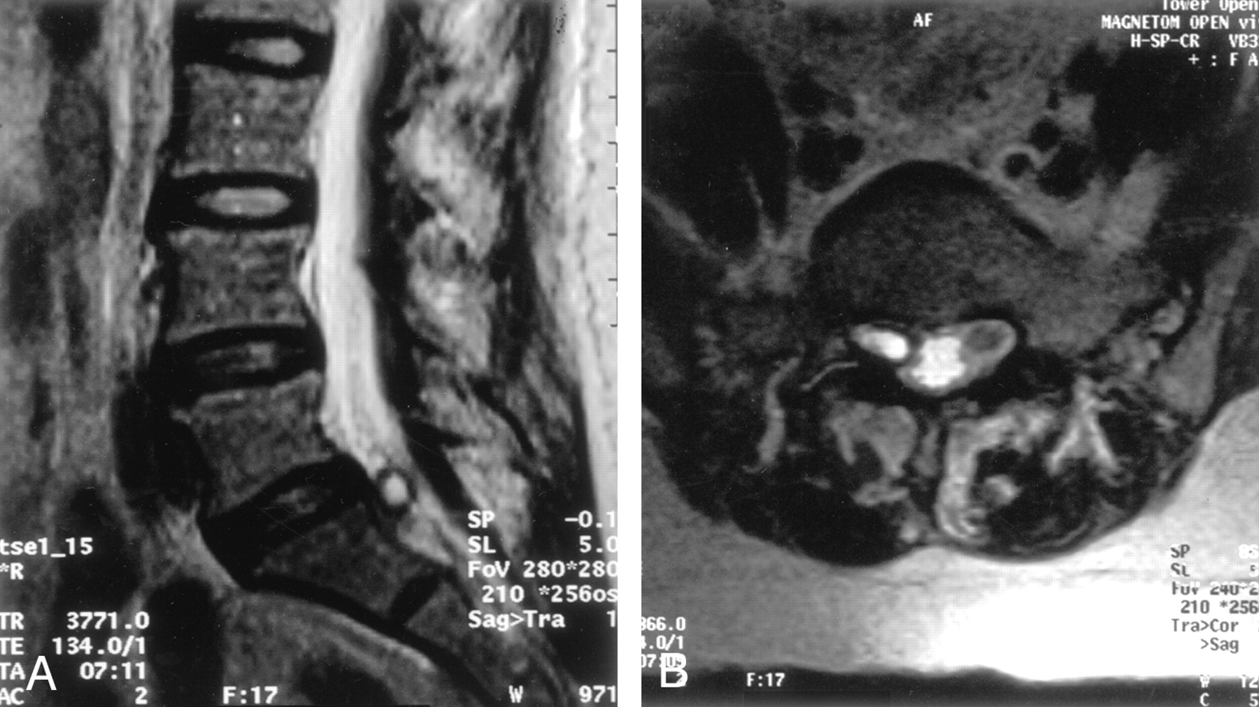
MR imaging of the lumbar spine performed 1 month after the onset of symptoms demonstrated facet osteoarthropathy with marked overgrowth of the facets but no remarkable canal stenosis at L5-S1. Grade I degenerative spondylolisthesis and mild bulging of the disk at L5-S1 were present, but these were present without notable canal stenosis. A synovial cyst was identified; this arose off the facet complex on the right at L5-S1 (Fig. 1) extending into the neural foramen and lateral recess of S1, causing compression at the right L5 and S1 nerve root. No disk disease was noted from T12 through L4-L5, and only mild facet osteoarthropathy was observed at L4-L5. with mild canal stenosis dorsolaterally.

Fig 1.
  • Download figure
  • Open in new tab
  • Download powerpoint
Fig 1.

T2-weighted fast spin-echo MR images.

A, Sagittal image (TR/TE, 3771/134) demonstrates a synovial cyst extending into the spinal canal at the L5-S1 level.

B, Axial image (5866/134) through the L5-S1 level demonstrates the synovial cyst arising from the facet complex on the right, extending into the lateral recess of S1 on the right side, and causing compression of the S1 nerve root.

Conservative medical treatment was initiated and consisted of physical therapy (mainly stretching), NSAID therapy, and orthopedic bracing. The patient reported gradual but substantial improvement of both pain and sensory symptoms over the next 12 months. Follow-up MR imaging was performed 18 months later. The images demonstrated continued, mild bulging at L5-S1 and facet osteoarthropathy with marked overgrowth of facets without canal stenosis at L5-S1. The previously noted synovial cyst was no longer present (Fig 2).

Fig 2.
  • Download figure
  • Open in new tab
  • Download powerpoint
Fig 2.

Follow-up T2-weighted fast spin-echo MR images demonstrate complete resolution of the previously noted synovial cyst. These follow-up images do not completely match the angulation or detail of the images in Figure 1. The follow-up examination was performed by using a low-field-strength (0.35-T) open magnet, and the images are less than optimal. The patient did not desire to enter a closed high-field-strength machine for additional studies.

A, Sagittal image (3500/120) through the lumbar spine.

B, Axial image through the L5-S1 lumbar spine (-10/15).

Discussion

Synovial cysts are most often described in extraspinal joints such as the knee, hip, and elbow. They are infrequently identified adjacent to the facet joints, and they account for an incidence of 0.002–0.8% depending on the institution and the diagnostic technique (2, 4). Among synovial cysts located in facet joints, 68.4% are found at L4-L5; 21.1%, at L5-S1; 5.2%, at L1-L2; and 5.2%, at L2-L3 (5). The incidence and location of synovial cysts at our institution is similar to that of the referenced studies. This was our first documented case of a spontaneously resolving synovial cyst of a facet joint.

Intraspinal synovial cysts generally result from facet joint arthrosis and degeneration that causes a capsular lesion and eventual herniation of the synovial membrane. Alternatively, synovial cysts can appear as complications of lumbar trauma, both a single major trauma and repeated microtraumatic events. Specifically, in this patient, the correlation between the synovial cyst of the facet joint and spondylolisthesis at the same lumbar level support the theory of functional instability (2) and repeated microtraumatic events leading to synovial cyst formation. The presence of spondylolisthesis in 33% of cases and facet joint hypermobilization in 60% of patients support the idea that hypermobility is an important etiologic factor in development of synovial cysts (6).

Explaining the spontaneous resolution of a lumbar synovial cyst is complicated. In this patient, who underwent conservative treatment with NSAIDs and mobilization, the synovial cyst resolved probably because of decreased inflammatory fluid production and a decrease in microtraumatic events. Another plausible explanation is that the physical therapy provided enough of a mechanical stimulus to cause the cyst to extrude its contents, followed by reabsorption of the cyst wall.

The management of symptomatic synovial cysts of the lumbar spine is controversial. No standards or guidelines have been established to lead the clinician toward one therapeutic technique or another. In this case, a conservative approach to treatment was implemented, starting with physical therapy (mainly stretching with and then without resistance to relieve perceived pain and tightness by patient), NSAID administration, and bracing. Clinical improvement noted by the patient and the physician encouraged the continued use of a conservative approach, which ultimately resulted in substantial improvement of the patient’s symptoms. This change was associated with complete resolution of the synovial cyst, as shown on the follow-up MR images. One explanation for incomplete symptomatic improvement even after spontaneous resolution of the intraspinal synovial cyst was the preexisting facet osteoarthropathy and the marked overgrowth of the facets at the same level as the previously noted synovial cyst. In addition, the grade I degenerative spondylolisthesis and the mild bulging of the L5-S1 disk probably contributed to the mild residual symptoms.

If conservative management had failed in this patient, other more invasive therapeutic interventions remained available; these included percutaneous cyst aspiration under CT control, methylprednisolone injections into the cyst under CT guidance, and cystectomy via a laminectomy or microneurosurgical flavectomy.

Conclusion

Although rare, synovial cyst formation and subsequent resolution in the clinical context of degenerated facet joints with or without acute or chronic trauma must be considered. Resolution of a lumbar synovial cyst at follow-up imaging can generally be attributed to decreased inflammatory fluid production and decreased microtraumatic events or mechanical irritation of the cyst wall that results in extrusion of cyst contents and resolution of the cyst capsule. Although no standard of care for treatment of a lumbar synovial cyst has been established, the general trend is for conservative management first followed by invasive treatment if conservative therapy fails.

References

  1. ↵
    Maezawa Y, Baba H, Uchida K, Furusawa N, Kubota C, Yoshizawa K. Spontaneous remission of a solitary intraspinal synovial cyst of the lumbar spine. Eur Spine J 2000;9:85–87
    PubMed
  2. ↵
    Mercader J, Gomez J, Cardenal C. Intraspinal synovial cyst: diagnosis by CT-follow-up and spontaneous remission. Neuroradiology 1985;27:346–348
    PubMed
  3. ↵
    Lyons MK, Atkinson JL, Wharen RE, Deen HG, Zimmerman RS, Lemens SM. Surgical evaluation and management of lumbar synovial cysts: the Mayo Clinic experience. J Neurosurg 2000;93:53–57
  4. ↵
    Trummer M, Flaschka G, Tillich M, Homann CN, Unger F, Eustacchio S. Diagnosis and surgical management of intraspinal synovial cysts: report of 19 cases. J Neurol Neurosurg Psychiatry 2001;70:74–77
    Abstract/FREE Full Text
  5. ↵
    Hsu KY, Zucherman JF, Shea WJ, Jeffrey RA. Lumbar intraspinal synovial and ganglion cysts (facet cysts): ten-year experience in evaluation and treatment. Spine 1995;20:80–89
    PubMed
  6. ↵
    Freidberg SR, Fellow T, Thomas CB, et al. Experience with symptomatic spinal epidural cysts. Neurosurgery 1994;34:989–993
    CrossRefPubMed
  • Received November 19, 2001.
  • Accepted after revision October 21, 2002.
  • Accepted after revision October 21, 2002.
  • Copyright © American Society of Neuroradiology
View Abstract
PreviousNext
Back to top

In this issue

American Journal of Neuroradiology: 24 (6)
American Journal of Neuroradiology
Vol. 24, Issue 6
1 Jun 2003
  • Table of Contents
  • Index by author
Advertisement
Print
Download PDF
Email Article

Thank you for your interest in spreading the word on American Journal of Neuroradiology.

NOTE: We only request your email address so that the person you are recommending the page to knows that you wanted them to see it, and that it is not junk mail. We do not capture any email address.

Enter multiple addresses on separate lines or separate them with commas.
Spontaneous Resolution of an Intraspinal Synovial Cyst
(Your Name) has sent you a message from American Journal of Neuroradiology
(Your Name) thought you would like to see the American Journal of Neuroradiology web site.
CAPTCHA
This question is for testing whether or not you are a human visitor and to prevent automated spam submissions.
Cite this article
Paul G. Swartz, F. Reed Murtagh
Spontaneous Resolution of an Intraspinal Synovial Cyst
American Journal of Neuroradiology Jun 2003, 24 (6) 1261-1263;

Citation Manager Formats

  • BibTeX
  • Bookends
  • EasyBib
  • EndNote (tagged)
  • EndNote 8 (xml)
  • Medlars
  • Mendeley
  • Papers
  • RefWorks Tagged
  • Ref Manager
  • RIS
  • Zotero
0 Responses
Respond to this article
Share
Bookmark this article
Spontaneous Resolution of an Intraspinal Synovial Cyst
Paul G. Swartz, F. Reed Murtagh
American Journal of Neuroradiology Jun 2003, 24 (6) 1261-1263;
del.icio.us logo Twitter logo Facebook logo Mendeley logo
  • Tweet Widget
  • Facebook Like
  • Google Plus One

Jump to section

  • Article
    • Abstract
    • Case Report
    • Discussion
    • Conclusion
    • References
  • Figures & Data
  • Info & Metrics
  • Responses
  • References
  • PDF

Related Articles

  • No related articles found.
  • PubMed
  • Google Scholar

Cited By...

  • Is Hypoglossal Nerve Palsy Caused by Craniocervical Junction Degenerative Disease an Underrecognized Entity?
  • Crossref
  • Google Scholar

This article has not yet been cited by articles in journals that are participating in Crossref Cited-by Linking.

More in this TOC Section

  • Management Outcomes For VO Spine Biopsy
  • Characteristics of SIH Type I Culprit Lesions
  • Advanced Imaging of Type 2 Spinal CSF Leaks
Show more Spine Imaging and Spine Image-Guided Interventions

Similar Articles

Advertisement

Indexed Content

  • Current Issue
  • Accepted Manuscripts
  • Article Preview
  • Past Issues
  • Editorials
  • Editor's Choice
  • Fellows' Journal Club
  • Letters to the Editor
  • Video Articles

Cases

  • Case Collection
  • Archive - Case of the Week
  • Archive - Case of the Month
  • Archive - Classic Case

More from AJNR

  • Trainee Corner
  • Imaging Protocols
  • MRI Safety Corner
  • Book Reviews

Multimedia

  • AJNR Podcasts
  • AJNR Scantastics

Resources

  • Turnaround Time
  • Submit a Manuscript
  • Submit a Video Article
  • Submit an eLetter to the Editor/Response
  • Manuscript Submission Guidelines
  • Statistical Tips
  • Fast Publishing of Accepted Manuscripts
  • Graphical Abstract Preparation
  • Imaging Protocol Submission
  • Evidence-Based Medicine Level Guide
  • Publishing Checklists
  • Author Policies
  • Become a Reviewer/Academy of Reviewers
  • News and Updates

About Us

  • About AJNR
  • Editorial Board
  • Editorial Board Alumni
  • Alerts
  • Permissions
  • Not an AJNR Subscriber? Join Now
  • Advertise with Us
  • Librarian Resources
  • Feedback
  • Terms and Conditions
  • AJNR Editorial Board Alumni

American Society of Neuroradiology

  • Not an ASNR Member? Join Now

© 2025 by the American Society of Neuroradiology All rights, including for text and data mining, AI training, and similar technologies, are reserved.
Print ISSN: 0195-6108 Online ISSN: 1936-959X

Powered by HighWire