Advertisement

Main menu

  • Home
  • Content
    • Current Issue
    • Accepted Manuscripts
    • Article Preview
    • Past Issue Archive
    • Video Articles
    • AJNR Case Collection
    • Case of the Week Archive
    • Case of the Month Archive
    • Classic Case Archive
  • Special Collections
    • Advancing NeuroMRI with High-Relaxivity Contrast Agents
    • AJNR Awards
    • Low-Field MRI
    • Alzheimer Disease
    • ASNR Foundation Special Collection
    • Photon-Counting CT
    • View All
  • Multimedia
    • AJNR Podcasts
    • AJNR SCANtastic
    • Trainee Corner
    • MRI Safety Corner
    • Imaging Protocols
  • For Authors
    • Submit a Manuscript
    • Submit a Video Article
    • Submit an eLetter to the Editor/Response
    • Manuscript Submission Guidelines
    • Statistical Tips
    • Fast Publishing of Accepted Manuscripts
    • Graphical Abstract Preparation
    • Imaging Protocol Submission
    • Author Policies
  • About Us
    • About AJNR
    • Editorial Board
    • Editorial Board Alumni
  • More
    • Become a Reviewer/Academy of Reviewers
    • Subscribers
    • Permissions
    • Alerts
    • Feedback
    • Advertisers
    • ASNR Home

User menu

  • Alerts
  • Log in

Search

  • Advanced search
American Journal of Neuroradiology

American Journal of Neuroradiology

ASHNR American Society of Functional Neuroradiology ASHNR American Society of Pediatric Neuroradiology ASSR
  • Alerts
  • Log in

Advanced Search

  • Home
  • Content
    • Current Issue
    • Accepted Manuscripts
    • Article Preview
    • Past Issue Archive
    • Video Articles
    • AJNR Case Collection
    • Case of the Week Archive
    • Case of the Month Archive
    • Classic Case Archive
  • Special Collections
    • Advancing NeuroMRI with High-Relaxivity Contrast Agents
    • AJNR Awards
    • Low-Field MRI
    • Alzheimer Disease
    • ASNR Foundation Special Collection
    • Photon-Counting CT
    • View All
  • Multimedia
    • AJNR Podcasts
    • AJNR SCANtastic
    • Trainee Corner
    • MRI Safety Corner
    • Imaging Protocols
  • For Authors
    • Submit a Manuscript
    • Submit a Video Article
    • Submit an eLetter to the Editor/Response
    • Manuscript Submission Guidelines
    • Statistical Tips
    • Fast Publishing of Accepted Manuscripts
    • Graphical Abstract Preparation
    • Imaging Protocol Submission
    • Author Policies
  • About Us
    • About AJNR
    • Editorial Board
    • Editorial Board Alumni
  • More
    • Become a Reviewer/Academy of Reviewers
    • Subscribers
    • Permissions
    • Alerts
    • Feedback
    • Advertisers
    • ASNR Home
  • Follow AJNR on Twitter
  • Visit AJNR on Facebook
  • Follow AJNR on Instagram
  • Join AJNR on LinkedIn
  • RSS Feeds

AJNR Awards, New Junior Editors, and more. Read the latest AJNR updates


Improved Turnaround Times | Median time to first decision: 12 days

Research ArticleNeurointervention

Cavernous Sinus Dural Fistulae Treated by Transvenous Approach through the Facial Vein: Report of Seven Cases and Review of the Literature

Alessandra Biondi, Dan Milea, Christophe Cognard, Giuseppe K. Ricciardi, Fabrice Bonneville and Rémy van Effenterre
American Journal of Neuroradiology June 2003, 24 (6) 1240-1246;
Alessandra Biondi
  • Find this author on Google Scholar
  • Find this author on PubMed
  • Search for this author on this site
Dan Milea
  • Find this author on Google Scholar
  • Find this author on PubMed
  • Search for this author on this site
Christophe Cognard
  • Find this author on Google Scholar
  • Find this author on PubMed
  • Search for this author on this site
Giuseppe K. Ricciardi
  • Find this author on Google Scholar
  • Find this author on PubMed
  • Search for this author on this site
Fabrice Bonneville
  • Find this author on Google Scholar
  • Find this author on PubMed
  • Search for this author on this site
Rémy van Effenterre
  • Find this author on Google Scholar
  • Find this author on PubMed
  • Search for this author on this site
  • Article
  • Figures & Data
  • Info & Metrics
  • Responses
  • References
  • PDF
Loading

Article Figures & Data

Figures

  • Fig 1.
    • Download figure
    • Open in new tab
    • Download powerpoint
    Fig 1.

    Dural AVF of the right cavernous sinus in a 45-year-old woman.

    A, Lateral view angiogram of the right common artery, obtained before embolization, shows a dural AVF. The inferior petrosal sinus is thrombosed at the level of its inferior portion.

    B, Oblique view angiogram of the internal carotid artery, obtained before embolization, shows a dural AVF with predominant venous drainage toward the SOV and facial vein (arrow).

    C, Anteroposterior view angiogram of the external carotid artery, obtained before embolization, shows a dural AVF with predominant venous drainage toward the SOV.

    D, Lateral view road map, obtained by injecting the internal carotid artery, shows intermediate steps of the retrograde catheterization through the facial vein up to the cavernous sinus. The tip of the microcatheter is in the angular vein. Facial vein (thick arrow), angular vein (short thin arrow), SOV inferior root (double arrows), SOV superior root (long thin arrow).

    E, Oblique view road map, obtained by injecting the external carotid artery, shows that the microcatheter (with the microguidewire) is in the angular vein before the SOV superior root, which is elongated and dilated. Facial vein (thick arrow), angular vein (short thin arrow), SOV inferior root (double arrows), SOV superior root (long thin arrow).

    F, Lateral view road map, obtained by injecting the internal carotid artery, shows intermediate steps of the retrograde catheterization through the facial vein up to the cavernous sinus. The microcatheter reaches the posterior part of the cavernous sinus, while the microguidewire is pushed up to the thrombosed inferior petrosal sinus.

    G, After positioning of coils within the cavernous sinus, lateral view angiogram of the right internal carotid artery shows complete occlusion of the dural fistula.

    H, After positioning of coils within the cavernous sinus, lateral view angiogram of the external carotid artery shows complete occlusion of the dural fistula.
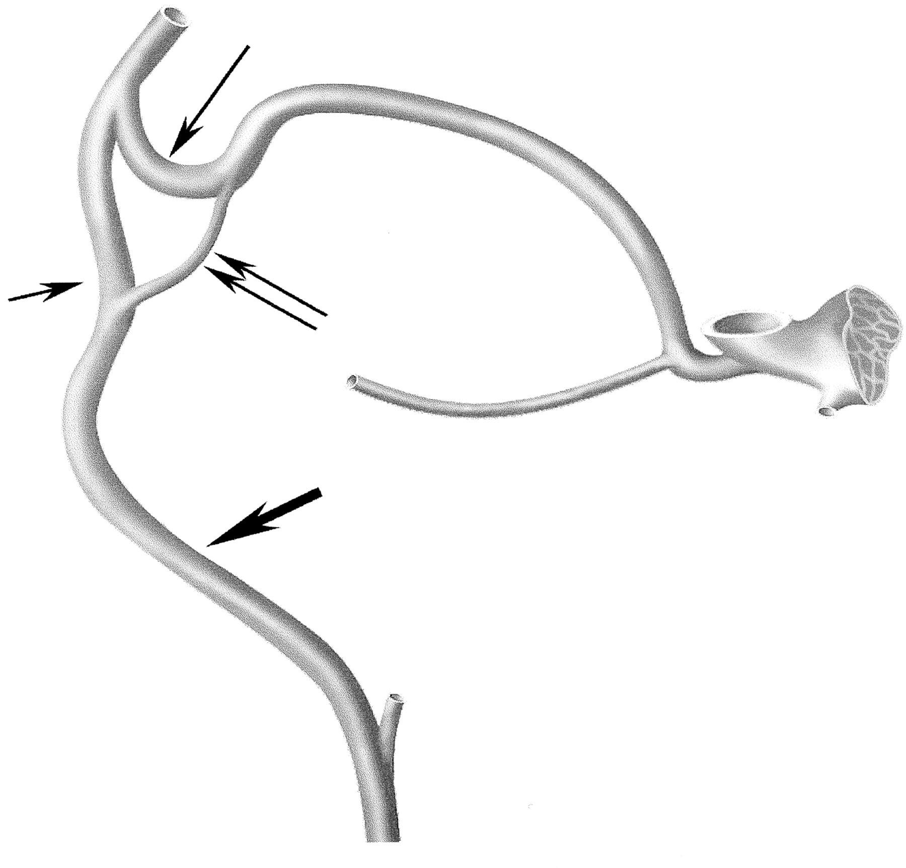
  • Fig 2.
    • Download figure
    • Open in new tab
    • Download powerpoint
    Fig 2.

    Schematic drawing of the venous system connecting the facial vein and the cavernous sinus. Facial vein (thick arrow), angular vein (short thin arrow), SOV inferior root (double arrows), SOV superior root (long thin arrow).

PreviousNext
Back to top

In this issue

American Journal of Neuroradiology: 24 (6)
American Journal of Neuroradiology
Vol. 24, Issue 6
1 Jun 2003
  • Table of Contents
  • Index by author
Advertisement
Print
Download PDF
Email Article

Thank you for your interest in spreading the word on American Journal of Neuroradiology.

NOTE: We only request your email address so that the person you are recommending the page to knows that you wanted them to see it, and that it is not junk mail. We do not capture any email address.

Enter multiple addresses on separate lines or separate them with commas.
Cavernous Sinus Dural Fistulae Treated by Transvenous Approach through the Facial Vein: Report of Seven Cases and Review of the Literature
(Your Name) has sent you a message from American Journal of Neuroradiology
(Your Name) thought you would like to see the American Journal of Neuroradiology web site.
CAPTCHA
This question is for testing whether or not you are a human visitor and to prevent automated spam submissions.
Cite this article
Alessandra Biondi, Dan Milea, Christophe Cognard, Giuseppe K. Ricciardi, Fabrice Bonneville, Rémy van Effenterre
Cavernous Sinus Dural Fistulae Treated by Transvenous Approach through the Facial Vein: Report of Seven Cases and Review of the Literature
American Journal of Neuroradiology Jun 2003, 24 (6) 1240-1246;

Citation Manager Formats

  • BibTeX
  • Bookends
  • EasyBib
  • EndNote (tagged)
  • EndNote 8 (xml)
  • Medlars
  • Mendeley
  • Papers
  • RefWorks Tagged
  • Ref Manager
  • RIS
  • Zotero
0 Responses
Respond to this article
Share
Bookmark this article
Cavernous Sinus Dural Fistulae Treated by Transvenous Approach through the Facial Vein: Report of Seven Cases and Review of the Literature
Alessandra Biondi, Dan Milea, Christophe Cognard, Giuseppe K. Ricciardi, Fabrice Bonneville, Rémy van Effenterre
American Journal of Neuroradiology Jun 2003, 24 (6) 1240-1246;
del.icio.us logo Twitter logo Facebook logo Mendeley logo
  • Tweet Widget
  • Facebook Like
  • Google Plus One

Jump to section

  • Article
    • Abstract
    • Methods
    • Results
    • Discussion
    • Conclusion
    • Acknowledgments
    • References
  • Figures & Data
  • Info & Metrics
  • Responses
  • References
  • PDF

Related Articles

  • No related articles found.
  • PubMed
  • Google Scholar

Cited By...

  • Cavernous Sinus Dural Arteriovenous Fistula: Treatment via the Transfemoral Transfacial Route
  • Endovascular Management of Intracranial Dural AVFs: Transvenous Approach
  • Intraprocedural Flat Panel Detector Rotational Angiography and an Image Fusion Technique for Delivery of a Microcatheter into the Targeted Shunt Pouch of a Dural Arteriovenous Fistula
  • Imaging-Guided Superior Ophthalmic Vein Access for Embolization of Dural Carotid Cavernous Fistulas: Report of 20 Cases and Review of the Literature
  • Intracranial Dural Arteriovenous Fistulae: Clinical Presentation and Management Strategies
  • Surgical access on the superior ophthalmic vein to the cavernous sinus dural fistula for embolization
  • Treatment of a traumatic carotid-cavernous fistula by the sole use of a flow diverting stent
  • A Safe and Efficacious Alternative: Sonographically Guided Internal Jugular Vein Puncture for Intracranial Endovascular Intervention
  • Transvenous Embolization of Intracranial Dural Arteriovenous Shunts through Occluded Venous Segments: Experience in 51 Patients
  • Complications Related to Percutaneous Transarterial Embolization of Intracranial Dural Arteriovenous Fistulas in 40 Patients
  • Diagnosis and treatment of dural carotid-cavernous fistulas: a consecutive series of 27 patients
  • Crossref
  • Google Scholar

This article has not yet been cited by articles in journals that are participating in Crossref Cited-by Linking.

More in this TOC Section

  • Prophylactic Endovascular Treatments for CBS
  • Smoking & Aneurysm Outcomes: A Meta-Analysis
  • “Buddy-wire anchoring” technique for TVE
Show more NEUROINTERVENTION

Similar Articles

Advertisement

Indexed Content

  • Current Issue
  • Accepted Manuscripts
  • Article Preview
  • Past Issues
  • Editorials
  • Editor's Choice
  • Fellows' Journal Club
  • Letters to the Editor
  • Video Articles

Cases

  • Case Collection
  • Archive - Case of the Week
  • Archive - Case of the Month
  • Archive - Classic Case

More from AJNR

  • Trainee Corner
  • Imaging Protocols
  • MRI Safety Corner
  • Book Reviews

Multimedia

  • AJNR Podcasts
  • AJNR Scantastics

Resources

  • Turnaround Time
  • Submit a Manuscript
  • Submit a Video Article
  • Submit an eLetter to the Editor/Response
  • Manuscript Submission Guidelines
  • Statistical Tips
  • Fast Publishing of Accepted Manuscripts
  • Graphical Abstract Preparation
  • Imaging Protocol Submission
  • Evidence-Based Medicine Level Guide
  • Publishing Checklists
  • Author Policies
  • Become a Reviewer/Academy of Reviewers
  • News and Updates

About Us

  • About AJNR
  • Editorial Board
  • Editorial Board Alumni
  • Alerts
  • Permissions
  • Not an AJNR Subscriber? Join Now
  • Advertise with Us
  • Librarian Resources
  • Feedback
  • Terms and Conditions
  • AJNR Editorial Board Alumni

American Society of Neuroradiology

  • Not an ASNR Member? Join Now

© 2025 by the American Society of Neuroradiology All rights, including for text and data mining, AI training, and similar technologies, are reserved.
Print ISSN: 0195-6108 Online ISSN: 1936-959X

Powered by HighWire