
Article Figures & Data
Figures
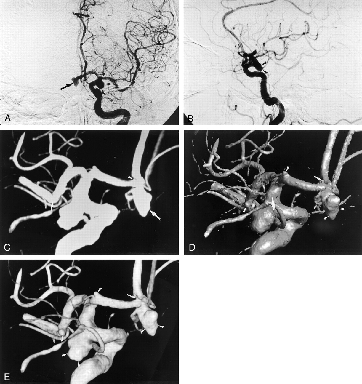
- Fig 1.
Multiple aneurysms (of one anterior communicating artery, two anterior cerebral arteries, one middle cerebral artery, and one posterior communicating artery) in a 73-year-old woman.
A, Anteroposterior and B, lateral 2D DSA images of the left internal carotid artery depict a 7-mm anterior communicating aneurysm (arrow in A) and a 9-mm posterior communicating aneurysm (arrowheads). The other aneurysms are not clearly shown on the 2D DSA images. On rotational DA images (not shown), one 1.5-mm aneurysm at the A1 segment was not detected by the two observers.
C–E, Three-dimensional DA images from behind reveal the five aneurysms (of one anterior communicating artery, two anterior cerebral arteries, one middle cerebral artery, and one posterior communicating artery). The MIP 3D image (C) does not clearly demonstrate a 1.5-mm aneurysm (arrowhead) at the A1 segment and a 2.5-mm aneurysm (small arrow) at the M1-M2 segment, which was not detected by the observers. Delineation of the shape of the anterior communicating aneurysm (large arrow) on the MIP 3D image is inferior to that on the SSD (D) and volume-rendering (E) 3D images because of lack of depth. On the SSD 3D image, the neck size of the posterior communicating aneurysm (large arrow in D) is overestimated compared with that on the volume-rendering 3D image (E). It is not clear whether the very small aneurysm (small arrow in D) at the A1 segment is an aneurysm or not. The two observers ranked it as ambiguous visualization. The other three aneurysms (arrowheads) are clearly demonstrated. On the volume-rendering image, the four aneurysms (arrowheads in E) were classified as sufficient visualization. However, the very small aneurysm (arrow in E) at the A1 segment was evaluated as ambiguous visualization. All the aneurysms were proved at surgery.
- Fig 2.
Left distal anterior cerebral artery aneurysm in a 69-year-old man.
A, Anteroposterior and B, lateral 2D DSA images of the left internal carotid artery depict a 19-mm aneurysm at the distal anterior cerebral artery. However, the relationship between the aneurysm and the adjacent arteries is not clearly demonstrated on this 2D DSA image and the rotational DA image (not shown). Regarding the relationship on rotational DA image, two observers ranked it as ambiguous visualization.
C–E, Three-dimensional DA images from behind depict a distal anterior cerebral aneurysm. MIP 3D image (C) does not demonstrate the relationship between the aneurysm and the two adjacent arteries (arrowheads) deriving from the aneurysm because of superimposition of the aneurysm and the arteries; the two observers classified this aneurysm as insufficient visualization on delineation of shape and relationship. The SSD (D) and volume-rendering (E) 3D images clearly show the shape and relationship (arrowheads), which were ranked as sufficient visualization by the two observers. Neurosurgeons chose to performed surgery on the basis of this additional information, and the relationship was confirmed during surgery.


Tables
Aneurysm Detection Score* Rotational DA (n = 30) MIP 3D DA (n = 30) SSD 3D DA (n = 30) Volume-Rendering 3D DA (n = 30) +2 24 (80) 25 (83) 27 (90) 29 (97) +1 4 (13) 3 (10) 3 (10) 1 (3) 0 2 (7) 2 (7) 0 (0) 0 (0) −1 0 (0) 0 (0) 0 (0) 0 (0) Mean score ± SD 1.73 ± 0.58 1.77 ± 0.57 1.90 ± 0.31 1.97 ± 0.18 Note.—Data are number of aneurysms. Numbers in parentheses are percentages.
* +2 indicates sufficient visualization; +1, ambiguous visualization; 0, poor visualization; −1, misinterpretation.
- TABLE 2:
Delineation of aneurysm neck at rotational DA and MIP, SSD, and volume-rendering 3D DA
Delineation of Neck Score* Rotational DA (n = 28) MIP 3D DA (n = 28) SSD 3D DA (n = 28) Volume-Rendering 3D DA (n = 28) +2 17 (61) 23 (82) 21 (75) 27 (96) +1 7 (25) 3 (10) 4 (14) 1 (4) 0 4 (14) 1 (4) 2 (7) 0 (0) −1 0 (0) 1 (4) 1 (4) 0 (0) Mean score ± SD 1.46 ± 0.74 1.71 ± 0.71 1.61 ± 0.79 1.96 ± 0.19 Note.—Data are number of aneurysms. Numbers in parentheses are percentages.
* +2 indicates sufficient visualization; +1, ambiguous visualization; 0, poor visualization; −1, misinterpretation.
- TABLE 3:
Delineation of aneurysm shape at rotational DA and MIP, SSD, and volume-rendering 3D DA
Delineation of Shape Score* Rotational DA (n = 28) MIP 3D DA (n = 28) SSD 3D DA (n = 28) Volume-Rendering 3D DA (n = 28) +2 16 (57) 18 (64) 24 (86) 27 (96) +1 8 (29) 6 (21) 4 (14) 1 (4) 0 4 (14) 4 (14) 0 (0) 0 (0) −1 0 (0) 0 (0) 0 (0) 0 (0) Mean score ± SD 1.43 ± 0.74 1.50 ± 0.75 1.86 ± 0.36 1.96 ± 0.19 Note.—Data are number of aneurysms. Numbers in parentheses are percentages.
* +2 indicates sufficient visualization; +1, ambiguous visualization; 0, poor visualization; −1, misinterpretation.
- TABLE 4:
Delineation of relationship between aneurysm and adjacent arteries at rotational DA and MIP, SSD, and volume-rendering 3D DA
Delineation of Location Score* Rotational DA (n = 28) MIP 3D DA (n = 28) SSD 3D DA (n = 28) Volume-Rendering 3D DA (n = 28) +2 12 (43) 21 (75) 26 (93) 27 (96) +1 10 (36) 6 (21) 2 (7) 1 (4) 0 6 (21) 1 (4) 0 (0) 0 (0) −1 0 (0) 0 (0) 0 (0) 0 (0) Mean score ± SD 1.21 ± 0.79 1.71 ± 0.54 1.93 ± 0.26 1.96 ± 0.19 Note.—Data are number of aneurysms. Numbers in parentheses are percentages.
* +2 indicates sufficient visualization; +1, ambiguous visualization; 0, poor visualization; −1, misinterpretation.
Aneurysm No. Site Size (mm) Treatment Additional Information Grade* 1 A1 4.7 Clip 0 2 A1 2 Wrap Aneurysm detection 1 3 A1 1.5 Wrap Aneurysm detection 1 4 A-com 10 Clip Delineation of neck, shape, and location 2(c) 5 A-com 8 Clip Delineation of neck and location 1 6 A-com 7 Clip Delineation of location 1 7 A-com 7 Clip 0 8 A-com 6 Clip Delineation of neck, shape, and location 1 9 A-com 6 Clip 0 10 A-com 5 Clip 0 11 Distal ACA 19 Clip Delineation of location 2(a,b) 12 Distal ACA 9 Clip 0 13 IC-PC 10 Coat Delineation of neck, shape, and location 2(a,b) 14 IC-PC 9 Clip Delineation of neck and location 1 15 IC-PC 8 Clip 0 16 IC-PC 3.5 Clip 0 17 M1–M2 18 Clip Delineation of neck 1 18 M1–M2 12 Clip Delineation of location 1 19 M1–M2 10 Clip Delineation of neck, shape, and location 2(c) 20 M1–M2 7 Clip 0 21 M1–M2 6 Clip 0 22 M1–M2 6 Clip Delineation of neck and location 1 23 M1–M2 5 Clip 0 24 M1–M2 4 Clip 0 25 M1–M2 3.6 Clip 0 26 M1–M2 3 Clip 0 27 M1–M2 2.5 Clip 0 28 M1–M2 4 Clip 0 29 BA top 6 Coil 0 30 BA-SCA 5 Coil Delineation of neck, shape, and location 2(b) Note.—A1 indicates A1 segment of the anterior cerebral artery; A-com, anterior communicating artery; ACA, the anterior cerebral artery; IC-PC, internal carotid-posterior communicating artery; M1–M2, M1–M2 segment of the middle cerebral artery; BA, basilar artery; BA-SCA, basilar-superior cerebellar artery; clip, clipping; wrap, wrapping; coil, coil embolization.
* Grade 2, provided further information that was helpful for surgery or embolization; grade 1, provided further information, but not helpful for surgery or embolization; grade 0, provided no further information. Reasons affecting treatment: (a) valuable information about whether treatment can be performed; (b) critical information about what treatment should be chosen when treatment can be performed; and (c) helpful information about manipulations of the aneurysm and adjacent arteries during surgery or endovascular treatment.
In this issue


Jump to section
Related Articles
Cited By...
- Artificial Intelligence-Based 3D Angiography for Visualization of Complex Cerebrovascular Pathologies
- Balloon-occluded retrograde transvenous obliteration of gastric varices using three-dimensional rotational angiography
- 3D Digital Subtraction Angiography of Intracranial Aneurysms: Comparison of Flat Panel Detector with Conventional Image Intensifier TV System Using a Vascular Phantom
- Superselective transcatheter arterial chemoembolisation of an unresectable hepatocellular carcinoma using three-dimensional rotational angiography
- Safety of Cerebral Digital Subtraction Angiography in Children: Complication Rate Analysis in 241 Consecutive Diagnostic Angiograms