
Article Figures & Data
Figures
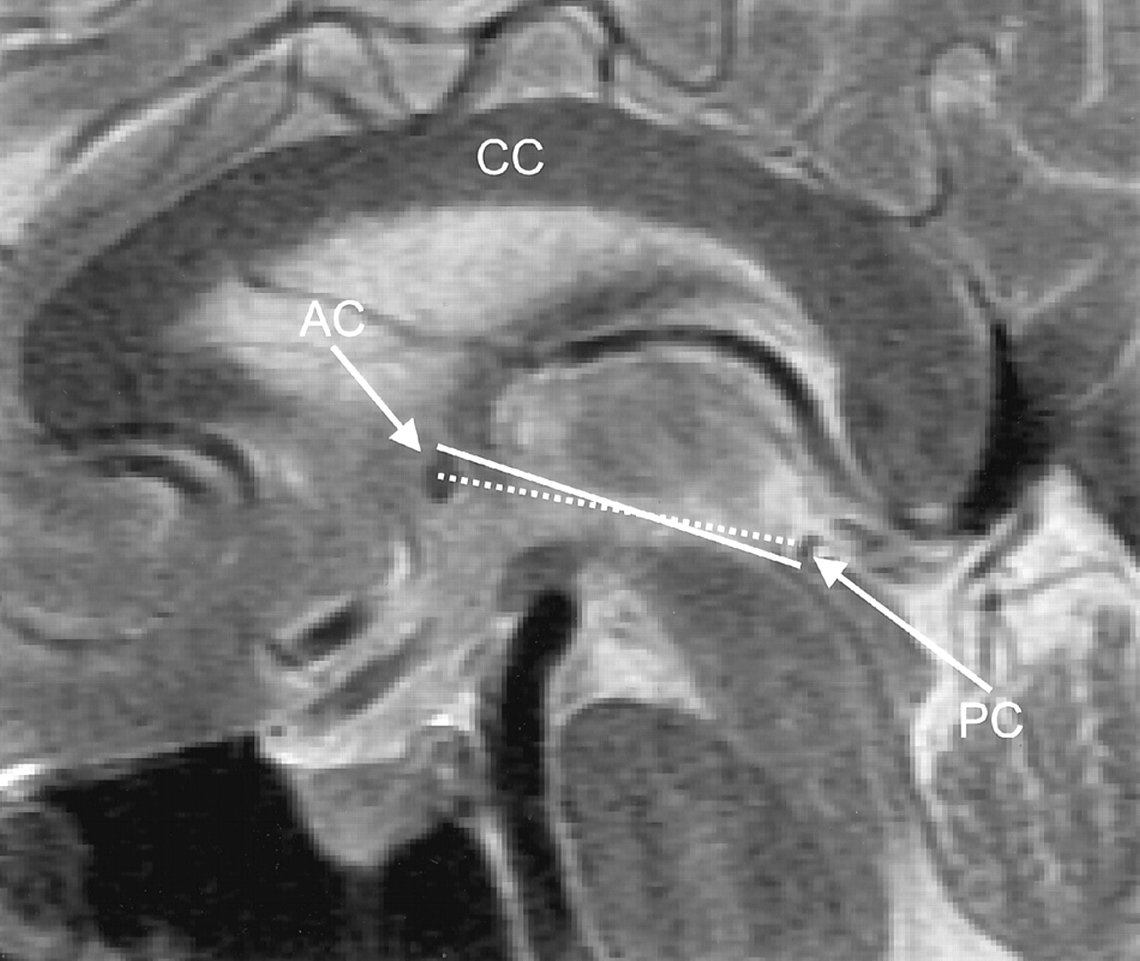
- Fig 1.
Midline sagittal FSE T2-weighted MR image (TR/TE, 3816/105eff; echo train length, 16; section thickness, 4 mm; matrix, 512 × 256; FOV, 20 cm). The solid line and dotted line correspond to the Talairach and Schaltenbrand AC-PC reference lines, respectively. AC indicates the anterior commissure; CC, corpus callosum; and PC, posterior commissure.
- Fig 2.
Sequential images from a single patient’s three-step clinical AC-PC protocol.
A, Coronal FGRE localizer image (6/1.6; flip angle, 20°; section thickness, 7 mm; matrix, 256 × 192; FOV, 24 cm) for roll prescription. The line indicates the plane of the image in B.
B, Roll-corrected axial oblique FGRE localizer image (8/1.6; flip angle, 20°; section thickness, 7 mm; matrix, 256 × 192; FOV, 24 cm) for yaw prescription. The thickest line indicates the plane of the image in C.
C, Roll- and yaw-corrected double oblique T2-weighted FSE image (3816/105eff; echo train length, 16; section thickness, 4 mm; skip, 1 mm; matrix, 512 × 256; FOV, 20 cm; NEX, 2; time, 1 minute 56 seconds) for pitch prescription. The thickest line indicates the Talairach reference plane used for subsequent triple oblique axial scans.
- Fig 3.
Measurements of canthomeatal angulation. Surface-rendered archetypal MNI brain (14) allows identification of the orbital canthus and the external acoustic meatus.
- Fig 4.
Requisite brain volume coverage: Talairach versus average axial sectioning for the archetypical MNI brain (14). Solid lines are Talairach referenced. Dotted lines correspond to an average patient head position oriented 15.1° counterclockwise from AC-PC. Measurements are in millimeters. Note the reduction in brain volume coverage afforded by Talairach obliquity compared with standard axial imaging.
- Fig 5.
Illustration of CATS functionality.
A, APE to determine the positions of the scalp and CC by examining intensities along the central column of pixels from a midline sagittal T2-weighted image.
B, Automated contours and a bisecting line on a 2-second axial oblique T1-weighted gradient-recalled echo image. Note that brightest point lies within the cross section of the superior sagittal sinus (SSS) as a result of entry-flow phenomenon.
C, Outline of the CC, triangle search mask, and Talairach AC-PC reference line on a midline sagittal T2-weighted image.
- Fig 6.
Violin plots of prescription errors. Technologist (Tech) and computer (CATS and SPM’99) methods are compared with physical alignment (PA). Spread narrowing indicates reduced image variability. (KLW, JLW, and WS are authors’ initials.)


Tables
Algorithm* N Mean Median Variance SD IQR Minimum Maximum Range Roll CS 126 −0.60 −0.59 13.47 3.67 3.95 −9.17 10.59 19.75 Tech 126 0.11 0.00 16.75 4.09 4.47 −13.46 12.35 25.81 CATS 126 −0.12 −0.15 11.49 3.39 5.11 −6.06 6.73 12.79 Yaw CS 126 −0.86 −0.94 22.38 4.73 6.20 −11.17 17.26 28.43 Tech 126 −0.26 −0.32 20.02 4.47 6.00 −9.48 18.51 28.00 CATS 126 0.01 −0.61 18.80 4.34 6.78 −8.49 16.21 24.70 Talairach pitch CS 124 15.16 15.11 104.23 10.21 12.97 −8.13 40.20 48.34 Tech 126 15.59 15.25 99.59 9.98 14.28 −18.78 38.85 57.64 CATS 121 13.86 14.04 111.03 10.54 14.66 −10.39 41.42 51.81 SPM’99 126 11.95 10.96 60.56 7.78 10.80 −4.40 36.04 40.45 Schaltenbrand pitch CS 124 9.35 8.63 107.62 10.37 13.60 −14.62 33.69 48.31 CATS 121 9.97 9.02 115.37 10.74 14.36 −13.82 38.97 52.78 * CS indicates criterion standard; Tech, technologist; IQR, interquartile range.
Algorithm* N Mean Median Variance SD IQR Minimum Maximum Range Roll PA 126 0.60 0.59 13.47 3.67 3.95 −10.59 9.17 19.75 Tech 126 0.71 0.78 3.42 1.85 1.99 −4.29 6.81 11.1 CATS 126 0.48 0.66 3.92 1.98 1.63 −8.21 9.37 17.6 Yaw PA 126 0.86 0.94 22.38 4.73 6.20 −17.26 11.17 28.43 Tech 126 0.59 0.63 0.65 0.81 0.97 −2.50 2.57 5.1 CATS 126 0.86 0.96 2.52 1.59 2.11 −2.40 4.73 7.1 Talairach pitch PA 124 −15.16 −15.11 104.23 10.21 12.97 −40.20 8.13 48.34 Tech 124 0.56 1.15 28.62 5.35 2.88 −24.62 20.68 45.3 CATS 120 −1.23 −1.11 18.52 4.30 3.60 −23.41 14.81 38.2 SPM’99 124 −3.14 −1.56 59.30 7.70 5.23 −37.96 9.88 47.8 Schaltenbrand pitch PA 124 −9.35 −8.63 107.62 10.37 13.60 −33.69 14.62 48.31 CATS 120 0.70 1.13 18.30 4.28 4.07 −20.41 15.48 35.9 * PA indicates physical alignment; Tech, technologist; IQR, interquartile range.


