
Article Figures & Data
Figures
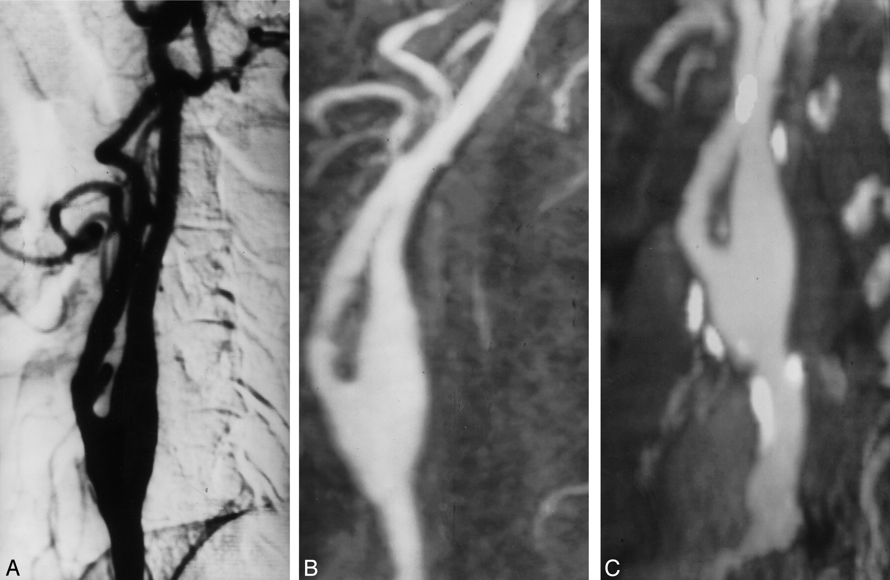
- Fig 1.
Grade 1 stenosis of the left internal carotid artery.
A, Oblique DSA image shows minimal stenosis of the left internal carotid artery.
B, Elliptic centric contrast-enhanced MR angiogram, in the same projection as that of the DSA image, depicts a good correlation with DSA.
C, Spiral CT angiogram shows that the calcium distribution (type 1) does not preclude a good evaluation of the vessel.
- Fig 2.
Grade 2 stenosis overestimated with elliptic centric contrast-enhanced MR angiography.
A, Oblique DSA image demonstrates a grade 2 stenosis in the left internal carotid artery.
B, Elliptic centric contrast-enhanced MR angiogram shows mild overestimation of the stenosis (grade 3).
C, Spiral CT angiogram shows good correlation with DSA.
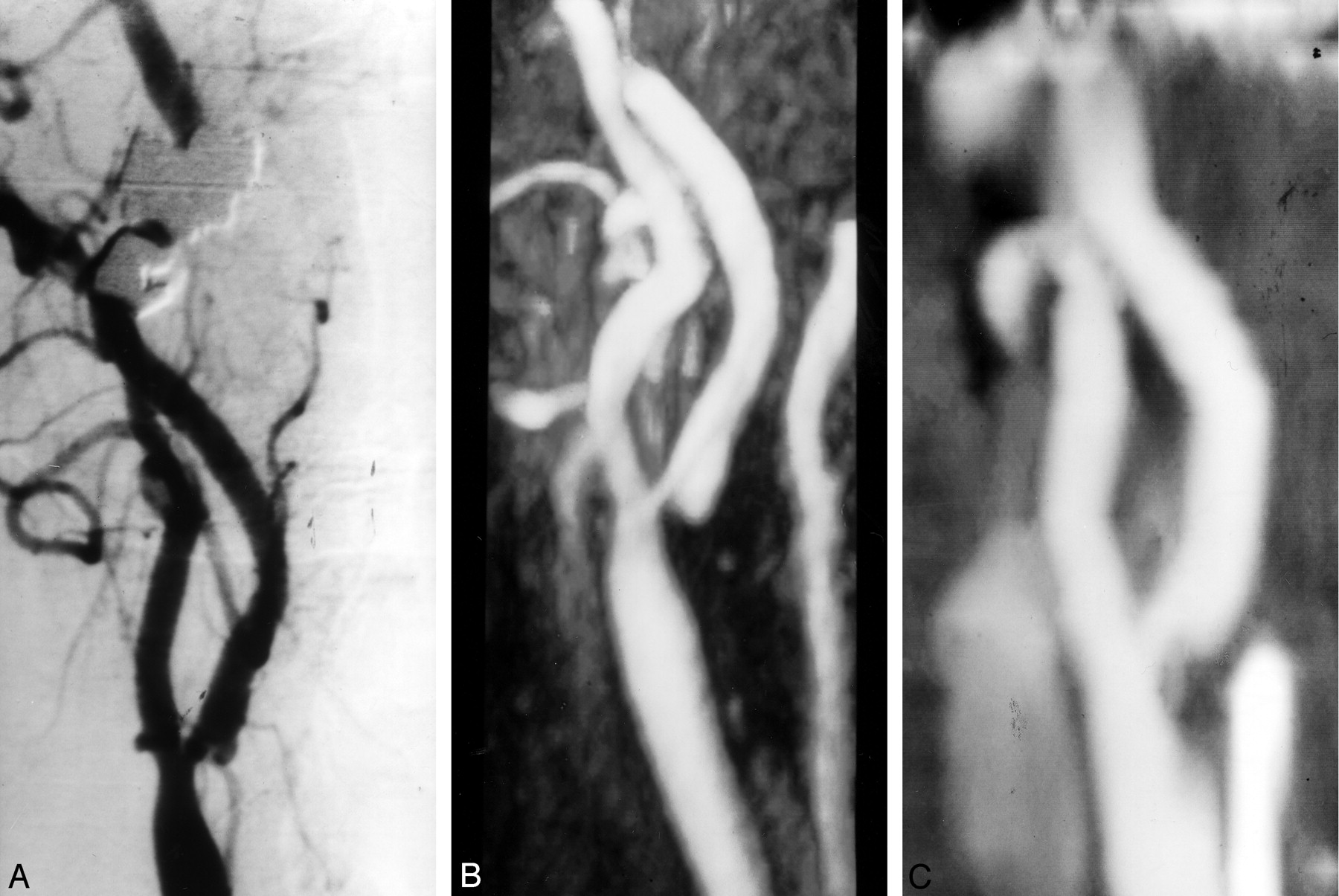
- Fig 3.
Grade 4 stenosis with good correlation in all techniques.
A, Oblique DSA image shows critical stenosis in the right internal carotid artery.
B, Elliptic centric contrast-enhanced MR angiogram depicts the same information.
C, Spiral CT angiogram shows a good correlation with the other imaging techniques. Notice a type 3 calcification in the wall of artery; however, it is sufficiently separated in the reconstruction image.
- Fig 4.
Grade 2 stenosis overestimated with spiral CT angiography.
A, Oblique DSA image shows grade 2 stenosis in the left internal carotid artery.
B, Elliptic centric contrast-enhanced MR angiogram shows the same findings.
C and D, Spiral CT angiograms show type 4 calcification. In this case, the stenosis was overestimated (grade 3) because of the difficulty in separating calcium from the contrast material.


Tables
Comparison of the degree of carotid stenosis with elliptic centric contrast-enhanced MR angiography, spiral CT angiography, and DSA
Degree of Stenosis Degree of Stenosis at DSA 0–49% 50–69% 70–99% Occlusion MR angiography 0–49% 32 0 0 0 50–69% 0 8 1 0 70–99% 0 2 34 0 Occlusion 0 0 0 3 CT angiography 0–49% 26 2 1 0 50–69% 6 7 8 0 70–99% 0 1 26 0 Occlusion 0 0 0 3 Note.—Data are number of arteries (n = 80).


