
Article Figures & Data
Figures
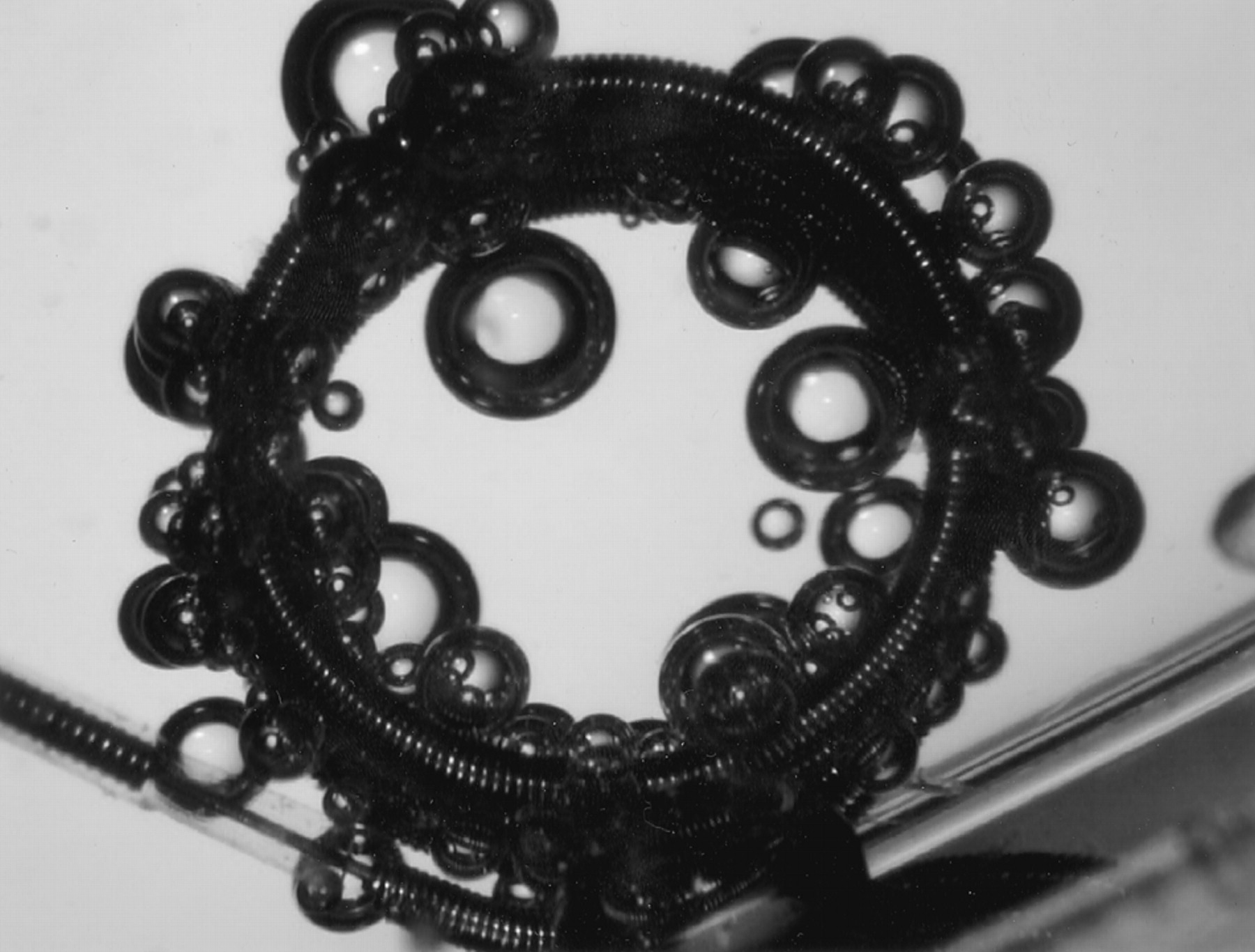
- Fig 1.
Depiction of the detachment process of a conventional coil (GDC-10, 4 mm × 10 cm) in saline. Many gas bubbles were generated from the coil and attached around the entire coil part. The bubbles varied in size.
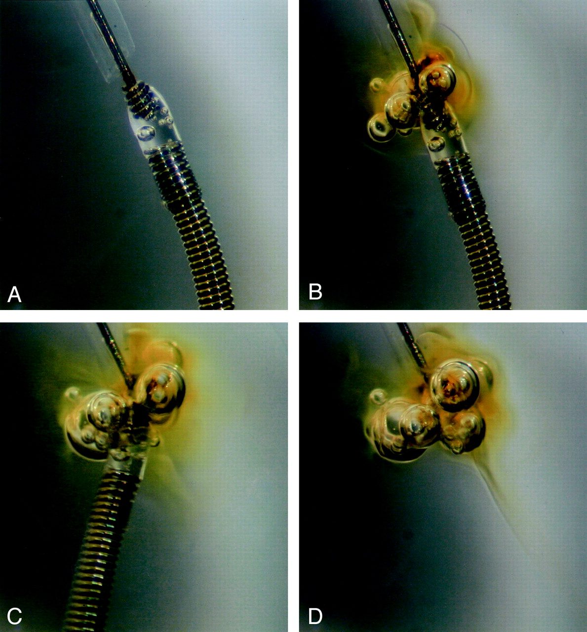
- Fig 2.
Depiction of detachment process of an insulated coil (GDC-10 SynerG, 6 mm × 6 cm) in heparinized human serum.
A, Before applying electric current, no gas bubbles surround the coil, and the detachment zone of the device is clearly seen with a transparent insulating plug.
B, After starting the electrolytic detachment process, gas bubbles are generated from the detachment zone, and a localized brownish discoloration is seen around the detachment zone.
C, The coil has just detached, and the bubbles enlarge.
D, After detachment, the coil moves out of the field, and the bubbles remain attached at the tip of the pusher wire and collected in the area of brownish discoloration. Because discoloration and collection of gas bubbles around this area were absent in the saline experiment, this brownish discoloration may be related to an electrolytically induced protein coagulation.
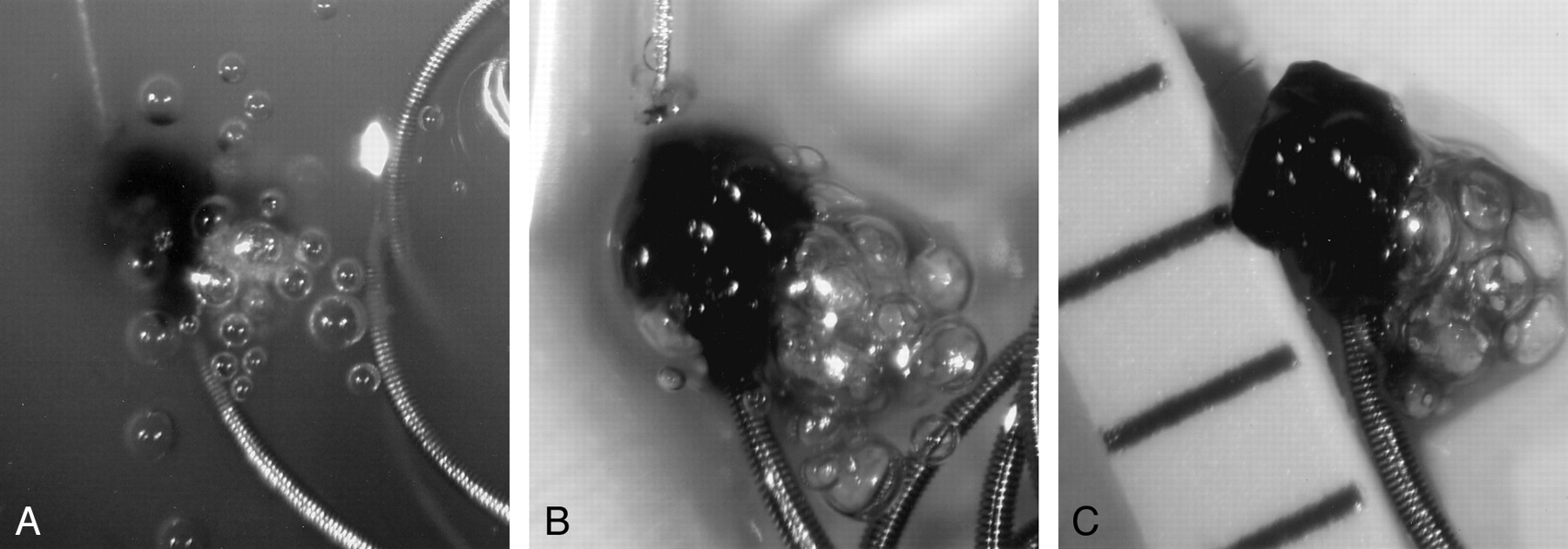
- Fig 3.
Depiction of the detachment process of an insulated coil (GDC-10 SynerG, 6 mm × 20 cm) in heparinized human whole blood.
A, During detachment, with the detachment zone dipped in the blood, a dark area of thrombus formation is clearly seen with many gas bubbles generated and floating to the surface level.
B, After detachment, the blood was gently irrigated by saline, and a lobulated thrombus is attached at the proximal tail of the coil part. Many gas bubbles remain in the thrombus.
C, After removal of the fluid, the bubble remains attached in the thrombus; the thrombus is 1.5 mm in diameter.
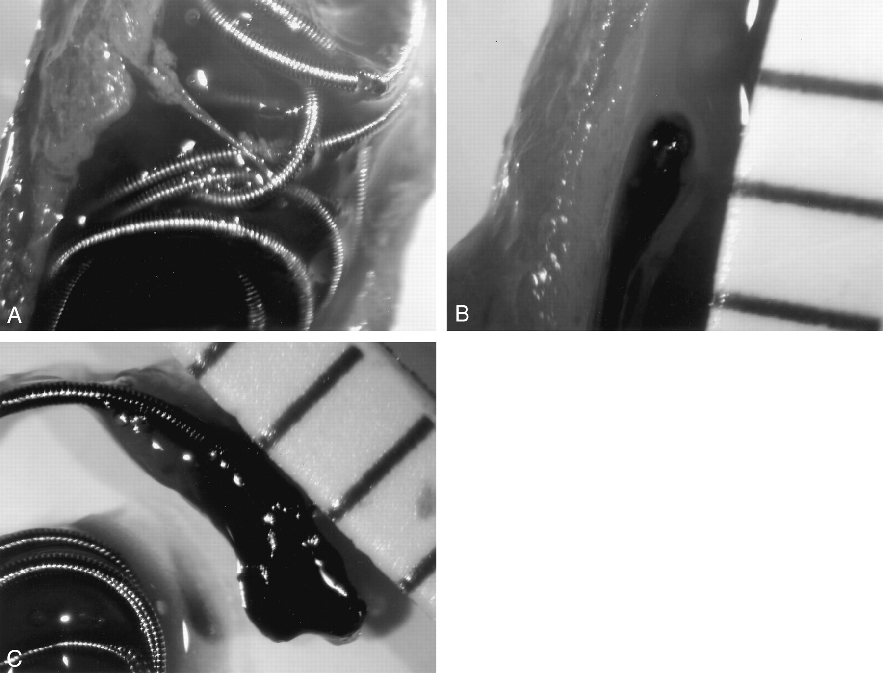
- Fig 4.
Depiction of the detachment process of a conventional coil (GDC-18, 8 mm × 20 cm) in the canine carotid artery.
A, Immediately after detachment of the coil, the segment of the artery is ligated and opened by use of a longitudinal arteriotomy. Without arteriotomy, multiple and tiny gas bubbles surround the coil.
B, A thrombus with elongated appearance is seen at the proximal tail of the detached coil.
C, After removal of the coil from the artery, the thrombus measures 3 mm in diameter.


Tables
Sizes and types of Guglielmi detachable coils used for in vitro and animal experiments
Experiments Conventional Coils Insulated Coils (SynerG) Saline GDC-10 (4 mm × 10 cm), GDC-10 (5 mm × 15 cm) GDC-10 (2 mm × 8 cm) Heparinized serum GDC-10 (5 mm × 15 cm), GDC-10 (6 mm × 6 cm), GDC-10 (6 mm × 6 cm) GDC-10 (5 mm × 8 cm), GDC-10 (10 mm × 30 cm) Heparinized blood GDC-10 (2 mm × 2 cm, soft), GDC-10 (3 mm × 8 cm), GDC-10 (4 mm × 10 cm), GDC-10 (6 mm × 20 cm), GDC-18(8 mm × 30 cm) GDC-10 (2 mm × 1 cm, Ultrasoft SR), GDC-10 (10 mm × 30 cm) Canine carotid artery GDC-10 (5 mm × 15 cm), GDC-18 (8 mm × 20 cm) Not used in animal experiment


