
Article Figures & Data
Figures
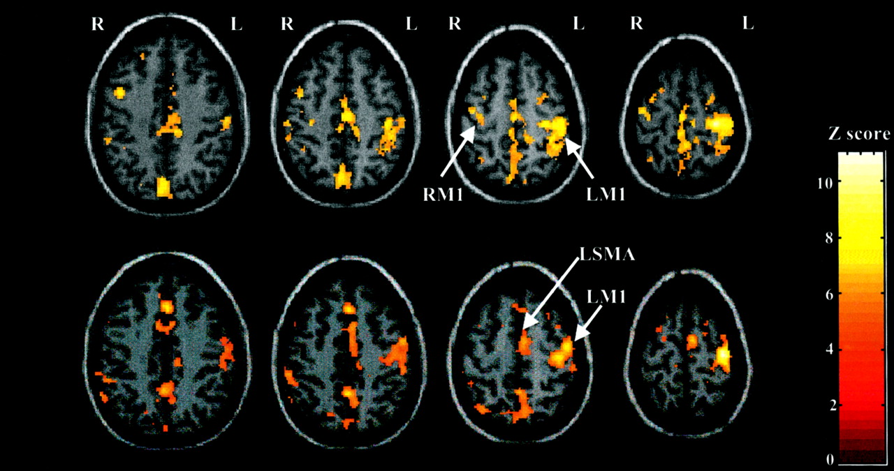
- Fig 1.
Motor activation during the visuomotor response task. Activation was superimposed onto normalized T1-weighted MR images by using SPM99. All regions displayed showed significant activation (P < .001 uncorrected for multiple comparisons). Top row, Individual SPM99 maps of a representative male subject show contralateral and ipsilateral M1 activation. Bottom row, Individual SPM99 maps of a representative female subject show similar degrees of left M1 and SMA activation.
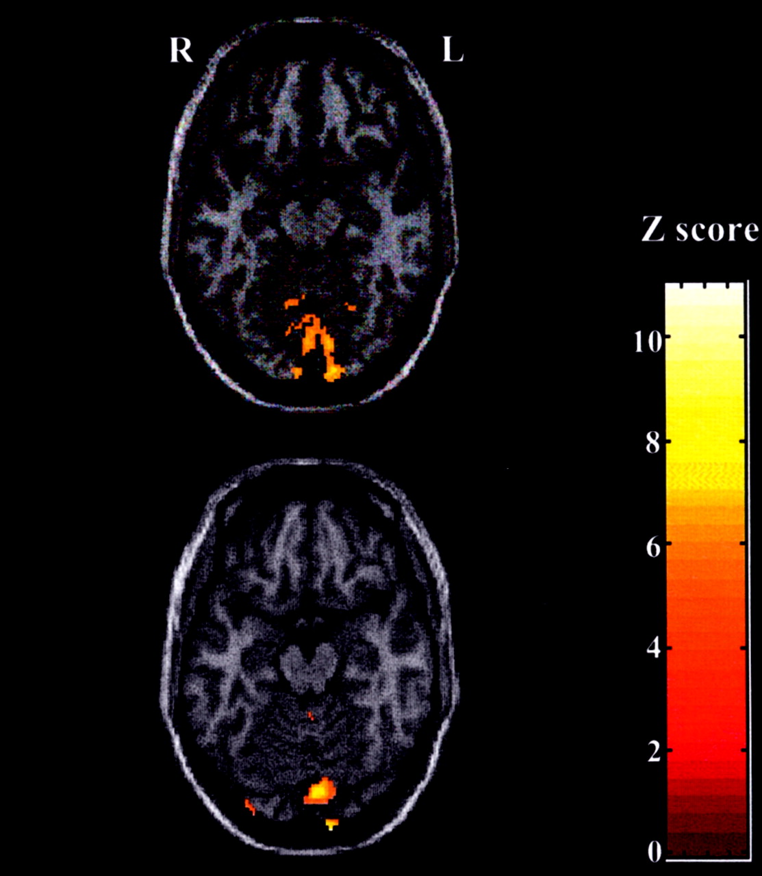
- Fig 2.
Visual activation during visuomotor response task. Activation was superimposed onto normalized T1-weighted MR images by using SPM99. Significant medial occipital cortex activation (P < .001 uncorrected for multiple comparisons) is demonstrated in a representative. Top, Image in male subject. Bottom, Image in female subject.
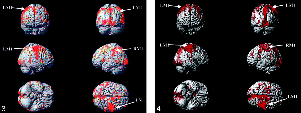
- Fig 3.
Surface-rendered activation in a representative female subject. Activation was superimposed on a T1-weighted by using the Montreal Neurologic Institute template provided by SPM99. All regions displayed showed significant activation (P < .001 uncorrected for multiple comparisons).
- Fig 4.
Surface-rendered activation in a representative male subject. Activation was superimposed on a T1-weighted by using the Montreal Neurologic Institute template provided by SPM99. All regions displayed showed significant activation (P < .001 uncorrected for multiple comparisons).
- Fig 5.
Random effects SPM99 of male versus female brain activation during visuomotor response task. Two-way t test (T = 3 .47, P < .001 uncorrected for multiple comparisons) shows no difference in brain activation at any defined specified site: RV, LV, LM1, LSMA, and LACA. However, men had a larger volume of activation in right inferior parietal lobule, right insula, and left thalamus.


Tables
- TABLE 1:
Age, RT, and number of voxels for each site activated on individual maps during the event-related paradigm
Subject No. Age (y) Mean RT, ms k Value RV LV LM1 LSMA LACA Men* 1 26 445.8 496 21 1 0 0 2 26 358.9 160 27 1864 0 1 3 26 407.27 10 146 521 0 14 4 31 441.8 41 275 31 0 0 5 33 260.6 1136 2405 2 0 0 6 42 478.3 0 7 0 0 0 7 53 327 129 68 14 605 0 8 58 505.2 81 84 28 0 0 9 70 417.27 10 14 1 0 0 10 70 431.26 1454 24 416 0 0 11 72 339.6 151 477 700 0 2 12 78 403 4 117 0 0 0 13 80 580.73 101 8 239 0 0 Women† 1 25 318.3 207 598 296 0 0 2 25 336.53 100 50 4283 0 0 3 26 481.4 14 7973 8 1 1 4 30 351.73 608 1095 911 0 0 5 38 350.5 3 7 19 0 0 6 49 447.53 253 172 282 0 0 7 54 445.9 628 655 1732 702 34 8 58 360 0 3 0 0 0 9 66 430.7 120 301 307 0 0 10 69 446.1 277 8 32 0 0 11 70 504.78 4 0 0 0 0 12 76 550.3 53 156 177 0 492 13 85 486 15 0 1 0 0 Note.—SPM99 was used. Uncorrected P < .001. LACA indicates the left anterior cingulate area; LM1, left primary somatomotor area; LSMA, left supplemental motor area; LV, left visual area; and RV, right visual area.
* Age, 51.15 years ± 21.34. RT, 415.1 ms ± 90.48.
† Age, 50.4 years ± 21.55 RT, 423.3 ms ± 76.67.
- TABLE 2:
Fixed-effects analysis: number of voxels in each location on the group map during event-related paradigm
Group LV RV LM1 LSMA LACA k Z k Z k Z k Z k Z Men, T =4.71 3602 7.0 1738 7.0 3050 6.9 246 6.9 212 6.23 Women, T = 4.73 2452 7.9 132 7.9 1342 7.82 1 4.75 1262 6.53 Note.—SPM99 was used. Corrected P < .05.
- TABLE 3:
Random-effects analysis: number of voxels in each location on the group map during event-related paradigm
Group LV RV LM1 LSMA LACA k Z k Z k Z k Z k Z Men, T = 3.93 583 4.47 367 4.11 144 3.58 0 0 0 0 Women, T = 3.93 294 3.61 51 3.96 132 3.68 37 4.64 1 3.09 Note.—SPM99 was used. Uncorrected P < .001.
Group RT, ms Mean Median SD Men 415.1 388 90.48 Women 423.25 385 76.67 Author and Year* Paradigm Sex Differences in RT Comments Botwinick and Brinley, 1962(6) Audio and visual SRT Young men > young women, elderly women > elderly men Elderly women were 8 years younger than elderly men Botwinick and Thompson, 1966 (7) Audio SRT Men = women Men and women were adequately equated by age (median ages: men, 78 y; women, 76 y) Botwinick and Storandt, 1974 (8) SRT Men = women Elderly men and elderly women were matched for age and education level Fozard et al, 1994 (9) Audio SRT and audio DRT Men > women Longitudinal study. Men were faster than women over 4-y follow-up Noble et al, 1964 (10) Visual CRT, emphasizing spatial and motor component Men > women Overall, men were faster than women, although women aged 71–87 y were slightly faster than men Landauer et al, 1980 (12) Visual CRT, emphasizing semantic and verbal component Men = women Women were faster in decision making, whereas men were faster in movement times Lahtela et al, 1985 (11) Visual CRT, emphasizing spatial and motor component Men > women In the sample of 2550, men were faster across all ages * Numbers in parentheses are reference citations.


