
Article Figures & Data
Figures
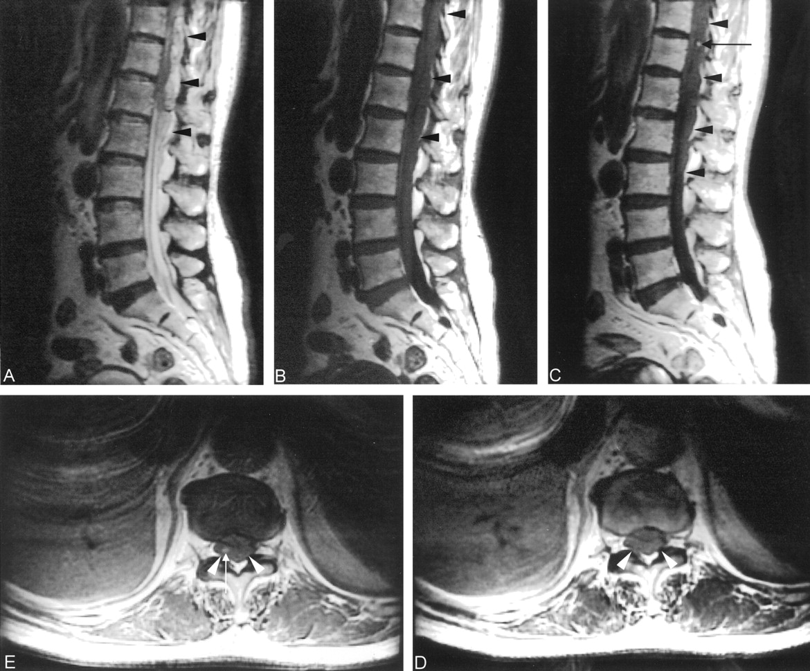
- Fig 1.
Case 1, a 69-year-old woman. MR images of the thoracolumbar spine, obtained 10 hours after sudden-onset severe back pain, show a large SEH in the dorsal area of T9–L3.
A, Sagittal T2-weighted (2000/80/2 [TR/TE/NEX]) image shows the hematoma as a heterogeneously hyperintense area (arrows).
B, Sagittal T1-weighted (400/20/2) image of the lumbar spine shows an isointense hematoma (arrowheads).
C, Contrast-enhanced sagittal T1-weighted image (400/20/2) shows an enhanced area (arrows) within the hematoma (arrowheads) at the level of T12.
D, Axial T1-weighted image (400/20/2), at the level of T12, shows an isointense hematoma (arrowheads).
E, Contrast-enhanced axial T1-weighted image (400/20/2), at the level of T12, shows an enhanced area (arrow) within the hematoma (arrowheads).
- Fig 2.
Case 2, an 82-year-old man. MR images of the cervical spine, obtained 4 hours after a fall, reveal a large SEH in the dorsal area of C5–C7.
A, Sagittal T2-weighted (2000/80/2) image shows a hyperintense epidural hematoma (arrowheads). A hypointense displaced dura (arrows) indicates the epidural location of the hematoma.
B, Sagittal proton density-weighted (2000/20/2) image shows a slightly hyperintense SEH (arrowheads).
C, Sagittal T1-weighted (400/20/2) image reveals the isointense SEH (arrowheads).
D, Contrast-enhanced sagittal T1-weighted (400/20/2) image shows multiple, enhanced spots (arrow) within the hematoma (arrowheads).
E, Contrast-enhanced axial T1-weighted (400/20/2) image shows spotty enhancement (arrows) at the level of C6.


Tables
Reported cases of contrast enhancement of SEH diagnosed on the basis of MR findings
Case Age (y)/Sex Time* Bleeding Diathesis Origin 1 31/male >1 week Yes (hemophilia A) Karen S. Caldemeyer et al, 1993 2 58/female <24 hours No (spontaneous) Chi-Jen Chen, 1997 3 60/female 5 hours No (spontaneous) Hiroshi Nawashiro, 2001 4 69/female 10 hours Yes (spontaneous) Case 1 5 82/male 4 hours Yes (trauma) Case 2 * The time interval between onset of symptoms and MR examination.
In this issue


Jump to section
Related Articles
Cited By...
- No citing articles found.