Advertisement

Main menu

  • Home
  • Content
    • Current Issue
    • Accepted Manuscripts
    • Article Preview
    • Past Issue Archive
    • Video Articles
    • AJNR Case Collection
    • Case of the Week Archive
    • Case of the Month Archive
    • Classic Case Archive
  • Special Collections
    • AJNR Awards
    • Low-Field MRI
    • Alzheimer Disease
    • ASNR Foundation Special Collection
    • Photon-Counting CT
    • View All
  • Multimedia
    • AJNR Podcasts
    • AJNR SCANtastic
    • Trainee Corner
    • MRI Safety Corner
    • Imaging Protocols
  • For Authors
    • Submit a Manuscript
    • Submit a Video Article
    • Submit an eLetter to the Editor/Response
    • Manuscript Submission Guidelines
    • Statistical Tips
    • Fast Publishing of Accepted Manuscripts
    • Graphical Abstract Preparation
    • Imaging Protocol Submission
    • Author Policies
  • About Us
    • About AJNR
    • Editorial Board
    • Editorial Board Alumni
  • More
    • Become a Reviewer/Academy of Reviewers
    • Subscribers
    • Permissions
    • Alerts
    • Feedback
    • Advertisers
    • ASNR Home

User menu

  • Alerts
  • Log in

Search

  • Advanced search
American Journal of Neuroradiology

American Journal of Neuroradiology

ASHNR American Society of Functional Neuroradiology ASHNR American Society of Pediatric Neuroradiology ASSR
  • Alerts
  • Log in

Advanced Search

  • Home
  • Content
    • Current Issue
    • Accepted Manuscripts
    • Article Preview
    • Past Issue Archive
    • Video Articles
    • AJNR Case Collection
    • Case of the Week Archive
    • Case of the Month Archive
    • Classic Case Archive
  • Special Collections
    • AJNR Awards
    • Low-Field MRI
    • Alzheimer Disease
    • ASNR Foundation Special Collection
    • Photon-Counting CT
    • View All
  • Multimedia
    • AJNR Podcasts
    • AJNR SCANtastic
    • Trainee Corner
    • MRI Safety Corner
    • Imaging Protocols
  • For Authors
    • Submit a Manuscript
    • Submit a Video Article
    • Submit an eLetter to the Editor/Response
    • Manuscript Submission Guidelines
    • Statistical Tips
    • Fast Publishing of Accepted Manuscripts
    • Graphical Abstract Preparation
    • Imaging Protocol Submission
    • Author Policies
  • About Us
    • About AJNR
    • Editorial Board
    • Editorial Board Alumni
  • More
    • Become a Reviewer/Academy of Reviewers
    • Subscribers
    • Permissions
    • Alerts
    • Feedback
    • Advertisers
    • ASNR Home
  • Follow AJNR on Twitter
  • Visit AJNR on Facebook
  • Follow AJNR on Instagram
  • Join AJNR on LinkedIn
  • RSS Feeds

AJNR Awards, New Junior Editors, and more. Read the latest AJNR updates

OtherHead and Neck Imaging

Solitary Fibrous Tumor of the Hypoglossal Nerve

Ma Lourdes S. Badion, C. C. Tchoyoson Lim, Jennifer Teo, Peck Leong Ong and Francis Hui
American Journal of Neuroradiology March 2003, 24 (3) 343-345;
Ma Lourdes S. Badion
  • Find this author on Google Scholar
  • Find this author on PubMed
  • Search for this author on this site
C. C. Tchoyoson Lim
  • Find this author on Google Scholar
  • Find this author on PubMed
  • Search for this author on this site
Jennifer Teo
  • Find this author on Google Scholar
  • Find this author on PubMed
  • Search for this author on this site
Peck Leong Ong
  • Find this author on Google Scholar
  • Find this author on PubMed
  • Search for this author on this site
Francis Hui
  • Find this author on Google Scholar
  • Find this author on PubMed
  • Search for this author on this site
  • Article
  • Figures & Data
  • Info & Metrics
  • Responses
  • References
  • PDF
Loading

Abstract

Summary: We report a case of solitary fibrous tumor (SFT) causing isolated hypoglossal nerve palsy. The neuroimaging appearance of the tumor was indistinguishable from that of schwannoma or meningioma. Immunohistochemical tests demonstrated strong reactivity for CD34 but an absence of staining for S100 and epithelial membrane antigen; this profile is indicative of an SFT. SFTs are mesenchymal tumors that can affect the dura-covered segments of cranial nerves. They may be considered in the differential diagnosis of an isolated cranial nerve palsy.

Isolated hypoglossal nerve (cranial nerve XII) palsy is an uncommon clinical presentation, and tumors of cranial nerve XII are rare. We describe a solitary fibrous tumor (SFT) of the hypoglossal nerves. This is a rare mesenchymal, nonmeningothelial tumor that may occur in the CNS. To our knowledge, this is the first report of an SFT causing a cranial nerve palsy.

Case Report

A 47-year-old woman noticed that she was having increasing difficulty in phonation for a year. It was gradually affecting her job and causing notable dysarthria. On examination, the muscles of the left half of her tongue were atrophied. With the sole exception of an inability of the tongue to reach the right inner cheek, no other cranial nerve deficits were noted. Results of a neurologic examination were normal, with no evidence of long-tract signs. No visible or palpable abnormality was present in the neck, nasopharyngeal, oropharyngeal and sublingual spaces.

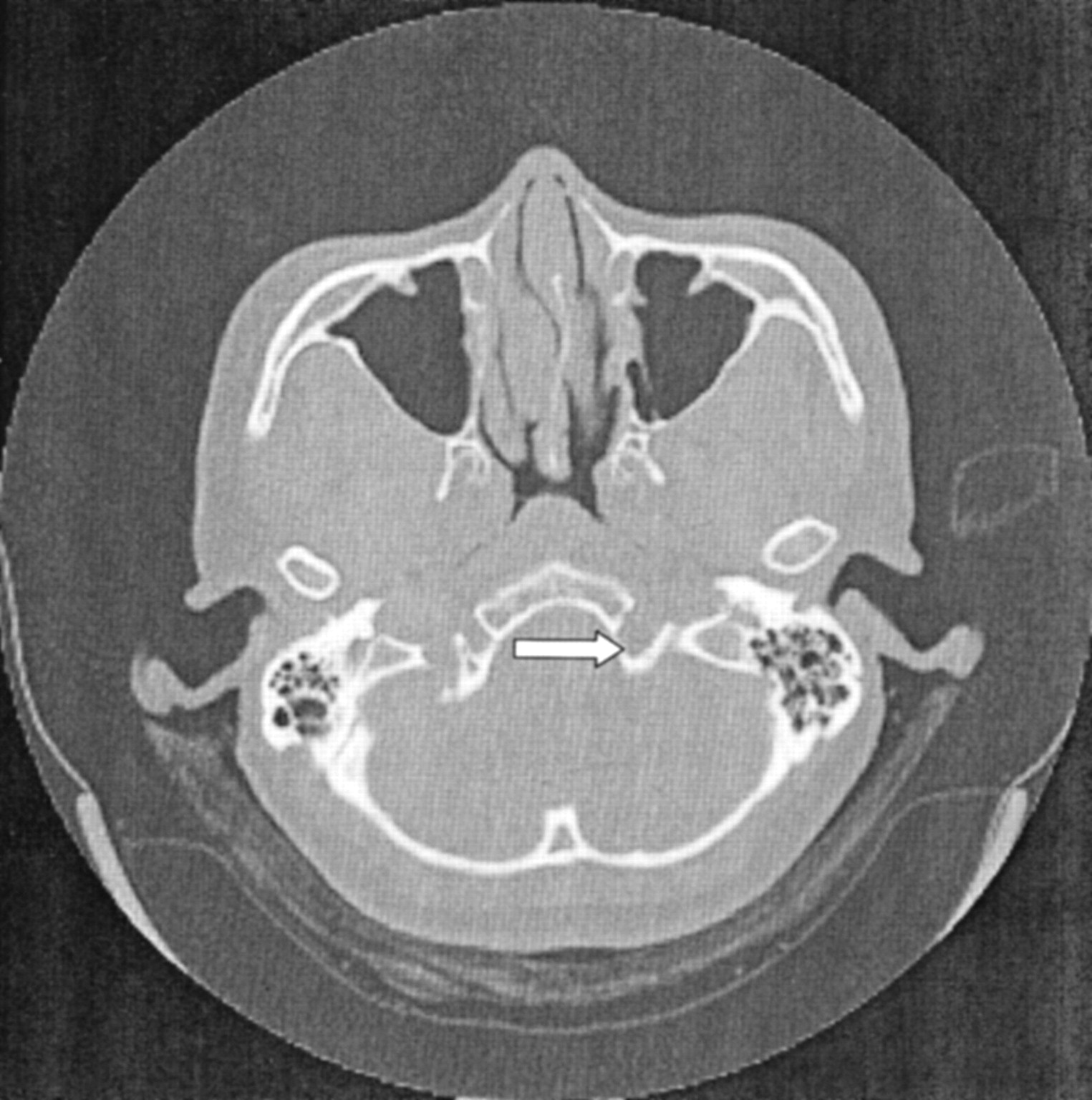
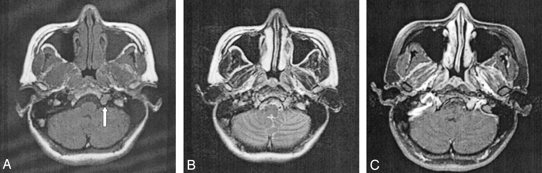
CT of the brain and skull base demonstrated a smooth widening of the left hypoglossal foramen with preservation of the cortical bone (Fig 1). No bony destruction was present in the rest of the skull base or in other cranial nerve foramina. MR imaging revealed the presence of a well-circumscribed, extra-axial tubular mass; this measured 1.5 × 0.7 × 1.5 cm and occupied the widened left hypoglossal canal. The tumor was homogeneously isointense relative to brain parenchyma on both T1- and T2-weighted images and showed uniform enhancement after the intravenous administration of contrast material (Fig 2). It did not extend into the cisternal or root entry-zone portions of cranial nerve XII. A schwannoma of the hypoglossal nerve was diagnosed with a differential diagnosis of meningioma. At surgery, a soft, fleshy, well-encapsulated tumor was found adherent to the left hypoglossal nerve along the lateral aspect. Complete surgical excision was achieved.

Fig 1.
  • Download figure
  • Open in new tab
  • Download powerpoint
Fig 1.

Axial CT scan of the skull base shows smooth, well-defined widening of the left hypoglossal canal (arrow). No bony destruction is demonstrated.

Fig 2.
  • Download figure
  • Open in new tab
  • Download powerpoint
Fig 2.

Axial MR images.

A, T1-weighted image reveals a well-circumscribed, elongated tumor occupying the hypoglossal canal (arrow). The T1 signal intensity is homogeneously isointense relative to gray matter.

B, On the corresponding T2-weighted image, the mass is also uniformly isointense relative to gray matter.

C, After the intravenous injection of contrast material, homogeneous enhancement of the tumor is observed.

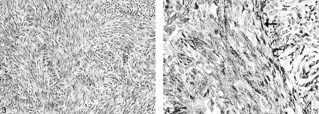
Microscopic examination showed a moderately cellular tumor composed of uniform, fibroblastlike spindle cells arranged in intersecting fascicles. Mitoses were rare, and necrosis was absent. A background of small, hyalinized blood vessels was noted. No staghorn vessels were evident (Fig 3). Immunohistochemical studies demonstrated strong diffuse positive staining of the tumor cells for CD34 (Fig. 4), but negative staining results for S100, epithelial membrane antigen (EMA), smooth-muscle actin, and desmin. This profile was indicative of SFT and excluded meningioma, nerve sheath tumor and smooth muscle tumor. An MIB-1 immunostaining assay was performed, and a labeling index of 2% was found. One year after surgery, the patient’s deficit stabilized with mild difficulty of phonation and left hemiatrophy of the tongue.

Fig 3.
  • Download figure
  • Open in new tab
  • Download powerpoint
Fig 3.

Uniform spindle cells are arranged in fascicles with thin bands of intercellular collagen (hematoxylin-eosin stain, original magnification ×100).

Fig 4.
  • Download figure
  • Open in new tab
  • Download powerpoint
Fig 4.

Tumor cells (arrow) stain positively for CD34. (original magnification ×200).

Discussion

The hypoglossal nerve is a pure motor nerve that innervates both the intrinsic and extrinsic muscles of the tongue. It is divided into five segments: medullary, cisternal, skull base, nasopharyngeal and oropharyngeal, carotid space, and sublingual. Different disorders affect each segment, and localizing a lesion to a particular segment allows the radiologist to narrow the differential diagnosis to palsy of cranial nerve XII (1).

Supranuclear hypoglossal nerve lesions do not result in denervation atrophy of the tongue muscles: Characteristic atrophy of the tongue is only seen when the nuclear or peripheral segments of the hypoglossal nerve are involved (2). The left and right hypoglossal nuclei are closely located together within the paramedian medulla and beneath the hypoglossal trigone of the fourth ventricle. Therefore, medullary lesions such as multiple sclerosis, syringobulbia, amyotrophic lateral sclerosis, and bulbar poliomyelitis often result in bilateral lower motor neuron palsy of the tongue. For lesions involving the cisternal course of cranial nerve XII, cranial nerve IX, X, and XI nerves are often also affected because they are in close spatial relationship to the hypoglossal nerve roots. In a series of 32 patients with paralysis of the hypoglossal nerve and tongue atrophy, Tommasi-Davenas et al (3) found only eight cases of isolated cranial nerve XII palsy without neurologic involvement of other the cranial nerves or the long tracts. In most cases, the cause was tumor, especially bony metastasis. In another study of nine cases of unilateral isolated hypoglossal nerve palsy, researchers found metastatic disease in three patients (4).

In its course distal to the base of skull, the nasopharyngeal, oropharyngeal, carotid, and sublingual segments of cranial nerve XII may be affected by vascular aneurysm; local infections; rheumatoid arthritis; surgical (eg, carotid endarterectomy) or accidental trauma; birth injuries; or tumors of the neck, salivary glands, or base of the tongue (2). In such situations, the abnormality is often apparent on clinical history taking or physical examination.

Our patient presented with clinical signs that clearly localized the lesion to the skull base segment of cranial nerve XII. Neuroimaging revealed a uniformly enhancing tumor of the hypoglossal nerve with expansion of the hypoglossal canal. The tumor was isointense relative to brain parenchyma on both T1- and T2-weighted images, and it showed fairly homogeneous contrast enhancement. These findings are nonspecific and may be due to a variety of neoplastic or inflammatory conditions.

In 1931, Klemperer and Rabin first described SFT in the pleura as a localized form of mesothelioma (5). Since then, SFT has been detected in different locations, including the pericardium, mediastinum, nasopharyngeal sinuses, thyroid, liver, mesentery, orbit, and nervous system. A review of the English-language literature revealed 16 cases of SFT affecting the CNS: nine cases were intracranial and seven were spinal (6–13). Of the intracranial tumors, all were meningeal based and grew to considerable size before causes symptoms of mass effect. To our knowledge, no case affecting the cranial or spinal nerves in isolation has been reported.

The histogenesis of SFT has been a matter of debate, but it is thought to arise from mesenchymal elements (14). This uncommon neoplasm consists of monomorphous sheets of spindle cells embedded in a collagenous matrix. It has no distinctive features on light microscopy; this feature makes it difficult to differentiate SFT from other spindle cell tumors such as fibrous meningioma, schwannoma, and neurofibroma. The histopathologic differential diagnosis also includes dura-based mesenchymal tumors such as hemangiopericytoma (HPC), fibroma, fibrosarcoma, and malignant fibrous histiocytoma (15–17).

The immunohistochemical features of SFT are, however, characteristic and essential in distinguishing SFT. Most important is the observation that more than 90% of SFTs contain cells that express an antigen recognized by CD34 (18). SFTs are S100- and EMA-negative, whereas meningiomas are EMA-positive and CD34-negative, and nerve sheath tumors are S100-positive and CD34-negative. In our case, the tumor was negative for EMA and S100 but strongly and diffusely positive for CD34m, consistent with SFT. A recent group (14) has suggested that SFTs are derived from CD34-positive dura-based fibroblasts. This may explain why the tumor in our patient affected only the dura-covered segment of the nerve in the hypoglossal canal; the cisternal segment, which lacks a dural covering, was spared.

Distinguishing SFT from HPC is especially important. HPC is the most common dura-based neoplasm of mesenchymal origin and accounts for 0.4% of all intracranial tumors (15). HPC is an aggressive tumor that tends to recur after resection, and it occasionally metastasizes extracranially (19). On the contrary, meningeal SFTs are slow growing, and to our knowledge, malignant meningeal SFTs have not been described (20). With the exception of hypervascularity and bone erosion, HPC may be indistinguishable from meningioma on neuroimages (8, 19). However, although HPCs may contain CD34-positive cells, distinguishing HPC from SFT is not difficult on the basis of the histopathologic findings: Unlike SFT, HPCs seldom show a fascicular arrangement of cells or the characteristic bands of intercellular collagen. Thus, the accurate diagnosis of SFT and its differentiation of SFT from HPC are important because it has implications for further treatment and the prognosis.

Conclusion

This is the first report of SFT involving a cranial nerve. Although rare, SFT may be considered in the neuroimaging differential diagnosis of tumors affecting the segments of cranial nerves covered by dura mater. Immunohistochemistry is important in differentiating SFT from the more common meningioma and schwannoma, which may mimic SFT both radiologically and histologically.

References

  1. ↵
    Thompson EO, Smoker WR. Hypoglossal nerve palsy: a segmental approach. Radiographics 1994;4:939–958
  2. ↵
    Brazis PW, Masdeu JC, Biller J. Cranial nerve XII: the hypoglossal nerve. In: Localization in Clinical Neurology. 4th ed. Philadelphia: Lippincott Williams & Wilkins;2001 :345–351
  3. ↵
    Tommasi-Davenas C, Vighetto A, Confavreux C, et al. Causes of paralysis of the hypoglossal nerve: apropos of 32 cases. Press Med 1990;19:864–868
  4. ↵
    Combarros O, Alvarez de Arcaya A, Berciano J. Isolated unilateral hypoglossal nerve palsy: nine cases. J Neurol 1998;245:98–100
    CrossRefPubMed
  5. ↵
    Klemperer P, Rabin CB. Primary neoplasms of the pleura. Arch Pathol 1931;11:385–412
  6. ↵
    Ahn JY, Shim JY, Yang WI, et al. Meningeal solitary fibrous tumor as an unusual cause of exophthalmos: case report and review of literature. Neurosurgery 2001;48:1362–1367
    PubMed
  7. Alston SR, Francel PC, Jane JA Jr. Solitary fibrous tumor of the spinal cord. Am J Surg Pathol 1997;21:477–483
    CrossRefPubMed
  8. ↵
    Brunori A, Cerasoli S, Donati R, et al. Solitary fibrous tumor of the meninges: two new cases and review of the literature. Surg Neurol 1999;51:636–640
    CrossRefPubMed
  9. Carneiro SS, Scheithauer BW, Nascimento AG, et al. Solitary fibrous tumor of the meninges: a lesion distinct from fibrous meningioma. A clinicopathologic and immunohistochemical study. Am J Clin Pathol 1996;106:217–224
    PubMed
  10. Kurtkaya O, Elmaci I, Sav A, et al. Solitary fibrous tumor: seventh reported case and review of literature. Spinal Cord 2001;39:57–60
    CrossRefPubMed
  11. Malek AM, Weller SJ, Price DL, et al. Solitary fibrous tumor presenting as a symptomatic intraspinal mass: case report. Neurosurgery 1997;40:844–847
    CrossRefPubMed
  12. Prayson RA, McMahon JT, Barnett GH. Solitary fibrous tumor of the meninges: case report and review of literature. J Neurosurg 1997;86:1049–1052
    PubMed
  13. ↵
    Vorster SJ, Prayson RA, Lee JH. Solitary fibrous tumor of the thoracic spine: case report and review of literature. J Neurosurg 2000;92:217–220
    PubMed
  14. ↵
    Cummings TJ, Burchette JL, McLendon RE. CD34 and dural fibroblasts: the relationship to solitary fibrous tumor and meningioma. Acta Neuropathol (Berl) 2001;102:349–354
    PubMed
  15. ↵
    Guthrie BL, Ebersold MJ, Scheithauer BJ, et al. Meningeal hemangiopericytoma: histopathological features, treatment and long-term follow-up of 44 cases. Neurosurgery 1989;25:514–522
    PubMed
  16. Okeda R, Mochizuki T, Terao E, et al. The origin of intracranial fibrosarcoma. Acta Neuropathol (Berl) 1980;52:223–230
    PubMed
  17. ↵
    Swamy KS, Shankar SK, Asha T, et al. Malignant fibrous histiocytoma arising from the meninges of the posterior fossa. Surg Neurol 1986;25:18–24
    PubMed
  18. ↵
    Goodlad JR, Fletcher CD. Solitary fibrous tumor arising at unusual sites: analysis of a series. Histopathology 1991;19:515–522
    PubMed
  19. ↵
    Chiechi MV, Smirniotopoulos JG, Mena H. Intracranial hemangiopericytomas: MR and CT features. AJNR Am J Neuroradiol 1996;17:1365–1371
    Abstract
  20. ↵
    Karamitopoulou E, Perentes E, Diamantis I, et al. Ki-67 immunoreactivity in human central nervous system tumors: a study with MIB 1 monoclonal antibody on archival material. Acta Neuropathol (Berl) 1994;87:47–54
    PubMed
  • Received October 22, 2002.
  • Accepted after revision October 25, 2002.
  • Copyright © American Society of Neuroradiology
View Abstract
PreviousNext
Back to top

In this issue

American Journal of Neuroradiology: 24 (3)
American Journal of Neuroradiology
Vol. 24, Issue 3
1 Mar 2003
  • Table of Contents
  • Index by author
Advertisement
Print
Download PDF
Email Article

Thank you for your interest in spreading the word on American Journal of Neuroradiology.

NOTE: We only request your email address so that the person you are recommending the page to knows that you wanted them to see it, and that it is not junk mail. We do not capture any email address.

Enter multiple addresses on separate lines or separate them with commas.
Solitary Fibrous Tumor of the Hypoglossal Nerve
(Your Name) has sent you a message from American Journal of Neuroradiology
(Your Name) thought you would like to see the American Journal of Neuroradiology web site.
CAPTCHA
This question is for testing whether or not you are a human visitor and to prevent automated spam submissions.
Cite this article
Ma Lourdes S. Badion, C. C. Tchoyoson Lim, Jennifer Teo, Peck Leong Ong, Francis Hui
Solitary Fibrous Tumor of the Hypoglossal Nerve
American Journal of Neuroradiology Mar 2003, 24 (3) 343-345;

Citation Manager Formats

  • BibTeX
  • Bookends
  • EasyBib
  • EndNote (tagged)
  • EndNote 8 (xml)
  • Medlars
  • Mendeley
  • Papers
  • RefWorks Tagged
  • Ref Manager
  • RIS
  • Zotero
0 Responses
Respond to this article
Share
Bookmark this article
Solitary Fibrous Tumor of the Hypoglossal Nerve
Ma Lourdes S. Badion, C. C. Tchoyoson Lim, Jennifer Teo, Peck Leong Ong, Francis Hui
American Journal of Neuroradiology Mar 2003, 24 (3) 343-345;
del.icio.us logo Twitter logo Facebook logo Mendeley logo
  • Tweet Widget
  • Facebook Like
  • Google Plus One

Jump to section

  • Article
    • Abstract
    • Case Report
    • Discussion
    • Conclusion
    • References
  • Figures & Data
  • Info & Metrics
  • Responses
  • References
  • PDF

Related Articles

  • No related articles found.
  • PubMed
  • Google Scholar

Cited By...

  • No citing articles found.
  • Crossref
  • Google Scholar

This article has not yet been cited by articles in journals that are participating in Crossref Cited-by Linking.

More in this TOC Section

  • FDG Uptake and Thyroid Cancer Post-BRAF Therapy
  • Normal Facial Nerve Enhancement on VIBE Sequence
  • Comprehensive Review of Inner Ear Anatomy on Photon-Counting CT
Show more Head and Neck Imaging

Similar Articles

Advertisement

Indexed Content

  • Current Issue
  • Accepted Manuscripts
  • Article Preview
  • Past Issues
  • Editorials
  • Editor's Choice
  • Fellows' Journal Club
  • Letters to the Editor
  • Video Articles

Cases

  • Case Collection
  • Archive - Case of the Week
  • Archive - Case of the Month
  • Archive - Classic Case

More from AJNR

  • Trainee Corner
  • Imaging Protocols
  • MRI Safety Corner

Multimedia

  • AJNR Podcasts
  • AJNR Scantastics

Resources

  • Turnaround Time
  • Submit a Manuscript
  • Submit a Video Article
  • Submit an eLetter to the Editor/Response
  • Manuscript Submission Guidelines
  • Statistical Tips
  • Fast Publishing of Accepted Manuscripts
  • Graphical Abstract Preparation
  • Imaging Protocol Submission
  • Evidence-Based Medicine Level Guide
  • Publishing Checklists
  • Author Policies
  • Become a Reviewer/Academy of Reviewers
  • News and Updates

About Us

  • About AJNR
  • Editorial Board
  • Editorial Board Alumni
  • Alerts
  • Permissions
  • Not an AJNR Subscriber? Join Now
  • Advertise with Us
  • Librarian Resources
  • Feedback
  • Terms and Conditions
  • AJNR Editorial Board Alumni

American Society of Neuroradiology

  • Not an ASNR Member? Join Now

© 2025 by the American Society of Neuroradiology All rights, including for text and data mining, AI training, and similar technologies, are reserved.
Print ISSN: 0195-6108 Online ISSN: 1936-959X

Powered by HighWire