
Article Figures & Data
Figures
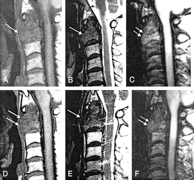
- Fig 1.
C2 metastasis in a 60-year-old male patient with renal cell carcinoma.
A, T1-weighted MR image (583/12 [TR/TE]) obtained before radiation therapy shows low signal intensity in the metastatic lesion (arrow).
B, T2-weighted MR image (3800/128) obtained before radiation therapy shows low signal intensity in the metastatic lesion (arrow).
C, Diffusion-weighted MR image (TR, 21.6; diffusion pulse length, 2 ms) obtained before radiation therapy shows slight hyperintensity (arrows) relative to normal vertebral bone marrow.
D, Follow-up T1-weighted MR image reveals persistent hypointensity (arrows) 1 month after therapy.
E, Follow-up T2-weighted MR image reveals persistent hypointensity (arrow) 1 month after therapy.
F, Follow-up diffusion-weighted MR image obtained 1 month after therapy shows iso- to hypointense signal (arrows). In a case of metastasis to the spine with clinical improvement, diffusion-weighted MR image shows decreased signal intensity change whereas conventional spin-echo T1- or T2-weighted MR images are not completely conclusive for monitoring the response.
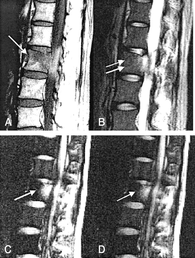
- Fig 2.
T2 metastasis in a 36-year-old female patient with invasive ductal carcinoma of the breast.
A, T1-weighted MR image (583/12) obtained before radiation therapy shows focal hypointense metastatic lesion (arrow) in right posterior portion of the vertebral body.
B, T2-weighted MR image (3800/128) obtained before radiation therapy shows focal hypointense metastatic lesion (arrow) in right posterior portion of the vertebral body.
C, Diffusion-weighted MR image (TR, 21.6; diffusion pulse length, 2 ms) obtained before radiation therapy reveals hyperintensity (arrows) relative to normal bone marrow.
D, Follow-up T1-weighted MR image shows persistent hypointense signal (arrow).
E, T2-weighted MR image reveals hyperintense signal (arrow).
F, Diffusion-weighted MR image shows hypointensity (arrow) relative to normal bone marrow 2 months after therapy.
G, Diffusion-weighted MR image obtained 3 months after therapy shows more hypointense change (arrows). Longer follow-up diffusion-weighted MR image shows more hypointense change in metastasis to the spine with clinical improvement.
- Fig 3.
L2 metastasis with no response to therapy in a 37-year-old female patient with hepatocellular carcinoma.
A, T1-weighted MR image (583/12) obtained before radiation therapy shows hypointense change (arrow).
B, T2-weighted MR image (3800/128) obtained before radiation therapy shows high signal intensity (arrows).
C, Diffusion-weighted MR image obtained before radiation therapy shows hyperintense metastatic tumor (arrow).
D, Diffusion-weighted MR image obtained after radiation therapy shows hyperintense metastatic tumor (arrow). This patient experienced persistent back pain and had no decreased uptake shown on a bone scan obtained 6 months after therapy. With metastasis to the spine without clinical improvement, persistent hyperintense bone marrow on follow-up diffusion-weighted MR images obtained after therapy is noted.
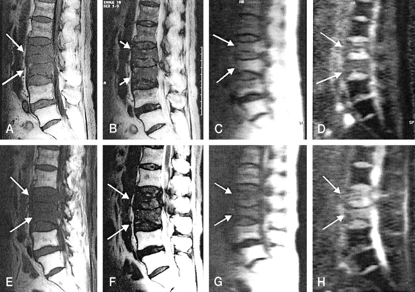
- Fig 4.
L2 and L3 metastasis in 70-year-old female patient with uterine cervix carcinoma.
A, T1-weighted MR image (583/12) obtained before radiation therapy shows low signal intensity in metastatic diseases (arrows). Hyperintense bone marrow caused by previous radiation therapy in the pelvic cavity can be seen.
B, T2-weighted MR image (3800/128) obtained before radiation therapy shows low signal intensity in metastatic diseases (arrows).
C, Diffusion-weighted MR image with high b value (650 s/mm2), obtained before therapy, shows slight high signal intensity in the metastatic disease (arrows).
D, Metastatic disease before therapy reveals high signal intensity on ADC maps (arrows). ADC value is (0.82 ± 0.03) × 10−3 mm2/s.
E, T1-weighted MR image (583/12) obtained after radiation therapy shows low signal intensity in metastatic diseases (arrows).
F, T2-weighted MR image (3800/128) obtained after radiation therapy shows low signal intensity in metastatic diseases (arrows).
G, Diffusion-weighted MR image with high b value (650 s/mm2), obtained after therapy, shows hypointensity in metastatic diseases (arrows).
H, ADC maps obtained after therapy reveal high signal intensity (arrows), and the ADC value ([1.33 ± 0.09] × 10−3 mm2/s) is increased. Normal bone marrow on ADC maps appear dark before therapy (as shown in D) and after therapy (as can be seen in this image) because of minimal diffusion of normal bone marrow. On ADC maps, ADC values after therapy are increased compared with those before therapy because of an increased extracellular volume fraction caused by necrosis of the tumor cells and bone marrow elements.


