
Article Figures & Data
Figures
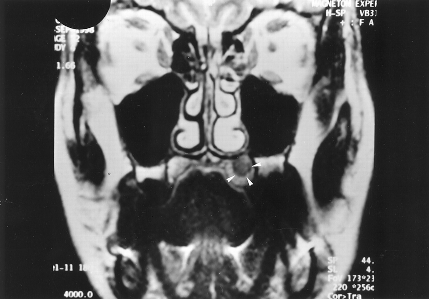
- Fig 1.
Coronal T2-weighted (TR/TE/NEX, 4000/99/4) MR image 1 year after initial presentation shows an oval, left palatine lesion (arrowheads) that is hyperintense relative to muscle.
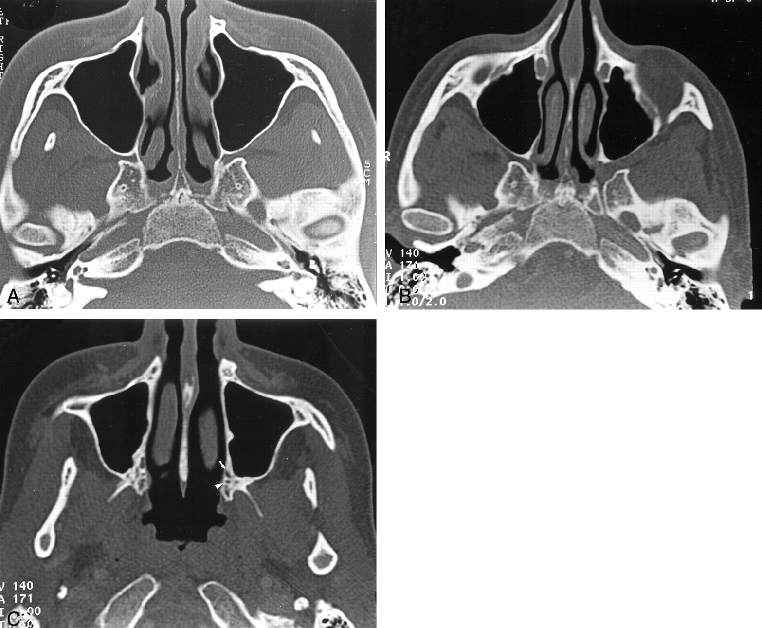
- Fig 2.
Axial bone-window CT scans.
A and B, Images obtained through the pterygopalatine fossa at initial presentation (A) and 14 months later (B) show widening of the left pterygopalatine fossa.
C, Image obtained through the palatine foramen at the same time as the image in B shows no abnormality of the left greater (arrow) and lesser (arrowhead) palatine foramina.
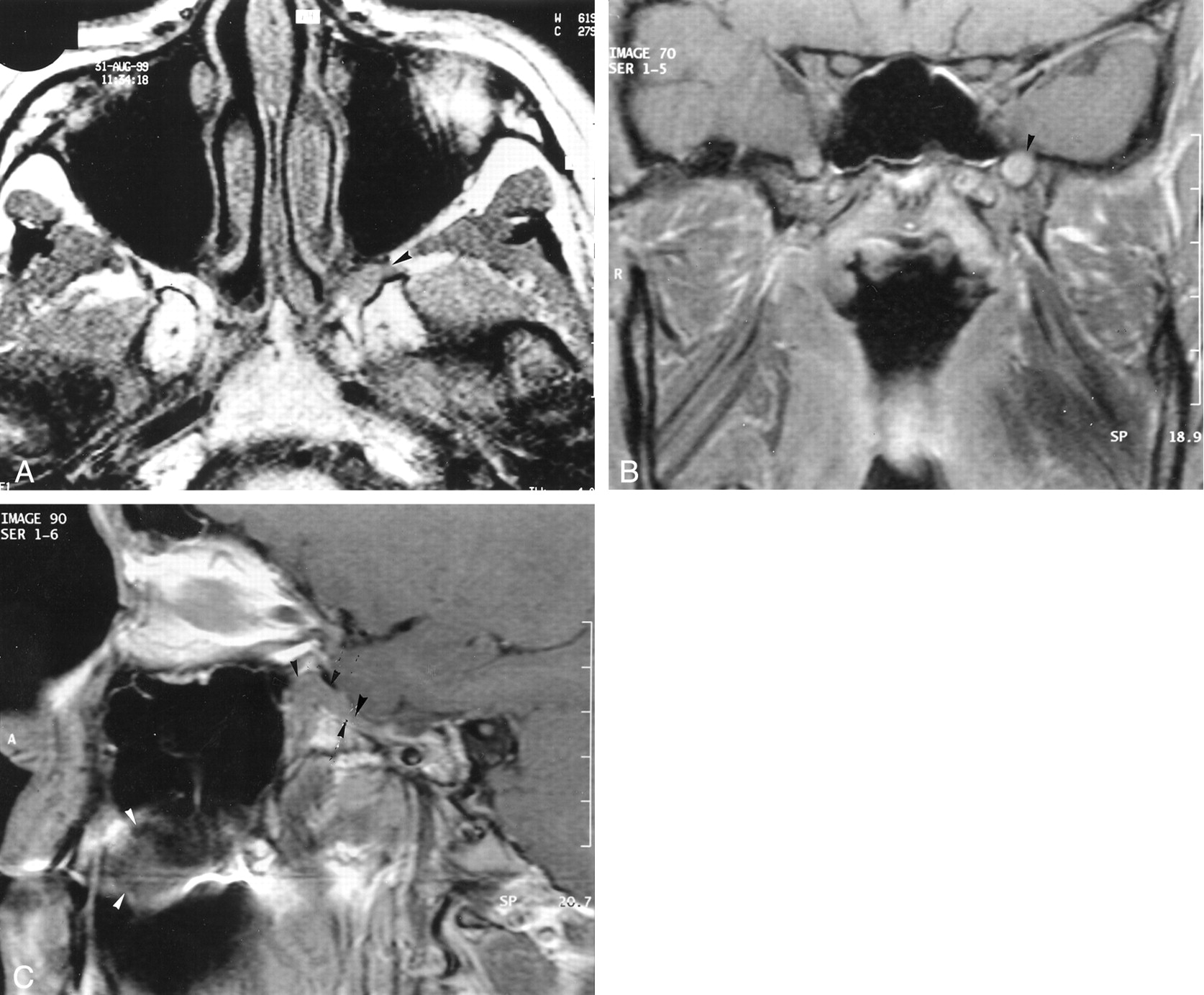
- Fig 3.
Coronal T1-weighted (550/15/4) MR images obtained at the same time.
A, Image obtained 14 months after the onset of symptoms shows a left subtle submucosal palatine mass that is isointense to muscle. Abnormal intermediate signal intensity of the surrounding bone (arrowhead) is depicted.
B, Image shows a discrete enhancement of the left palatine lesion (black arrowheads). Its margins are better delineated on this image. Intermediate signal intensity of the surrounding bone (white arrowhead) is depicted.
- Fig 4.
T1-weighted MR images.
A, Axial image (520/14/4) obtained 14 months after the onset of symptoms shows loss of the normal fat signal hyperintensity in the left pterygopalatine fossa (arrowhead). This finding represents infiltration of the latter by a lesion that is isointense relative to muscle.
B, Coronal enhanced image (520/14/3) obtained through the base of the skull. The left foramen rotundum is enlarged and infiltrated by a discrete, hyperintense, soft-tissue mass (arrowhead).
C, Sagittal enhanced image (520/14/3) obtained through the left pterygopalatine fossa shows the palate lesion (white arrowheads), which extends through a widened foramen rotundum into the pterygopalatine fossa (black arrowheads).
- Fig 5.
Photomicrographs (magnification ×100).
A, Well-organized clusters of eosinophilic granular cells (arrowhead) are depicted within haversian bone (hematein phloxine safranin).
B, At a higher magnification, the granular cells are distributed in small clusters. The cells have an elongated or spindle shape, with abundant cytoplasm and small basophilic nuclei (arrowhead). No high-grade nuclear atypia, necrosis, or mitoses are present (hematoxylin-eosin with S-100 protein).
- Fig 6.
Enhanced fat-saturated coronal T1-weighted MR image (520/14/3) obtained 6 months after the image in (Figure 3 shows enlargement of the lesion of the left pterygopalatine fossa arrowhead.


