
Article Figures & Data
Figures
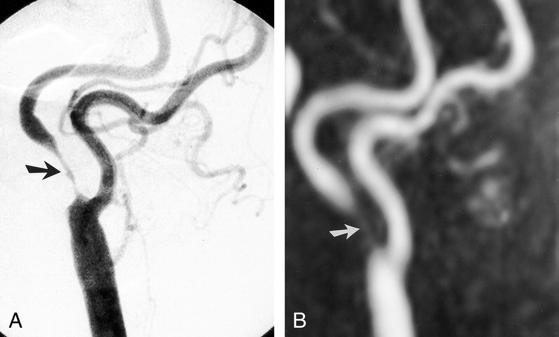
- Fig 1.
Agreement between findings at DSA and those at MR angiography.
A, DSA image of the right carotid bifurcation shows a focal severe stenosis (arrow) of the internal carotid artery.
B, The 3D contrast-enhanced MR angiographic MIP image (magnification factor, 2) shows the focal severe stenosis (arrow), correlating well with the DSA image.
- Fig 2.
Underestimation of stenoses.
A, DSA image of the left common carotid artery depicts a proximal moderate stenosis (straight arrow) and an extremely short distal stenosis (curved arrow) of the internal carotid artery.
B, 3D contrast-enhanced MR angiographic MIP image (magnification factor, 2) depicts the moderate proximal stenosis (straight arrow) and a short band of decreased enhancement (curved arrow) without clear definition of a stenosis.
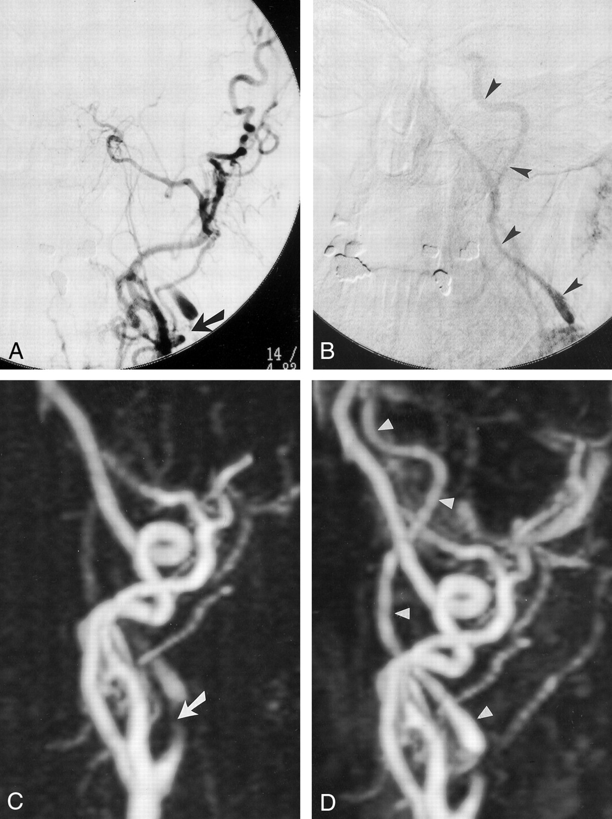
- Fig 3.
Carotid occlusions.
A and B, DSA images and, C and D, 3D contrast-enhanced MR angiographic MIP images (magnification factor, 1.5) of the left common carotid artery in the early (A and C) and delayed (B and D) phases demonstrate a stump (arrow) at the origin of the internal carotid artery and occlusion of the distal internal carotid artery.
- Fig 4.
A and B, DSA images of the left common carotid artery demonstrate an extremely severe stenosis (arrow) at the origin f the internal carotid artery in the early phase (A), with collapse of the distal lumen (“string” sign [arrowheads]) in the delayed phase (B).
C and D, The 3D contrast-enhanced MR angiographic MIP images (1.5 magnification factor) demonstrate the extremely severe stenosis (arrow in C) at the origin of the internal carotid artery and opacification of the collapsed distal lumen in only the delayed phase (arrowheads in D).


Tables
Agreement of Contrast-Enhanced MR Angiography with DSA in Evaluation of Carotid Artery Stenosis
Stenosis Grade DSA MRA Agreement (%) Estimation Over Under Mild (0–29%) 101 91 90 10 0 Moderate (30–69%) 38 26 68 6 6 Severe (70–99%) 73 68 93 2 3 Occlusion (100%) 28 28 100 0 0 Note.—MRA indicates MR angiography. Except for agreement, data are the number of arteries.
In this issue


Jump to section
Related Articles
Cited By...
- Appropriate Minimal Dose of Gadobutrol for 3D Time-Resolved MRA of the Supra-Aortic Arteries: Comparison with Conventional Single-Phase High-Resolution 3D Contrast-Enhanced MRA
- Detection of Carotid Artery Stenosis: A Comparison between 2 Unenhanced MRAs and Dual-Source CTA
- Molecular Imaging Changes with Cognition
- Multicenter, Intraindividual Comparison of Single-Dose Gadobenate Dimeglumine and Double-Dose Gadopentetate Dimeglumine for MR Angiography of the Supra-Aortic Arteries (the Supra-Aortic VALUE Study)
- 3D Computerized Occlusion Rating of Embolized Experimental Aneurysms Using Noninvasive 1.5T MR Imaging
- 2011 ASA/ACCF/AHA/AANN/AANS/ACR/ASNR/CNS/SAIP/SCAI/SIR/SNIS/SVM/SVS Guideline on the Management of Patients With Extracranial Carotid and Vertebral Artery Disease: A Report of the American College of Cardiology Foundation/American Heart Association Task Force on Practice Guidelines, and the American Stroke Association, American Association of Neuroscience Nurses, American Association of Neurological Surgeons, American College of Radiology, American Society of Neuroradiology, Congress of Neurological Surgeons, Society of Atherosclerosis Imaging and Prevention, Society for Cardiovascular Angiography and Interventions, Society of Interventional Radiology, Society of NeuroInterventional Surgery, Society for Vascular Medicine, and Society for Vascular Surgery
- 2011 ASA/ACCF/AHA/AANN/AANS/ACR/ASNR/CNS/SAIP/SCAI/SIR/SNIS/SVM/SVS Guideline on the Management of Patients With Extracranial Carotid and Vertebral Artery Disease: A Report of the American College of Cardiology Foundation/American Heart Association Task Force on Practice Guidelines, and the American Stroke Association, American Association of Neuroscience Nurses, American Association of Neurological Surgeons, American College of Radiology, American Society of Neuroradiology, Congress of Neurological Surgeons, Society of Atherosclerosis Imaging and Prevention, Society for Cardiovascular Angiography and Interventions, Society of Interventional Radiology, Society of NeuroInterventional Surgery, Society for Vascular Medicine, and Society for Vascular Surgery
- 2011 ASA/ACCF/AHA/AANN/AANS/ACR/ASNR/CNS/SAIP/SCAI/SIR/SNIS/SVM/SVS Guideline on the Management of Patients With Extracranial Carotid and Vertebral Artery Disease: A Report of the American College of Cardiology Foundation/American Heart Association Task Force on Practice Guidelines, and the American Stroke Association, American Association of Neuroscience Nurses, American Association of Neurological Surgeons, American College of Radiology, American Society of Neuroradiology, Congress of Neurological Surgeons, Society of Atherosclerosis Imaging and Prevention, Society for Cardiovascular Angiography and Interventions, Society of Interventional Radiology, Society of NeuroInterventional Surgery, Society for Vascular Medicine, and Society for Vascular Surgery Developed in Collaboration With the American Academy of Neurology and Society of Cardiovascular Computed Tomography
- Contrast-Enhanced MR Angiography Is Not More Accurate Than Unenhanced 2D Time-of-Flight MR Angiography for Determining >=70% Internal Carotid Artery Stenosis
- Diagnostic Accuracy of Magnetic Resonance Angiography for Internal Carotid Artery Disease: A Systematic Review and Meta-Analysis
- Carotid Stenosis Index Revisited With Direct CT Angiography Measurement of Carotid Arteries to Quantify Carotid Stenosis
- ACCF/SCAI/SVMB/SIR/ASITN 2007 Clinical Expert Consensus Document on Carotid Stenting: A Report of the American College of Cardiology Foundation Task Force on Clinical Expert Consensus Documents (ACCF/SCAI/SVMB/SIR/ASITN Clinical Expert Consensus Document Committee on Carotid Stenting)
- Comparison of image quality, diagnostic confidence and interobserver variability in contrast enhanced MR angiography and 2D time of flight angiography in evaluation of carotid stenosis.
- Assessment of CE-MRA for the rapid detection of supra-aortic vascular disease
- Measuring Carotid Stenosis on Contrast-Enhanced Magnetic Resonance Angiography: Diagnostic Performance and Reproducibility of 3 Different Methods
- Contrast-enhanced MR angiography for carotid disease: Diagnostic and potential clinical impact
- Preoperative Evaluation of Carotid Artery Stenosis: Comparison of Contrast-Enhanced MR Angiography and Duplex Sonography with Digital Subtraction Angiography
- Contrast-Enhanced Magnetic Resonance Angiography Carotid Arteries * Response
- Patient Selection for Carotid Endarterectomy: How Far Is Risk Modeling Applicable to the Individual?