
Article Figures & Data
Figures
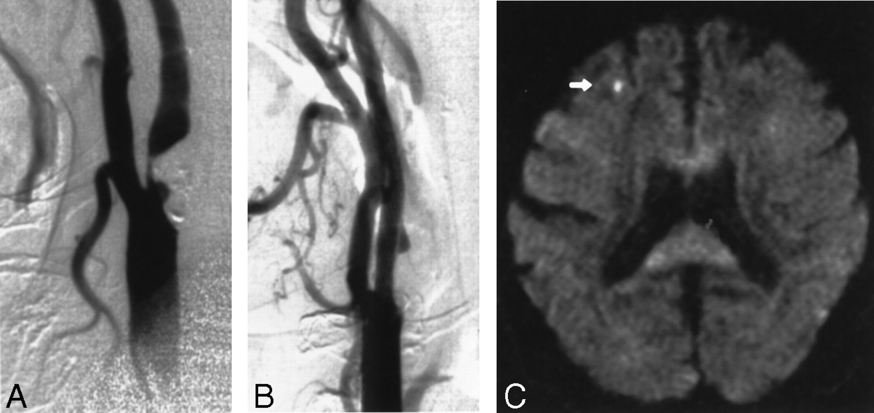
- Fig 1.
Images obtained in a 70-year-old man with an asymptomatic stenosis of the carotid artery.
A, Right anterior oblique angiogram shows a 94% stenosis of the right ICA.
B, Right anterior oblique angiogram shows the result after stent implantation.
C, Postprocedural axial diffusion-weighted MR image (6000/103/1) shows a new ipsilateral lesion (<5 mm) in the cortical territory of the MCA (arrow).
- Fig 2.
Images obtained in a 86-year-old man with a symptomatic stenosis of the carotid artery.
A, Left anterior oblique angiogram (transbrachial approach) shows an 87% stenosis of the left ICA.
B, Left anterior oblique angiogram shows the result after stent implantation.
C, Postprocedural axial diffusion-weighted MR image (6000/103/1) shows six new ipsilateral lesions (5–10 mm) in the cortical territory of the ACA (arrow).
- Fig 3.
Images obtained in a 73-year-old woman with a symptomatic stenosis of the carotid artery.
A, Left anterior oblique angiogram shows an 85% stenosis of the left ICA.
B, Left anterior oblique angiogram shows the result after stent implantation.
C, Postprocedural axial diffusion-weighted MR image (6000/103/1) shows eight new ipsilateral lesions (15–20 mm) in the cortical territory of the MCA (arrow).
D, Postprocedural axial T2-weighted MR image (5700/119/1) obtained at a corresponding level shows a new area of hyperintensity (arrow) that was not present in C.
- Fig 4.
Images obtained in a 76-year-old woman with a symptomatic stenosis of the carotid artery.
A, Left anterior oblique angiogram shows a 79% stenosis of the left ICA.
B, Left anterior oblique angiogram shows the result after stent implantation.
C, Lateral angiogram shows the intracranial circulation of the treated side after a procedure with a nonocclusive embolus in a branch of the MCA (arrow). The patient was symptomatic during the whole procedure. An angiogram of the intracranial circulation obtained after the infusion of 100,000 IU of urokinase into the ICA demonstrated complete disappearance of the embolus (not shown).
D, Postprocedural axial diffusion-weighted MR image (6000/103/1) shows eight new ipsilateral lesions (5–10 mm) in the cortical territory of the MCA (arrow).


Tables
Characteristics Patients P Value* All (n = 67) With Positive DW Findings (n = 47) With Negative DW Findings (n = 20) Demographic Sex .382 Male 47 (70) 31 (66) 16 (80) NA Female 20 (30) 16 (34) 4 (20) NA Age (y)† 67 ± 9.1 (44–86) 66 ± 9.3 (44–86) 70 ± 7.8 (57–86) .076 Medical history Hypertension 57 (85) 39 (83) 18 (90) .711 Coronary artery disease 45 (67) 30 (64) 15 (75) .412 Peripheral vascular disease 35 (52) 26 (55) 9 (45) .594 Diabetes mellitus 28 (42) 21 (45) 7 (35) .591 Hypocholesterolemia 53 (79) 39 (83) 14 (70) .325 Cardiac arrhythmia 20 (30) 14 (30) 6 (30) >.99 T2-weighted MR imaging Cerebral atrophy 46 (69) 31 (66) 15 (75) .571 Subcortical arteriosclerotic encephalopathy 58 (87) 41 (87) 17 (85) >.99 Cerebral infarction Ipsilateral 13 (19) 8 (17) 5 (25) .507 Contralateral 9 (13) 7 (15) 2 (10) .714 Note.—Data in parentheses are percentages, unless otherwise specified.
* NA indicates not applicable.
† Data are the mean ± SD. Data in parentheses are the range.
Characteristic Procedures P Value* All (n = 70) With Positive DW Findings (n = 50) With Negative DW Findings (n = 20) Symptoms Present 52 (75) 38 (76) 14 (70) .763 Present in the last 3 mo 42 (60) 30 (60) 12 (60) >.99 Absent 18 (25) 12 (24) 6 (30) .763 Cause .408 Atherosclerotic 63 (90) 45 (90) 18 (90) NA Surgery 6 (9) 5 (10) 1 (5) NA Irradiation 1 (1) 0 (0) 1 (5) NA Side .285 Right 40 (57) 31 (62) 9 (45) NA Left 30 (43) 19 (38) 11 (55) NA Grade (%)† 84 ± 7.9 (60–98) 83 ± 7.3 (60–98) 86 ± 9.3 (62–98) .131 Classification .493 0–29% 0 (0) 0 (0) 0 (0) NA 30–69% 2 (3) 1 (2) 1 (5) NA 70–99% 68 (97) 49 (98) 19 (95) NA Location .182 ICA at bifurcation 35 (50) 24 (48) 11 (55) NA Proximal ICA 32 (46) 25 (50) 7 (35) NA CCA 3 (4) 1 (2) 2 (10) NA Length .259 <1 cm 47 (67) 36 (72) 11 (55) NA >1 cm 23 (33) 14 (28) 9 (45) NA Morphology .796 Eccentric 37 (53) 27 (54) 10 (50) NA Concentric 33 (47) 23 (46) 10 (50) NA Ulcerations 17 (24) 14 (28) 3 (15) .359 Contralateral ICA occlusion 6 (9) 5 (10) 1 (5) .666 Note.—Data in parentheses are percentages, unless otherwise specified.
* NA indicates not applicable.
† Data are the mean ± SD. Data in parentheses are the range.
Detail Procedures P Value* All (n = 70) With Positive DW Findings (n = 50) With Negative DW Findings (n = 20) Access .194 Transfemoral 67 (96) 49 (98) 18 (90) NA Transbrachial 3 (4) 1 (2) 2 (10) NA Technique .107 OTW 61 (87) 46 (92) 15 (75) NA Long sheath 9 (13) 4 (8) 5 (25) NA Predilation 65 (93) 47 (94) 18 (90) .619 Type of Wallstent .551 Rolling membrane 10 (14) 6 (12) 4 (20) NA Easy 44 (63) 33 (66) 11 (55) NA OTW 16 (23) 11 (22) 5 (25) NA Location of stent .122 ICA 9 (13) 6 (12) 3 (15) NA ICA and/or CCA 59 (84) 44 (88) 15 (75) NA CCA 2 (3) 0 (0) 2 (10) NA Postdilation 61 (87) 44 (88) 17 (85) .708 Grade of remaining stenosis (%)† 5.8 ± 7.7 (0–29) 5.5 ± 6.8 (0–29) 6.6 ± 9.8 (0–29) .889 Note.—Data in parentheses are percentages, unless otherwise specified.
* NA indicates not applicable.
† Data are the mean ± SD. Data in parentheses are the range.
Characteristic Lesion Ipsilateral Preprocedural Contralateral Postprocedural Ipsilateral Contralateral Procedures with lesions* 10 (14 [7, 25]) 1 (1 [0, 7]) 20 (29 [19, 41]) 6 (9 [4, 17]) No. of lesions Total 24 1 52 7 Average 2.4 NA 2.6 1.2 Range 1–6 NA 1–8 1–2 Size <5 mm 10 (42) 0 (0) 36 (69) 6 (86) 5–10 mm 7 (29) 0 (0) 11 (21) 0 (0) >10 mm 7 (29) 1 (100) 5 (10) 1 (14) Location Vertical distribution Upper area of brain 15 (63) 1 (100) 27 (52) 4 (57) Middle area of brain 9 (38) 0 (0) 20 (39) 3 (43) Lower area of brain 0 (0) 0 (0) 5 (10) 0 (0) Horizontal distribution Cortical or subcortical area 24 (1000) 1 (100) 50 (96) 6 (86) Deep area 0 (0) 0 (0) 2 (4) 1 (14) Cerebral distribution Frontal lobe 0 (0) 0 (0) 4 (8) 3 (43) Parietal lobe 23 (96) 1 (100) 38 (73) 2 (29) Temporal lobe 1 (4) 0 (0) 5 (10) 0 (0) Occipital lobe 0 (0) 0 (0) 3 (6) 1 (14) Basal ganglia 0 (0) 0 (0) 1 (2) 0 (0) Thalamus 0 (0) 0 (0) 1 (2) 1 (14) Vascular distribution† ACA cortical branches 1 (4) 1 (100) 5 (10) 2 (29) ACA deep branches 0 (0) 0 (0) 1 (2) 0 (0) MCA cortical branches 23 (96) 0 (0) 42 (82) 3 (43) MCA deep branches 0 (0) 0 (0) 0 (0) 0 (0) PCA cortical branches 0 (0) 0 (0) 3 (6) 1 (14) PCA deep branches 0 (0) 0 (0) 1 (2) 1 (14) Note.—Total of procedures was 70. Data in parentheses are percentages, unless otherwise specified. NA indicates not applicable.
* Data in brackets are the 95% confidence intervals.
† PCA indicates posterior cerebral artery.


