Advertisement

Main menu

  • Home
  • Content
    • Current Issue
    • Accepted Manuscripts
    • Article Preview
    • Past Issue Archive
    • Video Articles
    • AJNR Case Collection
    • Case of the Week Archive
    • Case of the Month Archive
    • Classic Case Archive
  • Special Collections
    • AJNR Awards
    • Low-Field MRI
    • Alzheimer Disease
    • ASNR Foundation Special Collection
    • Photon-Counting CT
    • View All
  • Multimedia
    • AJNR Podcasts
    • AJNR SCANtastic
    • Trainee Corner
    • MRI Safety Corner
    • Imaging Protocols
  • For Authors
    • Submit a Manuscript
    • Submit a Video Article
    • Submit an eLetter to the Editor/Response
    • Manuscript Submission Guidelines
    • Statistical Tips
    • Fast Publishing of Accepted Manuscripts
    • Graphical Abstract Preparation
    • Imaging Protocol Submission
    • Author Policies
  • About Us
    • About AJNR
    • Editorial Board
    • Editorial Board Alumni
  • More
    • Become a Reviewer/Academy of Reviewers
    • Subscribers
    • Permissions
    • Alerts
    • Feedback
    • Advertisers
    • ASNR Home

User menu

  • Alerts
  • Log in

Search

  • Advanced search
American Journal of Neuroradiology

American Journal of Neuroradiology

ASHNR American Society of Functional Neuroradiology ASHNR American Society of Pediatric Neuroradiology ASSR
  • Alerts
  • Log in

Advanced Search

  • Home
  • Content
    • Current Issue
    • Accepted Manuscripts
    • Article Preview
    • Past Issue Archive
    • Video Articles
    • AJNR Case Collection
    • Case of the Week Archive
    • Case of the Month Archive
    • Classic Case Archive
  • Special Collections
    • AJNR Awards
    • Low-Field MRI
    • Alzheimer Disease
    • ASNR Foundation Special Collection
    • Photon-Counting CT
    • View All
  • Multimedia
    • AJNR Podcasts
    • AJNR SCANtastic
    • Trainee Corner
    • MRI Safety Corner
    • Imaging Protocols
  • For Authors
    • Submit a Manuscript
    • Submit a Video Article
    • Submit an eLetter to the Editor/Response
    • Manuscript Submission Guidelines
    • Statistical Tips
    • Fast Publishing of Accepted Manuscripts
    • Graphical Abstract Preparation
    • Imaging Protocol Submission
    • Author Policies
  • About Us
    • About AJNR
    • Editorial Board
    • Editorial Board Alumni
  • More
    • Become a Reviewer/Academy of Reviewers
    • Subscribers
    • Permissions
    • Alerts
    • Feedback
    • Advertisers
    • ASNR Home
  • Follow AJNR on Twitter
  • Visit AJNR on Facebook
  • Follow AJNR on Instagram
  • Join AJNR on LinkedIn
  • RSS Feeds

AJNR Awards, New Junior Editors, and more. Read the latest AJNR updates

Research ArticleNeurointervention

Endovascular Treatment Using Low-Power Ultraviolet Laser for Delayed Vasospasm in the Rabbit Carotid Artery Model

Kanji Nakai, Yuji Numaguchi, Thomas H. Foster, Katsuji Shima and Makoto Kikuchi
American Journal of Neuroradiology November 2002, 23 (10) 1725-1731;
Kanji Nakai
  • Find this author on Google Scholar
  • Find this author on PubMed
  • Search for this author on this site
Yuji Numaguchi
  • Find this author on Google Scholar
  • Find this author on PubMed
  • Search for this author on this site
Thomas H. Foster
  • Find this author on Google Scholar
  • Find this author on PubMed
  • Search for this author on this site
Katsuji Shima
  • Find this author on Google Scholar
  • Find this author on PubMed
  • Search for this author on this site
Makoto Kikuchi
  • Find this author on Google Scholar
  • Find this author on PubMed
  • Search for this author on this site
  • Article
  • Figures & Data
  • Info & Metrics
  • Responses
  • References
  • PDF
Loading

Article Figures & Data

Figures

  • Tables
  • Fig 1.
    • Download figure
    • Open in new tab
    • Download powerpoint
    Fig 1.

    Specially developed equipment used in this study.

    A, Photograph of optic fiber. The fiber can deliver laser energy and diffuse UV or visible light in a cylindrical pattern from a diffusing section. The diameter of the optical fiber is 0.49 mm (0.019 in). Scale bar, 5 mm.

    B, Illustration of endovascular UV irradiation. The blood was hand-flushed with saline to irradiate the entire arterial wall homogeneously with UV or visible light.

  • Fig 2.
    • Download figure
    • Open in new tab
    • Download powerpoint
    Fig 2.

    Bar graph shows the preventive effect of UV on the development of vasospasm for different irradiation time periods. Four groups are shown, for which the irradiation time was 0, 5, 10, or 20 seconds. UV irradiation with durations of 10 and 20 seconds significantly prevented the development of vasospasm (P < .001). Values represent the ratio of the relative diameter in each group on day 2 to that on day 0. The preventive effect seems to be close to maximum at an irradiation period of 10 seconds. Bars represent mean ± SD. *, P < .05 compared with no irradiation.

  • Fig 3.
    • Download figure
    • Open in new tab
    • Download powerpoint
    Fig 3.

    Bar graph shows values that represent the ratio of the RD in each group on each point to that on day 0 and represent mean ± SD.

    A, Preventive effect of UV on the development of vasospasm among different treatment groups on day 2. UV treatment significantly inhibited vasospasm (P < .001 compared with the VS + fiber group). Bar graph shows that the preventive effect on vasospasm was not due to manipulation (VS + fiber group) but to UV irradiation (VS + UV group). *, P < .05 compared with the VS + fiber group.

    B, Time course of %RD among different treatment groups.

  • Fig 4.
    • Download figure
    • Open in new tab
    • Download powerpoint
    Fig 4.

    Bar graph shows the preventive effect of UV or visible light on the development of vasospasm. Values represent the ratio of the RD in each group on each point to that on day 0 and represent mean ± SD. Although visible light significantly prevented vasospasm (P = .045), UV light was more effective. *, P < .05; **, P < .01 compared with the no irradiation (VS + fiber) group.
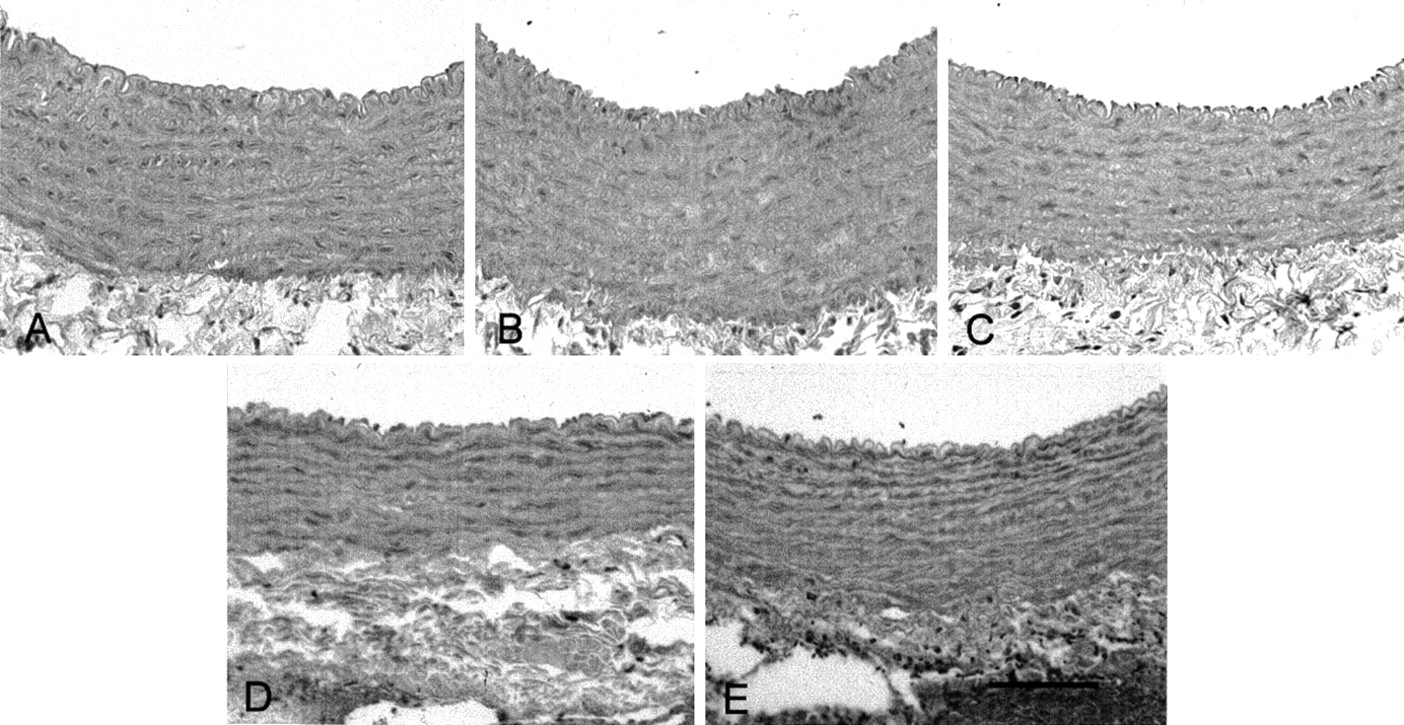
  • Fig 5.
    • Download figure
    • Open in new tab
    • Download powerpoint
    Fig 5.

    Photomicrographs show cross sections of rabbit CCAs on day 2. Hematoxylin and eosin stain was used. Placement of autologous blood around CCAs induced folding of the endothelial surface, the corrugation of the internal elastic lamina, contraction of smooth muscle cells, and thickening of the arterial wall compared with no blood placement. In the UV-treated vessel, there is less folding of the internal elastic lamina and the smooth muscle cells appear relaxed. Scale bar, 100 μm.

    A, VS group.

    B, VS + fiber group.

    C, Wrap group.

    D, Wrap + UV group.

    E, VS + UV group.

Tables

  • Figures
  • Summary of physiological parameters

    GroupMABPPCO2PO2
    Wrap79.0 ± 12.734.4 ± 2.488.3 ± 9.9
    Wrap + UV (10 s)70.6 ± 11.736.0 ± 3.096.6 ± 13.5
    VS80.2 ± 10.936.8 ± 2.077.5 ± 8.0
    VS + fiber71.3 ± 6.833.4 ± 1.889.9 ± 9.8
    VS + UV (5 s)79.2 ± 11.033.3 ± 3.186.8 ± 0.8
    VS + UV (10 s)75.2 ± 9.534.2 ± 1.692.7 ± 8.4
    VS + UV (20 s)75.0 ± 9.835.0 ± 2.288.5 ± 13.6
    VS + 442 nm71.9 ± 9.635.3 ± 1.988.5 ± 12.0
    Correlation coefficient−0.1210.0170.285
    • Note.—All values are mean ± SD. MABP indicates mean arterial blood pressure; PCO2, partial pressure of carbon dioxide; PO2, partial pressure of oxygen; UV, ultraviolet; VS, vasospasm.

PreviousNext
Back to top

In this issue

American Journal of Neuroradiology: 23 (10)
American Journal of Neuroradiology
Vol. 23, Issue 10
1 Nov 2002
  • Table of Contents
  • Index by author
Advertisement
Print
Download PDF
Email Article

Thank you for your interest in spreading the word on American Journal of Neuroradiology.

NOTE: We only request your email address so that the person you are recommending the page to knows that you wanted them to see it, and that it is not junk mail. We do not capture any email address.

Enter multiple addresses on separate lines or separate them with commas.
Endovascular Treatment Using Low-Power Ultraviolet Laser for Delayed Vasospasm in the Rabbit Carotid Artery Model
(Your Name) has sent you a message from American Journal of Neuroradiology
(Your Name) thought you would like to see the American Journal of Neuroradiology web site.
CAPTCHA
This question is for testing whether or not you are a human visitor and to prevent automated spam submissions.
Cite this article
Kanji Nakai, Yuji Numaguchi, Thomas H. Foster, Katsuji Shima, Makoto Kikuchi
Endovascular Treatment Using Low-Power Ultraviolet Laser for Delayed Vasospasm in the Rabbit Carotid Artery Model
American Journal of Neuroradiology Nov 2002, 23 (10) 1725-1731;

Citation Manager Formats

  • BibTeX
  • Bookends
  • EasyBib
  • EndNote (tagged)
  • EndNote 8 (xml)
  • Medlars
  • Mendeley
  • Papers
  • RefWorks Tagged
  • Ref Manager
  • RIS
  • Zotero
0 Responses
Respond to this article
Share
Bookmark this article
Endovascular Treatment Using Low-Power Ultraviolet Laser for Delayed Vasospasm in the Rabbit Carotid Artery Model
Kanji Nakai, Yuji Numaguchi, Thomas H. Foster, Katsuji Shima, Makoto Kikuchi
American Journal of Neuroradiology Nov 2002, 23 (10) 1725-1731;
del.icio.us logo Twitter logo Facebook logo Mendeley logo
  • Tweet Widget
  • Facebook Like
  • Google Plus One

Jump to section

  • Article
    • Abstract
    • Methods
    • Results
    • Discussion
    • Conclusion
    • Acknowledgments
    • Footnotes
    • References
  • Figures & Data
  • Info & Metrics
  • Responses
  • References
  • PDF

Related Articles

  • No related articles found.
  • PubMed
  • Google Scholar

Cited By...

  • No citing articles found.
  • Crossref
  • Google Scholar

This article has not yet been cited by articles in journals that are participating in Crossref Cited-by Linking.

More in this TOC Section

  • Contour Neurovascular System: Five Year Follow Up
  • Effect of SARS-CoV2 on Endovascular Thrombectomy
  • Flow diversion for distal circulation aneurysms
Show more NEUROINTERVENTION

Similar Articles

Advertisement

Indexed Content

  • Current Issue
  • Accepted Manuscripts
  • Article Preview
  • Past Issues
  • Editorials
  • Editor's Choice
  • Fellows' Journal Club
  • Letters to the Editor
  • Video Articles

Cases

  • Case Collection
  • Archive - Case of the Week
  • Archive - Case of the Month
  • Archive - Classic Case

More from AJNR

  • Trainee Corner
  • Imaging Protocols
  • MRI Safety Corner
  • Book Reviews

Multimedia

  • AJNR Podcasts
  • AJNR Scantastics

Resources

  • Turnaround Time
  • Submit a Manuscript
  • Submit a Video Article
  • Submit an eLetter to the Editor/Response
  • Manuscript Submission Guidelines
  • Statistical Tips
  • Fast Publishing of Accepted Manuscripts
  • Graphical Abstract Preparation
  • Imaging Protocol Submission
  • Evidence-Based Medicine Level Guide
  • Publishing Checklists
  • Author Policies
  • Become a Reviewer/Academy of Reviewers
  • News and Updates

About Us

  • About AJNR
  • Editorial Board
  • Editorial Board Alumni
  • Alerts
  • Permissions
  • Not an AJNR Subscriber? Join Now
  • Advertise with Us
  • Librarian Resources
  • Feedback
  • Terms and Conditions
  • AJNR Editorial Board Alumni

American Society of Neuroradiology

  • Not an ASNR Member? Join Now

© 2025 by the American Society of Neuroradiology All rights, including for text and data mining, AI training, and similar technologies, are reserved.
Print ISSN: 0195-6108 Online ISSN: 1936-959X

Powered by HighWire