Advertisement

Main menu

  • Home
  • Content
    • Current Issue
    • Accepted Manuscripts
    • Article Preview
    • Past Issue Archive
    • Video Articles
    • AJNR Case Collection
    • Case of the Week Archive
    • Case of the Month Archive
    • Classic Case Archive
  • Special Collections
    • AJNR Awards
    • Low-Field MRI
    • Alzheimer Disease
    • ASNR Foundation Special Collection
    • Photon-Counting CT
    • View All
  • Multimedia
    • AJNR Podcasts
    • AJNR SCANtastic
    • Trainee Corner
    • MRI Safety Corner
    • Imaging Protocols
  • For Authors
    • Submit a Manuscript
    • Submit a Video Article
    • Submit an eLetter to the Editor/Response
    • Manuscript Submission Guidelines
    • Statistical Tips
    • Fast Publishing of Accepted Manuscripts
    • Graphical Abstract Preparation
    • Imaging Protocol Submission
    • Author Policies
  • About Us
    • About AJNR
    • Editorial Board
    • Editorial Board Alumni
  • More
    • Become a Reviewer/Academy of Reviewers
    • Subscribers
    • Permissions
    • Alerts
    • Feedback
    • Advertisers
    • ASNR Home

User menu

  • Alerts
  • Log in

Search

  • Advanced search
American Journal of Neuroradiology

American Journal of Neuroradiology

ASHNR American Society of Functional Neuroradiology ASHNR American Society of Pediatric Neuroradiology ASSR
  • Alerts
  • Log in

Advanced Search

  • Home
  • Content
    • Current Issue
    • Accepted Manuscripts
    • Article Preview
    • Past Issue Archive
    • Video Articles
    • AJNR Case Collection
    • Case of the Week Archive
    • Case of the Month Archive
    • Classic Case Archive
  • Special Collections
    • AJNR Awards
    • Low-Field MRI
    • Alzheimer Disease
    • ASNR Foundation Special Collection
    • Photon-Counting CT
    • View All
  • Multimedia
    • AJNR Podcasts
    • AJNR SCANtastic
    • Trainee Corner
    • MRI Safety Corner
    • Imaging Protocols
  • For Authors
    • Submit a Manuscript
    • Submit a Video Article
    • Submit an eLetter to the Editor/Response
    • Manuscript Submission Guidelines
    • Statistical Tips
    • Fast Publishing of Accepted Manuscripts
    • Graphical Abstract Preparation
    • Imaging Protocol Submission
    • Author Policies
  • About Us
    • About AJNR
    • Editorial Board
    • Editorial Board Alumni
  • More
    • Become a Reviewer/Academy of Reviewers
    • Subscribers
    • Permissions
    • Alerts
    • Feedback
    • Advertisers
    • ASNR Home
  • Follow AJNR on Twitter
  • Visit AJNR on Facebook
  • Follow AJNR on Instagram
  • Join AJNR on LinkedIn
  • RSS Feeds

AJNR Awards, New Junior Editors, and more. Read the latest AJNR updates

OtherHead and Neck Imaging

Radiologic and Cytologic Findings in a Case of Thyrolipoma

Gulgan Demirpolat, Bunyamin Guney, Recep Savas, Goksel Tuncay, Hadaver Alper and Rifat Nuri Sener
American Journal of Neuroradiology November 2002, 23 (10) 1640-1641;
Gulgan Demirpolat
  • Find this author on Google Scholar
  • Find this author on PubMed
  • Search for this author on this site
Bunyamin Guney
  • Find this author on Google Scholar
  • Find this author on PubMed
  • Search for this author on this site
Recep Savas
  • Find this author on Google Scholar
  • Find this author on PubMed
  • Search for this author on this site
Goksel Tuncay
  • Find this author on Google Scholar
  • Find this author on PubMed
  • Search for this author on this site
Hadaver Alper
  • Find this author on Google Scholar
  • Find this author on PubMed
  • Search for this author on this site
Rifat Nuri Sener
  • Find this author on Google Scholar
  • Find this author on PubMed
  • Search for this author on this site
  • Article
  • Figures & Data
  • Info & Metrics
  • Responses
  • References
  • PDF
Loading

Abstract

Summary: Thyrolipomas are rare capsulated mass lesions containing fat and thyroid tissue. We herein report the case of a 60-year-old man who had goitre for 10 years, with rapid enlargement of the gland during the last several months before presentation. Sonography, scintigraphy, CT, MR imaging, and sonography-guided fine needle aspiration were performed. Sonography and scintigraphy revealed an unusual mass of the thyroid. The diagnosis of thyrolipoma was based on CT and MR imaging findings and fine needle aspiration cytology.

Thyrolipomas, also called adenolipomas, are rare capsulated mass lesions containing fat and thyroid tissue. They are benign and usually biologically inactive neoplasms of the thyroid gland (1–5). We report a case of thyrolipoma and discuss briefly the differential diagnosis of fat-containing thyroid lesions, with emphasis on imaging findings.

Case Report

A 60-year-old man was admitted with dyspnea, expectoration, and fatigue. A physical examination revealed signs of chronic obstructive pulmonary disease and diffuse goitre with a soft swelling in the midline of the neck. The patient had goitre for 10 years, and a rapid enlargement of the gland was noted during the last several months before presentation. He had a history of having received antituberculosis treatment five times between 1960 and 1987. While he was hospitalized in another hospital 3 years previously, for chronic obstructive pulmonary disease, he was found to have mild renal insufficiency and gout. At admittance to our hospital, he had a white blood count of 9.9 × 10³/mm3, a blood creatinine level of 4.12 mg/dL, and a urea level of 149 mg/dL. Thyroid and thyroid-stimulating hormone levels were normal. Sonography of the thyroid gland revealed diffuse enlargement of the gland with retrosternal extension and a few solid and cystic nodules, measuring 1 to 1.5 cm in the left lobe. The soft mass in the midline was confined to the thyroid gland and was almost iso-echoic with no distinguished margins, but it caused more attenuation than the normal thyroid gland. No enlarged lymph nodes were detected. Thyroid scintigraphy revealed a large hypoactive mass in the midline. Unenhanced CT of the thorax and neck revealed a mass with distinct margins and predominantly fat attenuation in the thyroid gland (Fig 1). The hypoattenuated mass contained some high attenuation areas isoattenuated with the normal thyroid gland. CT of the thorax showed left upper lobe atelectasis, fibrosis, and cicatricial emphysema in the left upper lobe, which were considered to be sequelae to previous tuberculous infection. MR images of the neck confirmed the fatty nature of the mass (Fig 2). Fine needle aspiration was performed under ultrasonographic guidance and revealed thyroid epithelial cells and fat cells containing mesenchymal elements, supporting the diagnosis of thyrolipoma. A skin biopsy was negative for amyloid deposition.

Fig 1.
  • Download figure
  • Open in new tab
  • Download powerpoint
Fig 1.

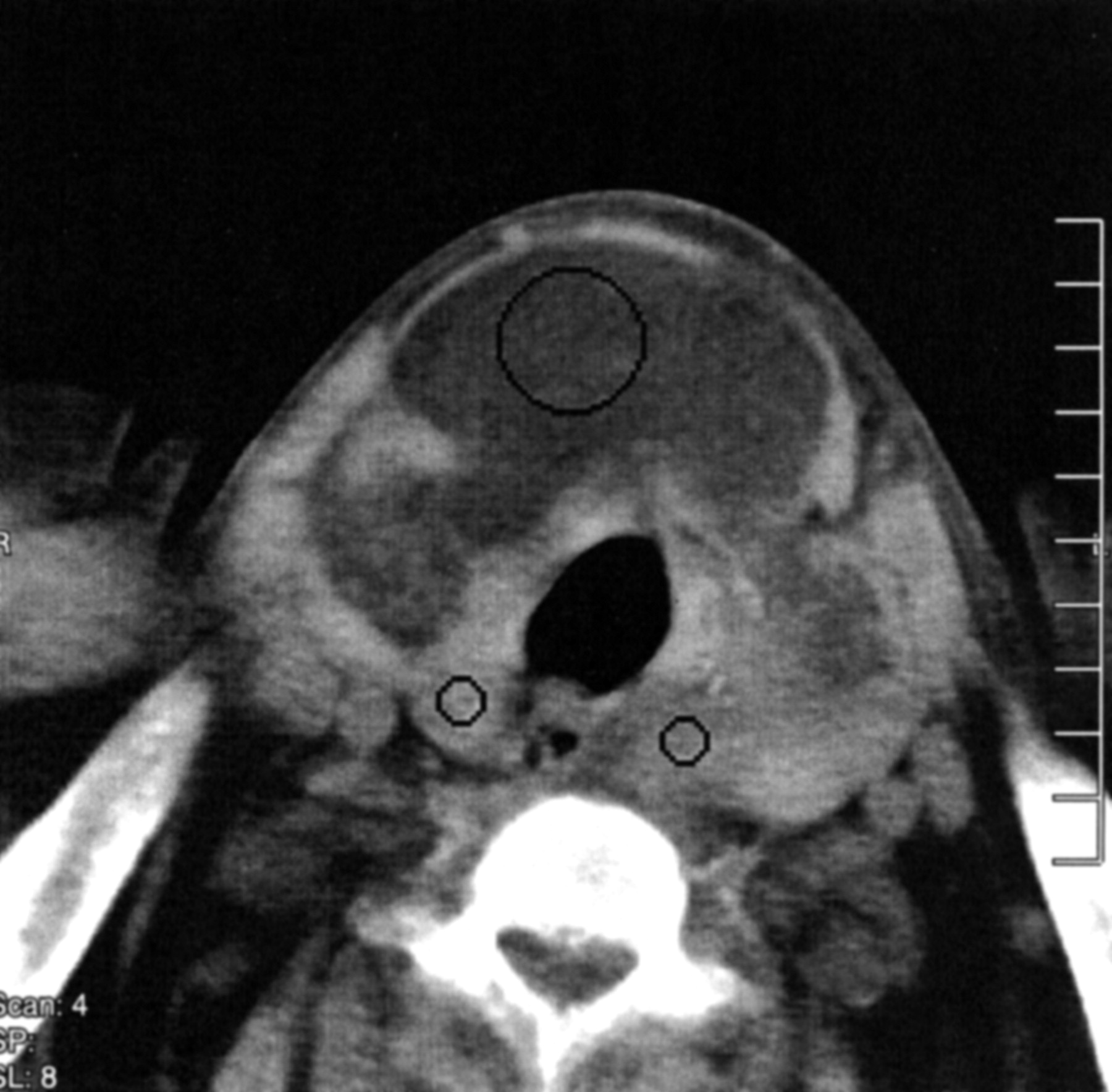
Unenhanced axial view CT scan shows a well-circumscribed, predominantly fatty mass (Hounsfield unitsH. = −60) of the thyroid gland. The mass causes enlargement of the isthmus and extends to both right and left lobes. Small portions of the right and left lobes have a soft tissue attenuation (Hounsfield units = +50), consistent with normal tyroid tissue.

Fig 2.
  • Download figure
  • Open in new tab
  • Download powerpoint
Fig 2.

Fast spin-echo T1-weighted MR image (1104/12 [TR/TE]; acquisition, 2; section thickness, 5 mm). The thyrolipoma is isointense with subcutaneous fat.

Discussion

Small amounts of adipose tissue are rarely seen in normal thyroid glands adjacent to the capsule, adjacent to surrounding vessels, or in connective tissue septa. They have been described in rare conditions such as adenomas, papillary carcinoma, amyloid goitres, nodular hyperplasia, and Graves’ disease (1, 2).

Fine needle aspiration cytology of these conditions may reveal adipocytes and follicular cells. Thyrolipomas differ from diffuse lipomatosis and amyloid goitre with adipose tissue in that they are well circumscribed and capsulated mass lesions (1, 2).

The origin of adipose tissue in the thyroid gland is unclear. Some authors think that adipose tissue is included in the thyroid gland during embryogenesis and that thyrolipoma is a true neoplasm with proliferation of fatty tissue (3). Other authors suggest metaplastic origin from fibroblasts (3). Trites (4), who reported a case of thyrolipoma associated with thymolipoma and a pharyngeal lipoma, noted that the thymus, thyroid, and aryepiglottic folds arise from the primitive foregut and added that the simultaneous appearance of these tumors suggested a disturbance in the development of the primitive foregut. Breek at al (5) reported a case of thyrolipoma and thymolipoma and agreed with the hypothesis presented by Trites.

Our patient had diffuse enlargement of the thyroid gland. His condition was euthyroid, and no enlarged lymph nodes or clinical signs of malignancy were found. Because the patient had a history of tuberculosis, amyloidosis was suspected to be the cause of renal insufficiency. Diffuse enlargement of the thyroid gland could also be explained with amyloid goitre, but CT disclosed a well-circumscribed fatty mass in the thyroid gland instead of diffuse lipomatosis as seen in association with amyloid goitre. Amyloid goitre is always associated with amyloid deposition elsewhere in the body (2). Skin biopsy of our patient did not disclose amyloid deposition. Fine needle aspiration cytology of the mass confirmed the presence of adipocytes and normal follicular cells.

Conclusion

Sonography and scintigraphy are widely performed for imaging the thyroid gland and are usually sufficient to make a diagnosis when combined with laboratory findings and fine needle aspiration cytology. Fatty tissue infiltration or fatty masses may be iso-echoic and cannot be differentiated from normal thyroid gland on ultrasonograms. For the current patient, sonography and scintigraphy were not diagnostic, and the diagnosis of thyrolipoma was based on the CT and MR imaging appearances, fine needle aspiration cytology, and exclusion of other disorders.

References

  1. ↵
    Laforga JB, Vierna J Adenoma of thyroid gland containing fat (thyrolipoma): report of a case. J Laryngol Otol 1996;110:1088–1089
    PubMed
  2. ↵
    Hjorth L, Thomsen LB, Nielsen VT. Adenolipoma of the thyroid gland. Histopathology 1986;10:91–96
    CrossRefPubMed
  3. ↵
    Rosai J, Carcangiu ML, Delellis RA. Tumors of the thyroid gland. In: Atlas of Tumor Pathology. Washington: Armed Forces Institute of Pathology;1992
  4. ↵
    Trites AE. Thyrolipoma, thymolipoma and pharyngeal lipoma: a syndrome. Can Med Assoc J 1996;95:1254–1259
  5. ↵
    Breek JK, Vallaeys JH, Rutsaert RR. Simultaneous presentation of a thyrolipoma and a thymolipoma in a young man. Eur J Surg 1997;163:941–943
    PubMed
  • Received January 4, 2002.
  • Accepted after revision May 6, 2002.
  • Copyright © American Society of Neuroradiology
View Abstract
PreviousNext
Back to top

In this issue

American Journal of Neuroradiology: 23 (10)
American Journal of Neuroradiology
Vol. 23, Issue 10
1 Nov 2002
  • Table of Contents
  • Index by author
Advertisement
Print
Download PDF
Email Article

Thank you for your interest in spreading the word on American Journal of Neuroradiology.

NOTE: We only request your email address so that the person you are recommending the page to knows that you wanted them to see it, and that it is not junk mail. We do not capture any email address.

Enter multiple addresses on separate lines or separate them with commas.
Radiologic and Cytologic Findings in a Case of Thyrolipoma
(Your Name) has sent you a message from American Journal of Neuroradiology
(Your Name) thought you would like to see the American Journal of Neuroradiology web site.
CAPTCHA
This question is for testing whether or not you are a human visitor and to prevent automated spam submissions.
Cite this article
Gulgan Demirpolat, Bunyamin Guney, Recep Savas, Goksel Tuncay, Hadaver Alper, Rifat Nuri Sener
Radiologic and Cytologic Findings in a Case of Thyrolipoma
American Journal of Neuroradiology Nov 2002, 23 (10) 1640-1641;

Citation Manager Formats

  • BibTeX
  • Bookends
  • EasyBib
  • EndNote (tagged)
  • EndNote 8 (xml)
  • Medlars
  • Mendeley
  • Papers
  • RefWorks Tagged
  • Ref Manager
  • RIS
  • Zotero
0 Responses
Respond to this article
Share
Bookmark this article
Radiologic and Cytologic Findings in a Case of Thyrolipoma
Gulgan Demirpolat, Bunyamin Guney, Recep Savas, Goksel Tuncay, Hadaver Alper, Rifat Nuri Sener
American Journal of Neuroradiology Nov 2002, 23 (10) 1640-1641;
del.icio.us logo Twitter logo Facebook logo Mendeley logo
  • Tweet Widget
  • Facebook Like
  • Google Plus One

Jump to section

  • Article
    • Abstract
    • Case Report
    • Discussion
    • Conclusion
    • References
  • Figures & Data
  • Info & Metrics
  • Responses
  • References
  • PDF

Related Articles

  • No related articles found.
  • PubMed
  • Google Scholar

Cited By...

  • Airway obstruction secondary to large thyroid adenolipoma
  • Crossref
  • Google Scholar

This article has not yet been cited by articles in journals that are participating in Crossref Cited-by Linking.

More in this TOC Section

  • FDG Uptake and Thyroid Cancer Post-BRAF Therapy
  • Normal Facial Nerve Enhancement on VIBE Sequence
  • Comprehensive Review of Inner Ear Anatomy on Photon-Counting CT
Show more Head and Neck Imaging

Similar Articles

Advertisement

Indexed Content

  • Current Issue
  • Accepted Manuscripts
  • Article Preview
  • Past Issues
  • Editorials
  • Editor's Choice
  • Fellows' Journal Club
  • Letters to the Editor
  • Video Articles

Cases

  • Case Collection
  • Archive - Case of the Week
  • Archive - Case of the Month
  • Archive - Classic Case

More from AJNR

  • Trainee Corner
  • Imaging Protocols
  • MRI Safety Corner

Multimedia

  • AJNR Podcasts
  • AJNR Scantastics

Resources

  • Turnaround Time
  • Submit a Manuscript
  • Submit a Video Article
  • Submit an eLetter to the Editor/Response
  • Manuscript Submission Guidelines
  • Statistical Tips
  • Fast Publishing of Accepted Manuscripts
  • Graphical Abstract Preparation
  • Imaging Protocol Submission
  • Evidence-Based Medicine Level Guide
  • Publishing Checklists
  • Author Policies
  • Become a Reviewer/Academy of Reviewers
  • News and Updates

About Us

  • About AJNR
  • Editorial Board
  • Editorial Board Alumni
  • Alerts
  • Permissions
  • Not an AJNR Subscriber? Join Now
  • Advertise with Us
  • Librarian Resources
  • Feedback
  • Terms and Conditions
  • AJNR Editorial Board Alumni

American Society of Neuroradiology

  • Not an ASNR Member? Join Now

© 2025 by the American Society of Neuroradiology All rights, including for text and data mining, AI training, and similar technologies, are reserved.
Print ISSN: 0195-6108 Online ISSN: 1936-959X

Powered by HighWire