
Article Figures & Data
Figures
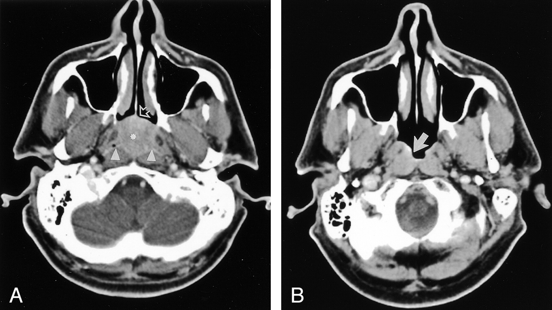
- Fig 1.
Case 1. Images in a 43-year-old man with a 3-week history of epistaxis caused by SEP of the nasopharynx.
A, Contrast-enhanced axial CT scan shows an inhomogeneous mass (asterisk) that fills the entire postnasal space, anteriorly abuts the posterior nasal septum (arrow), and posteriorly obliterates bilateral pharyngeal recesses (arrowheads).
B, Nine months after local radiation therapy, this contrast-enhanced CT scan shows minimal residual disease (arrow).
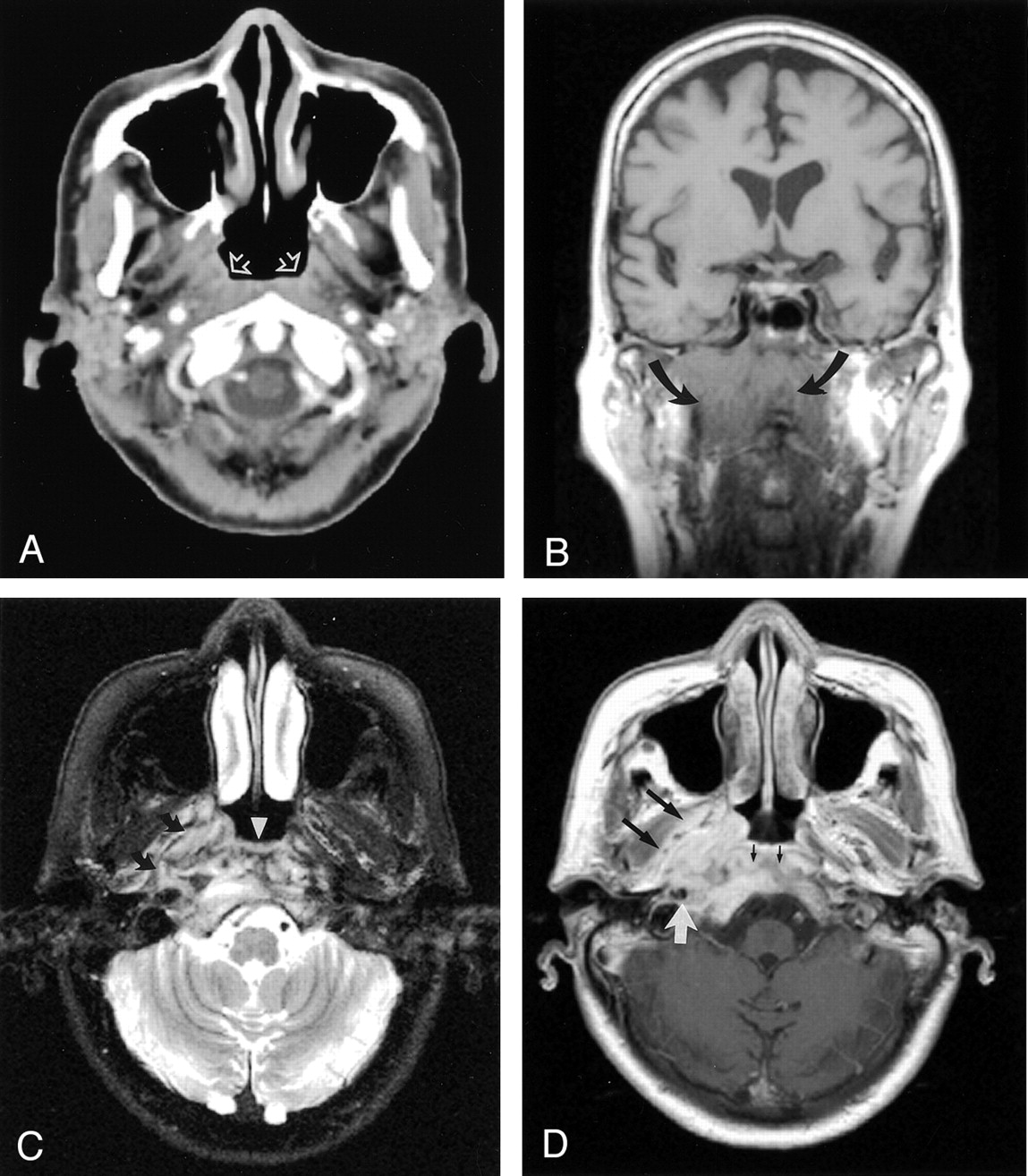
- Fig 2.
Case 2. Images in a 66-year-old woman with nasopharyngeal extramedullary plasmacytoma who presented with a 9-month history of headache.
A, Contrast-enhanced axial CT scan shows subtle asymmetry of the bilateral fossae of Rosenmuller (arrows).
B, Coronal T1-weighted spin-echo MR image (TR/TE, 500/2) shows a diffuse isointense mass infiltrating the mucosal and submucosal regions (arrows).
C, Axial T2-weighted fast spin-echo MR image (5400/99) shows a hyperintense lesion at the posterior pharyngeal wall (arrowhead) infiltrating the right parapharyngeal region (arrows).
D, Contrast-enhanced T1-weighted spin-echo image (490/18) shows moderate-to-marked enhancement of the lesion and involvements of right medial pterygoid muscle (large black arrows), right carotid space (white arrow), clivus, and prevertebral musculature (small black arrows).
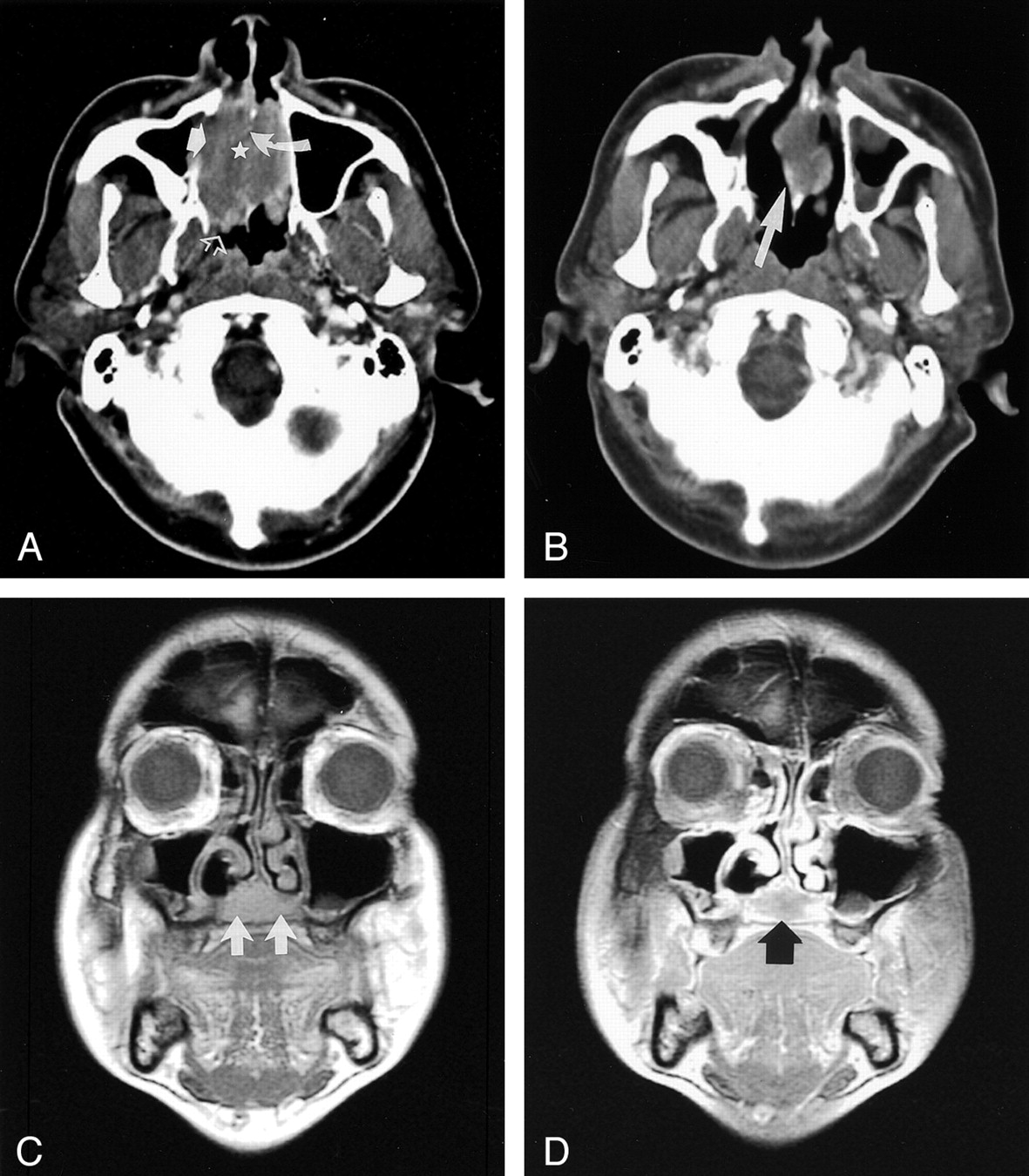
- Fig 3.
Case 3. Images in a 63-year-old man with a 3-month history of nasal blockage caused by nasal extramedullary plasmacytoma.
A, Contrast-enhanced axial CT scan shows a large soft-tissue mass (star) filling the entire nasal cavity, with erosion of the nasal septum (curved solid arrow) and right medial maxillary wall (straight solid arrow) extending posteriorly to the nasopharynx (open arrow).
B, Eight-month follow-up contrast-enhanced CT scan shows a considerable decrease in the size of the tumor (arrow) after regional radiation therapy. This finding indicates partial remission.
C, Ten-month follow-up coronal T1-weighted spin-echo MR image (470/19) shows a new isointense area in the hard palate (arrows) that suggests recurrence.
D, Coronal T2-weighted fast spin-echo image (5000/93) shows moderate inhomogeneous enhancement of the tumor (arrow).


In this issue


Jump to section
Related Articles
Cited By...
- No citing articles found.