
Article Figures & Data
Figures
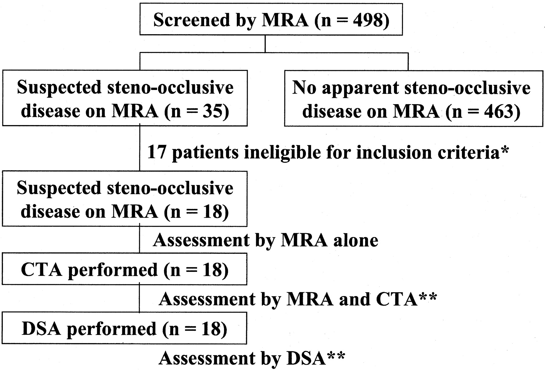
- Fig 1.
Flow diagram shows the enrollment and assessment of patients. * indicates the criteria for inclusion into the study, which were the availability of MR angiograms of good quality for diagnostic purposes, the acquisition of informed consent from the patients, and the absence of a history of brain surgery or risk factors such as heart failure. ** indicates cases in which one radiologist judged the quality of the CT angiograms and digital subtraction angiograms as excellent or good for diagnostic purposes. The results of the assessments of MR angiograms and CT angiograms were compared with those of DSA.
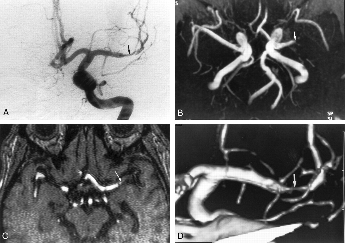
- Fig 2.
Images obtained in a 76-year-old man with mild stenosis of the left middle cerebral artery.
A, Anteroposterior angiogram of the left carotid artery shows mild stenoses at the left M1 segment (arrows).
B, Selective MIP image (35/9.6; flip angle, 25°) of MR angiogram in the anteroposterior projection depicts hypointensity that simulates severe stenosis (arrow).
C, Coronal view MPR image (35/9.6; flip angle, 25°) of MR angiogram also shows stenoses of more than 50% at the M1 segment. Two observers overestimated stenosis in this segment.
D, CT angiogram also shows mild stenoses at the M1 segment (arrows). The reviewers correctly interpreted stenosis in this segment.
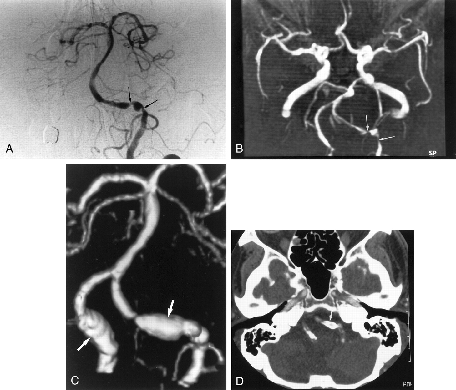
- Fig 3.
Images obtained in a 47-year-old man with severe stenosis of the left middle cerebral artery.
A, Anteroposterior angiogram of the left carotid artery shows severe stenosis at the M1 portion of the left middle cerebral artery (arrow).
B, MR angiogram (35/9.6; flip angle, 25°) shows the lesion as an occlusion (arrow).
C, On axial source image (35/9.6; flip angle, 25°), the M1 segment was also interpreted as occlusion (arrow).
D, CT angiogram reveals severe stenosis at the M1 portion (arrow). Two reviewers correctly interpreted this segment as being severely stenotic.
- Fig 4.
Images obtained in a 76-year-old man with severe stenosis of the left vertebral artery.
A, Anteroposterior angiogram of left vertebral artery shows severe stenoses in the intracranial segment of the left vertebral artery (arrows).
B, MR angiogram (35/9.6; flip angle, 25°) also depicts severe stenoses in the left vertebral artery (arrows).
C, 3D CT angiogram shows aneurysm-like dilatation of both vertebral arteries (arrows), which correspond to the calcification of the vessel wall.
D, Axial source image shows no apparent lumen in the stenotic artery because of circumferential calcification. The segment was interpreted as being occluded (arrow). Finally, this segment was interpreted as being severe stenotic on the basis of MR angiographic findings.


Tables
MRA alone and combined use of MRA and CTA versus conventional DSA
Results at MR Angiography Alone and Combined Use of MR Angiography and CT Angiography* Results at Conventional DSA Total (n = 198) 0%–29% (n = 170) 30%–49% (n = 10) 50%–69% (n = 8) 70%–99% (n = 5) 100% (n = 5) 0%–29% 142/166 1/0 0/0 0/0 0/0 143/166 30%–49% 15/4 5/9 0/0 0/0 0/0 20/13 50%–69% 9/0 3/1 4/7 0/0 0/0 16/8 70%–99% 4/0 1/0 4/1 4/5 0/0 13/6 100% 0/0 0/0 0/0 1/0 5/5 6/5 * (MRA images alone)/(MRA images plus CTA images).


