Advertisement
  • Main menu
  • User menu
  • Search
  • English ▼
    • English
    • Afrikaans
    • Albanian
    • Amharic
    • Arabic
    • Armenian
    • Azerbaijani
    • Basque
    • Belarusian
    • Bengali
    • Bosnian
    • Bulgarian
    • Catalan
    • Cebuano
    • Chichewa
    • Chinese (Simplified)
    • Chinese (Traditional)
    • Corsican
    • Croatian
    • Czech
    • Danish
    • Dutch
    • Esperanto
    • Estonian
    • Filipino
    • Finnish
    • French
    • Frisian
    • Galician
    • Georgian
    • German
    • Greek
    • Gujarati
    • Haitian Creole
    • Hausa
    • Hawaiian
    • Hebrew
    • Hindi
    • Hmong
    • Hungarian
    • Icelandic
    • Igbo
    • Indonesian
    • Irish
    • Italian
    • Japanese
    • Javanese
    • Kannada
    • Kazakh
    • Khmer
    • Korean
    • Kurdish (Kurmanji)
    • Kyrgyz
    • Lao
    • Latin
    • Latvian
    • Lithuanian
    • Luxembourgish
    • Macedonian
    • Malagasy
    • Malay
    • Malayalam
    • Maltese
    • Maori
    • Marathi
    • Mongolian
    • Myanmar (Burmese)
    • Nepali
    • Norwegian
    • Pashto
    • Persian
    • Polish
    • Portuguese
    • Punjabi
    • Romanian
    • Russian
    • Samoan
    • Scottish Gaelic
    • Serbian
    • Sesotho
    • Shona
    • Sindhi
    • Sinhala
    • Slovak
    • Slovenian
    • Somali
    • Spanish
    • Sudanese
    • Swahili
    • Swedish
    • Tajik
    • Tamil
    • Telugu
    • Thai
    • Turkish
    • Ukrainian
    • Urdu
    • Uzbek
    • Vietnamese
    • Welsh
    • Xhosa
    • Yiddish
    • Yoruba
    • Zulu

Main menu

  • Home
  • Content
    • Current Issue
    • Accepted Manuscripts
    • Article Preview
    • Past Issue Archive
    • Video Articles
    • AJNR Case Collection
    • Case of the Week Archive
    • Case of the Month Archive
    • Classic Case Archive
  • Special Collections
    • Advancing NeuroMRI with High-Relaxivity Contrast Agents
    • AJNR Awards
    • Low-Field MRI
    • Alzheimer Disease
    • ASNR Foundation Special Collection
    • Photon-Counting CT
    • View All
  • Multimedia
    • AJNR Podcasts
    • AJNR SCANtastic
    • Trainee Corner
    • MRI Safety Corner
    • Imaging Protocols
  • For Authors
    • Submit a Manuscript
    • Submit a Video Article
    • Submit an eLetter to the Editor/Response
    • Manuscript Submission Guidelines
    • Statistical Tips
    • Fast Publishing of Accepted Manuscripts
    • Graphical Abstract Preparation
    • Imaging Protocol Submission
    • Author Policies
  • About Us
    • About AJNR
    • Editorial Board
    • Editorial Board Alumni
  • More
    • Become a Reviewer/Academy of Reviewers
    • Subscribers
    • Permissions
    • Alerts
    • Feedback
    • Advertisers
    • ASNR Home

User menu

  • Alerts
  • Log in

Search

  • Advanced search
American Journal of Neuroradiology

American Journal of Neuroradiology

ASHNR American Society of Functional Neuroradiology ASHNR American Society of Pediatric Neuroradiology ASSR
  • Alerts
  • Log in

Advanced Search

English ▼
  • English
  • Afrikaans
  • Albanian
  • Amharic
  • Arabic
  • Armenian
  • Azerbaijani
  • Basque
  • Belarusian
  • Bengali
  • Bosnian
  • Bulgarian
  • Catalan
  • Cebuano
  • Chichewa
  • Chinese (Simplified)
  • Chinese (Traditional)
  • Corsican
  • Croatian
  • Czech
  • Danish
  • Dutch
  • Esperanto
  • Estonian
  • Filipino
  • Finnish
  • French
  • Frisian
  • Galician
  • Georgian
  • German
  • Greek
  • Gujarati
  • Haitian Creole
  • Hausa
  • Hawaiian
  • Hebrew
  • Hindi
  • Hmong
  • Hungarian
  • Icelandic
  • Igbo
  • Indonesian
  • Irish
  • Italian
  • Japanese
  • Javanese
  • Kannada
  • Kazakh
  • Khmer
  • Korean
  • Kurdish (Kurmanji)
  • Kyrgyz
  • Lao
  • Latin
  • Latvian
  • Lithuanian
  • Luxembourgish
  • Macedonian
  • Malagasy
  • Malay
  • Malayalam
  • Maltese
  • Maori
  • Marathi
  • Mongolian
  • Myanmar (Burmese)
  • Nepali
  • Norwegian
  • Pashto
  • Persian
  • Polish
  • Portuguese
  • Punjabi
  • Romanian
  • Russian
  • Samoan
  • Scottish Gaelic
  • Serbian
  • Sesotho
  • Shona
  • Sindhi
  • Sinhala
  • Slovak
  • Slovenian
  • Somali
  • Spanish
  • Sudanese
  • Swahili
  • Swedish
  • Tajik
  • Tamil
  • Telugu
  • Thai
  • Turkish
  • Ukrainian
  • Urdu
  • Uzbek
  • Vietnamese
  • Welsh
  • Xhosa
  • Yiddish
  • Yoruba
  • Zulu
  • Home
  • Content
    • Current Issue
    • Accepted Manuscripts
    • Article Preview
    • Past Issue Archive
    • Video Articles
    • AJNR Case Collection
    • Case of the Week Archive
    • Case of the Month Archive
    • Classic Case Archive
  • Special Collections
    • Advancing NeuroMRI with High-Relaxivity Contrast Agents
    • AJNR Awards
    • Low-Field MRI
    • Alzheimer Disease
    • ASNR Foundation Special Collection
    • Photon-Counting CT
    • View All
  • Multimedia
    • AJNR Podcasts
    • AJNR SCANtastic
    • Trainee Corner
    • MRI Safety Corner
    • Imaging Protocols
  • For Authors
    • Submit a Manuscript
    • Submit a Video Article
    • Submit an eLetter to the Editor/Response
    • Manuscript Submission Guidelines
    • Statistical Tips
    • Fast Publishing of Accepted Manuscripts
    • Graphical Abstract Preparation
    • Imaging Protocol Submission
    • Author Policies
  • About Us
    • About AJNR
    • Editorial Board
    • Editorial Board Alumni
  • More
    • Become a Reviewer/Academy of Reviewers
    • Subscribers
    • Permissions
    • Alerts
    • Feedback
    • Advertisers
    • ASNR Home
  • Follow AJNR on Twitter
  • Visit AJNR on Facebook
  • Follow AJNR on Instagram
  • Join AJNR on LinkedIn
  • RSS Feeds

AJNR Awards, New Junior Editors, and more. Read the latest AJNR updates


Improved Turnaround Times | Median time to first decision: 12 days

Research ArticleBRAIN

Frequencies Contributing to Functional Connectivity in the Cerebral Cortex in “Resting-state” Data

Dietmar Cordes, Victor M. Haughton, Konstantinos Arfanakis, John D. Carew, Patrick A. Turski, Chad H. Moritz, Michelle A. Quigley and M. Elizabeth Meyerand
American Journal of Neuroradiology August 2001, 22 (7) 1326-1333;
Dietmar Cordes
  • Find this author on Google Scholar
  • Find this author on PubMed
  • Search for this author on this site
Victor M. Haughton
  • Find this author on Google Scholar
  • Find this author on PubMed
  • Search for this author on this site
Konstantinos Arfanakis
  • Find this author on Google Scholar
  • Find this author on PubMed
  • Search for this author on this site
John D. Carew
  • Find this author on Google Scholar
  • Find this author on PubMed
  • Search for this author on this site
Patrick A. Turski
  • Find this author on Google Scholar
  • Find this author on PubMed
  • Search for this author on this site
Chad H. Moritz
  • Find this author on Google Scholar
  • Find this author on PubMed
  • Search for this author on this site
Michelle A. Quigley
  • Find this author on Google Scholar
  • Find this author on PubMed
  • Search for this author on this site
M. Elizabeth Meyerand
  • Find this author on Google Scholar
  • Find this author on PubMed
  • Search for this author on this site
  • Article
  • Figures & Data
  • Info & Metrics
  • Responses
  • References
  • PDF
Loading

Article Figures & Data

Figures

  • fig 1.
    • Download figure
    • Open in new tab
    • Download powerpoint
    fig 1.

    Consecutive coronal fMR maps of voxels activated by a text-listening task (top) and of the voxels functionally connected with a 2 × 2-seed voxel ROI (crosshairs) within the auditory cortex during resting-state (bottom). Task activation is evident in the region of the primary and association auditory cortices in the superior temporal lobe. Many of the voxels functionally connected with the seed voxels in the left auditory cortex (crosshairs) have a similar distribution. All maps are representative and are from a single subject. The color scale for the task-activation map refers to z scores (red z, 3.5; yellow z, 5.5), and that for the resting-state map refers to correlation coefficients (red, 0.5; yellow, 0.7). The MR pulse sequence parameters are 2000/50/82 (TR/TE/flip angle) for fMR and 400/50/50 for fcMR.fig 2. Consecutive coronal images showing task activation for the visual task (top) and connectivity for the visual cortex in resting-state (bottom). The task produces activation in the striate cortex region. The voxels with connectivity to the seed voxel (crosshairs) appear to lie in similar locations in the striate cortex region. The color scale for the task-activation map refers to z scores (red z, 3.5; yellow z, 5.5), and that for the resting-state map refers to correlation coefficients (red, 0.6; yellow, 0.8). The MR pulse sequence parameters are as in figure 1.fig 3. A selected coronal fMR map of voxels activated by the bilateral finger-tapping task (top) and of voxels functionally connected to an ROI of 2 × 2-seed voxels (crosshairs) in the sensorimotor cortex in a resting acquisition (bottom). The sensorimotor cortex is identified in both the fMR and the fcMR imaging. Not shown are voxels within the dentate nucleus of the cerebellum that were identified with fMR but not with the functional connectivity study. The color scale for the task-activation map refers to z scores (red z, 3.5; yellow z, 5.5), and that for the resting-state map refers to correlation coefficients (red, 0.62; yellow, 0.8). The MR pulse sequence parameters are as in figure 1

  • fig 4.
    • Download figure
    • Open in new tab
    • Download powerpoint
    fig 4.

    A selected axial fMR map of voxels activated by the bilateral finger-tapping task (top) and of the voxels functionally connected to an ROI of 2 × 2-seed voxels (crosshairs) in the sensorimotor cortex in a resting-state acquisition (bottom). The sensorimotor cortex is identified in both the fMR and the fcMR maps. The SMA is not activated by the paradigm, but connectivity is clearly seen. The color scale for the task-activation map refers to z scores (red z, 3.5; yellow z, 5.5), and that for the resting-state map refers to correlation coefficients (red, 0.62; yellow, 0.8). The MR pulse sequence parameters are as in figure 1

  • fig 5.
    • Download figure
    • Open in new tab
    • Download powerpoint
    fig 5.

    Spectral analysis of the average correlation coefficient for interregional connectivity in the auditory cortex (same subject illustrated in figure 1). Only frequency components between 0 and 0.05 Hz contribute significantly to the correlation coefficient.fig 6. Spectral analysis of the average correlation coefficient for interregional connectivity in the striate cortex (same subject illustrated in figure 2). Only frequency components less than 0.05 Hz contribute significantly to the correlation coefficient
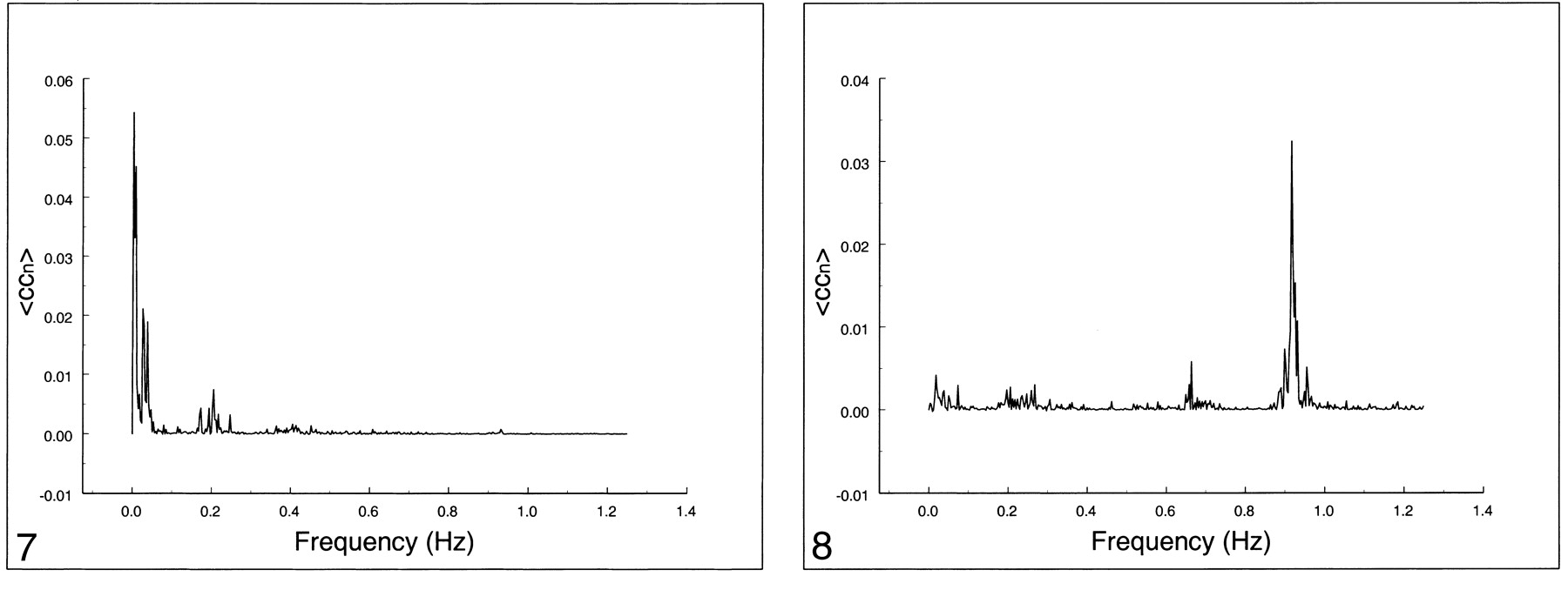
  • fig 7.
    • Download figure
    • Open in new tab
    • Download powerpoint
    fig 7.

    Spectral analysis of the average correlation coefficient for interregional connectivity in the motor cortex (same subject illustrated in figure 3). Only the same low-frequency components contribute significantly to the correlation coefficient.fig 8. Spectral analysis of the average correlation coefficient obtained using a seed voxel in the right internal carotid artery. Low frequencies do not predominate. The main peak occurs at the cardiac frequency (0.93 Hz); the first harmonic is aliased to 0.65 Hz. A respiratory band is seen at 0.2 Hz

  • fig 9.
    • Download figure
    • Open in new tab
    • Download powerpoint
    fig 9.

    Spectral analysis of the average correlation coefficient from a seed voxel in the left jugular vein. Note the spread over many frequencies up to 0.4 Hz and the cardiac peaks at 0.93 Hz and 0.65 Hz.fig 10. Spectral analysis of the average correlation coefficient obtained using a seed voxel in the left ventricle. Note the spread over many frequencies. The cardiac bands show a large spread compared with other tissue. Low-frequency peaks are visible at 0.03 Hz

  • fig 11.
    • Download figure
    • Open in new tab
    • Download powerpoint
    fig 11.

    Contribution of low frequencies (0 to 0.1 Hz), respiratory frequencies (0.1 to 0.5 Hz), and cardiac frequencies to the cross-correlation coefficient in different ROIs in the same subject. Low-frequency components clearly dominate, contributing to functional connectivity in auditory, visual, and motor cortices. For blood vessels and cerebrospinal fluid, low-frequency components are present, but cardiac and respiratory noise sources are the main contributors

PreviousNext
Back to top

In this issue

American Journal of Neuroradiology: 22 (7)
American Journal of Neuroradiology
Vol. 22, Issue 7
1 Aug 2001
  • Table of Contents
  • Index by author
Advertisement
Print
Download PDF
Email Article
Cite this article
0 Responses
Respond to this article
Share
Bookmark this article
Frequencies Contributing to Functional Connectivity in the Cerebral Cortex in “Resting-state” Data
Dietmar Cordes, Victor M. Haughton, Konstantinos Arfanakis, John D. Carew, Patrick A. Turski, Chad H. Moritz, Michelle A. Quigley, M. Elizabeth Meyerand
American Journal of Neuroradiology Aug 2001, 22 (7) 1326-1333;
del.icio.us logo Twitter logo Facebook logo Mendeley logo
  • Tweet Widget

Jump to section

  • Article
    • Abstract
    • Methods
    • Results
    • Discussion
    • Conclusion
    • Footnotes
    • References
  • Figures & Data
  • Info & Metrics
  • Responses
  • References
  • PDF

Related Articles

  • No related articles found.
  • PubMed
  • Google Scholar

Cited By...

  • Disrupted resting-state fMRI coherence at vasomotor frequency links carotid artery stenosis to functional connectivity loss
  • Cognitive reserve linked to network-specific brain-ventricle coupling modes
  • The cerebellar components of the human language network
  • Temporal and Spatial Scales of Resting-state Human Cortical Activity Throughout Lifespan
  • Large-scale Signal Propagation Modes in the Human Brain
  • Functional divergence between the two cerebral hemispheres contributes to human fluid intelligence
  • Precision fMRI reveals that the language network exhibits adult-like left-hemispheric lateralization by 4 years of age
  • Theoretical proposal for restoration of hand motor function utilizing plasticity of motor-cortical interhemispheric interaction
  • Younger adult brain utilizes interhemispheric strategy of recruiting ipsilateral dorsal premotor cortex for complex finger movement, but not aging brain
  • Reliability of brain metrics derived from a Time-Domain Functional Near-Infrared Spectroscopy System
  • Transient destabilization of whole brain dynamics induced by DMT
  • Event Integration and Temporal Differentiation: How Hierarchical Knowledge Emerges in Hippocampal Subfields through Learning
  • Exploring the neuronal and systemic physiological contributions to spontaneous cerebral fluctuations - Insights from functional MRI in The Maastricht Study
  • Traumatic brain injury disrupts state-dependent functional cortical connectivity in a mouse model
  • Event integration and temporal differentiation: how hierarchical knowledge emerges in hippocampal subfields through learning
  • Model connectivity: leveraging the power of encoding models to overcome the limitations of functional connectivity
  • Comparing data-driven physiological denoising approaches for resting-state fMRI: Implications for the study of aging
  • Single Ventricle Reconstruction III: Brain Connectome and Neurodevelopmental Outcomes: Design, Recruitment, and Technical Challenges of a Multicenter, Observational Neuroimaging Study
  • Optimizing network neuroscience computation of individual differences in human spontaneous brain activity for test-retest reliability
  • Consolidation-dependent behavioral integration of sequences related to mPFC neural overlap and hippocampal-cortical connectivity
  • Low-dimensional organization of global brain states of reduced consciousness
  • Model-based whole-brain perturbational landscape of neurodegenerative diseases
  • Data-driven discovery of canonical large-scale brain dynamics
  • Ultra-slow fMRI fluctuations in the fourth ventricle as a marker of drowsiness
  • BOLD cofluctuation 'events are predicted from static functional connectivity
  • Callosal Fiber Length Scales with Brain Size According to Functional Lateralization, Evolution, and Development
  • Callosal fiber length scales with brain size according to functional lateralization, evolution, and development
  • The universal language network: A cross-linguistic investigation spanning 45 languages and 12 language families
  • Differential tracking of linguistic vs. mental state content in naturalistic stimuli by language and Theory of Mind (ToM) brain networks
  • Chemogenetic activation of Locus Coeruleus Noradrenergic Neurons Modulates the Default Mode Network
  • Functional connectome of arousal and motor brainstem nuclei in living humans by 7 Tesla resting-state fMRI
  • Brain stimulation boosts perceptual learning by altering sensory GABAergic plasticity and functional connectivity
  • External drivers of BOLD signals non-stationarity
  • Study on acupuncture in the treatment of painful diabetic peripheral neuropathy based on rs-fMRI: a protocol for systematic review and meta-analysis
  • Developmental coupling of cerebral blood flow and fMRI fluctuations in youth
  • Frequency-specific coactivation patterns in resting-state and their alterations in schizophrenia: an fMRI study
  • Rare long-range cortical connections enhance information processing
  • Predicting behavior through dynamic modes in resting-state fMRI data
  • Neural dynamics of parametrically modulated human mechanical pain determined using whole-brain quantitative perfusion imaging
  • Cingulo-opercular control network and disused motor circuits joined in standby mode
  • Visual processing speed is linked to functional connectivity between right frontoparietal and visual networks
  • Turbulent-like dynamics in the human brain
  • Atlas55+: Brain Functional Atlas of Resting-state Networks for Late Adulthood
  • Attention network modulation via tRNS correlates with attention gain
  • Cingulo-Opercular Control Network Supports Disused Motor Circuits in Standby Mode
  • Predicting Sex from Resting-State fMRI Across Multiple Independent Acquired Datasets
  • Noise-driven multistability versus deterministic chaos in phenomenological semi-empirical models of whole-brain activity
  • Modular and state-relevant connectivity in high-frequency resting-state BOLD fMRI data: An independent component analysis
  • Dynamical Mechanisms of Interictal Resting-State Functional Connectivity in Epilepsy
  • Altered Thalamocortical Connectivity in Six-Week Old Infants at High Familial Risk for Autism Spectrum Disorder
  • Low variability of dynamical functional connectivity in cerebellar networks
  • The Developing Human Connectome Project: typical and disrupted perinatal functional connectivity
  • Data augmentation based on dynamical systems for the classification of brain states
  • Dynamical informational structures characterize the different human brain states of wakefulness and deep sleep
  • Studying the precuneus reveals structure-function-affect correlation in long-term meditators
  • Predicting the fMRI signal fluctuation with echo-state neural networks trained on vascular network dynamics
  • A robust deep neural network for denoising task-based fMRI data: An application to working memory and episodic memory
  • Increased soluble amyloid-beta causes early aberrant brain network hypersynchronisation in a mature-onset mouse model of amyloidosis
  • No evidence for functional distinctions across fronto-temporal language regions in their temporal receptive windows
  • A dynamical systems framework to uncover the drivers of large-scale cortical activity
  • Early maltreatment experience increases resting-state functional connectivity in the theory of mind (ToM) network
  • The (dis)similarities between neural networks based upon functional connectivity, representational similarity, and univariate analyses
  • Group ICA for Identifying Biomarkers in Schizophrenia: 'Adaptive Networks via Spatially Constrained ICA Show More Sensitivity to Group Differences than Spatio-temporal Regression
  • Hyperconnectivity of the default mode network in multiorgan dysfunction syndrome
  • Modular preprocessing pipelines can reintroduce artifacts into fMRI data
  • Assessment of Functional Connectome Construction Strategies in Neurodegeneration
  • Interaction information along lifespan of the resting brain dynamics reveals a major redundant role of the default mode network
  • Interhemispheric Integration for Complex Behaviors, Absent the Corpus Callosum in Normal Ontogeny
  • Study on Linear Combination of Long Memory Processes Corrupted by Additive Noises for fMRI Time Series Analysis
  • What the structural-functional connectome reveals about brain aging: The key role of the fronto-striatal-thalamic circuit and the rejuvenating impact of physical activity
  • The processing of dynamic faces in the human brain: Support for an integrated neural framework of face processing
  • Domain-general brain regions do not track linguistic input as closely as language-selective regions
  • The Neuro Bureau ADHD-200 Preprocessed Repository
  • Assessing Variations in Areal Organization for the Intrinsic Brain: From Fingerprints to Reliability
  • Disruption of periaqueductal grey-default mode network functional connectivity predicts persistent post-traumatic headache in mild traumatic brain injury
  • Functional connectivity networks with and without global signal correction
  • Crossref
  • Google Scholar

This article has not yet been cited by articles in journals that are participating in Crossref Cited-by Linking.

More in this TOC Section

  • Predictors of Reperfusion in Patients with Acute Ischemic Stroke
  • Enhanced Axonal Metabolism during Early Natalizumab Treatment in Relapsing-Remitting Multiple Sclerosis
  • Progression of Microstructural Damage in Spinocerebellar Ataxia Type 2: A Longitudinal DTI Study
Show more Brain

Similar Articles

Advertisement

Indexed Content

  • Current Issue
  • Accepted Manuscripts
  • Article Preview
  • Past Issues
  • Editorials
  • Editor's Choice
  • Fellows' Journal Club
  • Letters to the Editor
  • Video Articles

Cases

  • Case Collection
  • Archive - Case of the Week
  • Archive - Case of the Month
  • Archive - Classic Case

More from AJNR

  • Trainee Corner
  • Imaging Protocols
  • MRI Safety Corner
  • Book Reviews

Multimedia

  • AJNR Podcasts
  • AJNR Scantastics

Resources

  • Turnaround Time
  • Submit a Manuscript
  • Submit a Video Article
  • Submit an eLetter to the Editor/Response
  • Manuscript Submission Guidelines
  • Statistical Tips
  • Fast Publishing of Accepted Manuscripts
  • Graphical Abstract Preparation
  • Imaging Protocol Submission
  • Evidence-Based Medicine Level Guide
  • Publishing Checklists
  • Author Policies
  • Become a Reviewer/Academy of Reviewers
  • News and Updates

About Us

  • About AJNR
  • Editorial Board
  • Editorial Board Alumni
  • Alerts
  • Permissions
  • Not an AJNR Subscriber? Join Now
  • Advertise with Us
  • Librarian Resources
  • Feedback
  • Terms and Conditions
  • AJNR Editorial Board Alumni

American Society of Neuroradiology

  • Not an ASNR Member? Join Now

© 2025 by the American Society of Neuroradiology All rights, including for text and data mining, AI training, and similar technologies, are reserved.
Print ISSN: 0195-6108 Online ISSN: 1936-959X

Powered by HighWire
Email this Article

Thank you for your interest in spreading the word on American Journal of Neuroradiology.

NOTE: We only request your email address so that the person you are recommending the page to knows that you wanted them to see it, and that it is not junk mail. We do not capture any email address.

Enter multiple addresses on separate lines or separate them with commas.
Frequencies Contributing to Functional Connectivity in the Cerebral Cortex in “Resting-state” Data
(Your Name) has sent you a message from American Journal of Neuroradiology
(Your Name) thought you would like to see the American Journal of Neuroradiology web site.
CAPTCHA
This question is for testing whether or not you are a human visitor and to prevent automated spam submissions.
Cite this article
Dietmar Cordes, Victor M. Haughton, Konstantinos Arfanakis, John D. Carew, Patrick A. Turski, Chad H. Moritz, Michelle A. Quigley, M. Elizabeth Meyerand
Frequencies Contributing to Functional Connectivity in the Cerebral Cortex in “Resting-state” Data
American Journal of Neuroradiology Aug 2001, 22 (7) 1326-1333;

Citation Manager Formats

  • BibTeX
  • Bookends
  • EasyBib
  • EndNote (tagged)
  • EndNote 8 (xml)
  • Medlars
  • Mendeley
  • Papers
  • RefWorks Tagged
  • Ref Manager
  • RIS
  • Zotero

We use cookies on this site to enhance your user experience. By clicking any link on this page you are giving your consent for us to set cookies.