
Article Figures & Data
Figures
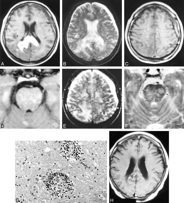
- fig 1.
Case 1.
A, Contrast-enhanced T1-weighted axial image (spin-echo sequence with parameters 600/14 [TR/TE]) shows an enhanced lesion from the splenium of the corpus callosum extending to the right parietal white matter. There is a small area of necrosis within the mass (arrow). Nodular and multiple punctate enhancements are seen in the white matter (arrowhead).
B, T2-weighted image (fast spin-echo sequence with parameters of 4000/96 [TR/TE]) shows a hyperintense mass (arrow) and surrounding edema.
C and D, Contrast-enhanced T1-weighted images show multiple punctate enhancements in the pons and cerebral white matter bilaterally.
E and F, T2-weighted axial image shows patchy hyperintense areas in the pons and cerebral white matter bilaterally (arrows).
G, Stereotactic biopsy specimen from the right parietal lobe shows small lymphoid cells, microglia, and macrophages, which were prominent in the perivascular space (arrow).
H, Contrast-enhanced T-weighted image 6 months after radiation therapy shows that the mass has almost disappeared.
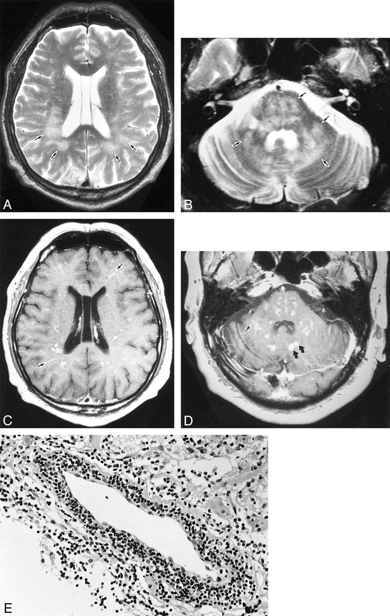
- fig 2.
Case 2. A and B, T2-weighted images (fast spin-echo sequence with parameters of 4500/96 [TR/TE]) show diffuse hyperintense lesions in the white matter bilaterally, as well as in the pons, middle cerebellar peduncle, and cerebellar hemispheres (arrows). C and D, Contrast-enhanced T1-weighted images (spin-echo sequence with parameters 600/14 [TR/TE]) show multiple punctate and linear enhancements scattered both in the white matter bilaterally (that appear to reside along the intramedullary vessels) and in the pons and cerebellar hemispheres (arrows indicate linear enhancements). Nodular enhancement also is shown (curved arrows). E, Brain stereotactic biopsy specimen from the right occipital lobe shows inflammatory destruction of the cerebral cortex with mononuclear cell infiltration. The infiltrating cells consist of polyclonal small lymphoid cells that include nuclear atypia predominantly in the perivascular space (arrows)
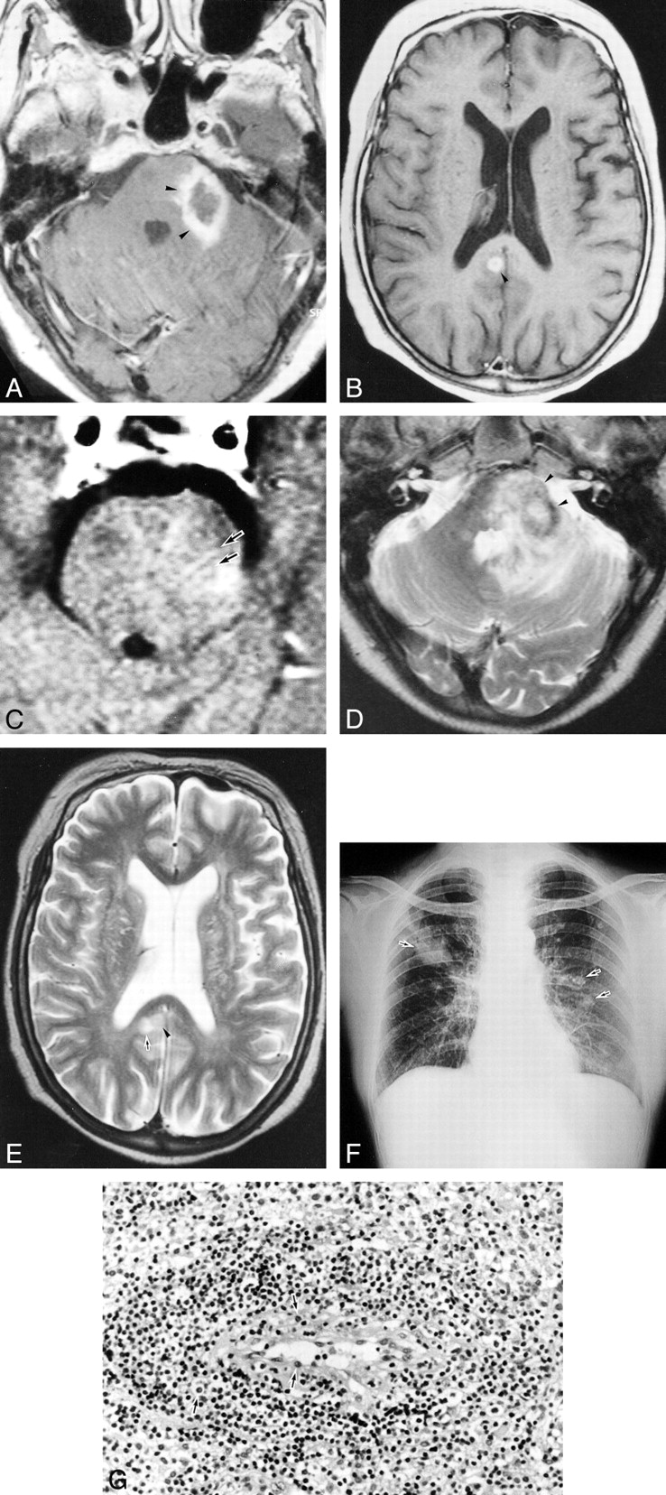
- fig 3.
Case 4.
A, T2-weighted image (fast spin-echo sequence with parameters of 3500/96 [TR/TE]) shows diffuse hyperintense areas in the cerebral white matter bilaterally. Cerebral sulci are enlarged bilaterally, indicating cerebral atrophy.
B, Contrast-enhanced T1-weighted image (spin-echo sequence with parameters of 600/14 [TR/TE]) shows multiple punctate and linear (arrows) enhancements in the white matter bilaterally and nodular enhancement in left parietal lobe (curved arrows).
- fig 4.
Case 3.
A and B, Contrast-enhanced T1-weighted images (spin-echo sequence with parameters of 600/14 [TR/TE]) show ringlike enhancements in the left middle cerebral peduncle and pons and in the right cingulate gyrus (arrowheads).
C, The slices of 4-mm thickness show linear enhancements in the pons (arrows).
D and E, T2-weighted MR images (fast spin-echo sequence with parameters of 4500/96 [TR/TE]) show that the ringlike, enhanced lesions are hypointense to isointense with gray matter (arrowheads). There are hyperintense areas surrounding these lesions, suggesting perifocal edema (arrow).
F, Chest radiograph shows multiple nodular shadows in both lung fields (arrows).
G, Open brain biopsy specimen from the left middle cerebellar peduncle shows perivascular lymphoid-cell infiltration with partial nuclear atypia (arrows).


Tables

Summary of MR findings in patients with LG of the brain
Extended
In this issue


Jump to section
Related Articles
Cited By...
- Imaging of Lymphomas Involving the CNS: An Update-Review of the Full Spectrum of Disease with an Emphasis on the World Health Organization Classifications of CNS Tumors 2021 and Hematolymphoid Tumors 2022
- CLIPPERS and its mimics: evaluation of new criteria for the diagnosis of CLIPPERS
- Perivascular enhancement in anti-MOG antibody demyelinating disease of the CNS
- Fatal lymphomatoid granulomatosis with primary CNS-involvement in an immunocompetent 80-year-old woman
- Neuroimaging of Rapidly Progressive Dementias, Part 2: Prion, Inflammatory, Neoplastic, and Other Etiologies
- A 54-year-old woman with progressive gait disturbance and MRI abnormalities
- A 52-year-old man with progressive left-sided weakness and white matter disease