Advertisement

Main menu

  • Home
  • Content
    • Current Issue
    • Accepted Manuscripts
    • Article Preview
    • Past Issue Archive
    • Video Articles
    • AJNR Case Collection
    • Case of the Week Archive
    • Case of the Month Archive
    • Classic Case Archive
  • Special Collections
    • AJNR Awards
    • Low-Field MRI
    • Alzheimer Disease
    • ASNR Foundation Special Collection
    • Photon-Counting CT
    • View All
  • Multimedia
    • AJNR Podcasts
    • AJNR SCANtastic
    • Trainee Corner
    • MRI Safety Corner
    • Imaging Protocols
  • For Authors
    • Submit a Manuscript
    • Submit a Video Article
    • Submit an eLetter to the Editor/Response
    • Manuscript Submission Guidelines
    • Statistical Tips
    • Fast Publishing of Accepted Manuscripts
    • Graphical Abstract Preparation
    • Imaging Protocol Submission
    • Author Policies
  • About Us
    • About AJNR
    • Editorial Board
    • Editorial Board Alumni
  • More
    • Become a Reviewer/Academy of Reviewers
    • Subscribers
    • Permissions
    • Alerts
    • Feedback
    • Advertisers
    • ASNR Home

User menu

  • Alerts
  • Log in

Search

  • Advanced search
American Journal of Neuroradiology

American Journal of Neuroradiology

ASHNR American Society of Functional Neuroradiology ASHNR American Society of Pediatric Neuroradiology ASSR
  • Alerts
  • Log in

Advanced Search

  • Home
  • Content
    • Current Issue
    • Accepted Manuscripts
    • Article Preview
    • Past Issue Archive
    • Video Articles
    • AJNR Case Collection
    • Case of the Week Archive
    • Case of the Month Archive
    • Classic Case Archive
  • Special Collections
    • AJNR Awards
    • Low-Field MRI
    • Alzheimer Disease
    • ASNR Foundation Special Collection
    • Photon-Counting CT
    • View All
  • Multimedia
    • AJNR Podcasts
    • AJNR SCANtastic
    • Trainee Corner
    • MRI Safety Corner
    • Imaging Protocols
  • For Authors
    • Submit a Manuscript
    • Submit a Video Article
    • Submit an eLetter to the Editor/Response
    • Manuscript Submission Guidelines
    • Statistical Tips
    • Fast Publishing of Accepted Manuscripts
    • Graphical Abstract Preparation
    • Imaging Protocol Submission
    • Author Policies
  • About Us
    • About AJNR
    • Editorial Board
    • Editorial Board Alumni
  • More
    • Become a Reviewer/Academy of Reviewers
    • Subscribers
    • Permissions
    • Alerts
    • Feedback
    • Advertisers
    • ASNR Home
  • Follow AJNR on Twitter
  • Visit AJNR on Facebook
  • Follow AJNR on Instagram
  • Join AJNR on LinkedIn
  • RSS Feeds

AJNR Awards, New Junior Editors, and more. Read the latest AJNR updates

Research ArticleHead and Neck Imaging

Postoperative Complications in Otospongiosis: Usefulness of MR Imaging

Anne-Sophie Rangheard, Kathlyn Marsot-Dupuch, Alexander S. Mark, Bernard Meyer and Jean-Michel Tubiana
American Journal of Neuroradiology June 2001, 22 (6) 1171-1178;
Anne-Sophie Rangheard
  • Find this author on Google Scholar
  • Find this author on PubMed
  • Search for this author on this site
Kathlyn Marsot-Dupuch
  • Find this author on Google Scholar
  • Find this author on PubMed
  • Search for this author on this site
Alexander S. Mark
  • Find this author on Google Scholar
  • Find this author on PubMed
  • Search for this author on this site
Bernard Meyer
  • Find this author on Google Scholar
  • Find this author on PubMed
  • Search for this author on this site
Jean-Michel Tubiana
  • Find this author on Google Scholar
  • Find this author on PubMed
  • Search for this author on this site
  • Article
  • Figures & Data
  • Info & Metrics
  • Responses
  • References
  • PDF
Loading

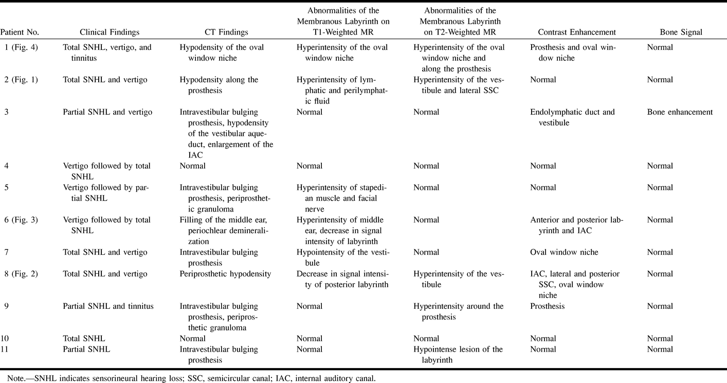
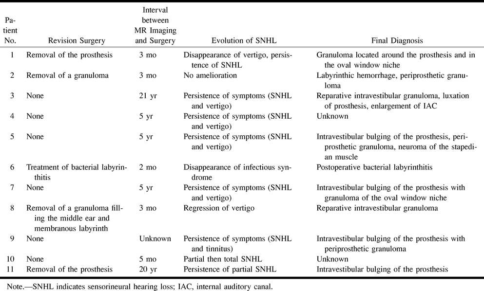
Article Figures & Data

Figures

  • Tables
  • fig 1.
    • Download figure
    • Open in new tab
    • Download powerpoint
    fig 1.

    58 year-old woman with postoperative hemorrhagic labyrinthitis 2 months after left-sided stapedectomy.

    A, Coronal CT scan of left temporal bone shows a hypodensity along a well-located prosthesis (arrow). The lateral semicircular canal is normal.

    B, Axial T1-weighted MR image shows abnormal signal intensity of the left lateral semicircular canal, as compared with right ear.

    C, Axial T2-weighted MR image (2500/90) shows normal hyperintensity of the lateral semicircular canal (arrow).

    D, Contrast-enhanced axial T1-weighted MR image (500/15) shows no change in the signal intensity of the labyrinth.

    E, Contrast-enhanced coronal T1-weighted MR image shows an enhancing mass engulfing the prosthesis (arrow). Note the normal enhancement of the tympanic portion of the facial nerve (arrowhead) under the abnormal lateral semicircular canal.

    F, Follow-up axial T1-weighted MR image at 1 year shows disappearance of the high signal intensity of the lateral semicircular canal.

  • fig 2.
    • Download figure
    • Open in new tab
    • Download powerpoint
    fig 2.

    58 year-old man referred for SNHL and vertigo 2 days after stapedectomy with a reparative intravestibular granuloma, confirmed by surgery.

    A, Coronal CT scan of the left temporal bone shows a soft tissue mass filling the oval window niche (arrow) and surrounding the tympanic portion of the facial nerve and the prosthesis.

    B, Noncontrast T1-weighted MR image (500/15) reveals slight high signal intensity of the vestibule (arrow).

    C, Axial contrast-enhanced T1-weighted MR image shows an enhancing mass of the oval window niche (arrowhead) spreading within the vestibule (single arrow) and the posterior (double arrows) and lateral (triple arrows) semicircular canals.

  • fig 3.
    • Download figure
    • Open in new tab
    • Download powerpoint
    fig 3.

    39 year-old woman referred for SNHL and vertigo 2 months after stapedectomy complicated by suppurative labyrinthitis.

    A, Coronal CT scan shows complete filling of the middle ear and oval window niche, enlargement of the oval window (double arrows), osseous erosion of the promontory and the lateral semicircular canal (single arrow), and enlargement of the vestibule and superior semicircular canal (arrowhead).

    B, Contrast-enhanced coronal T1-weighted MR image shows enhancement of an inflammatory mass of the epitympanum in communication with the lateral semicircular canal (arrow). Note associated abnormal enhancement of the vestibule, superior semicircular canal (arrowhead), and internal auditory canal.

  • fig 4.
    • Download figure
    • Open in new tab
    • Download powerpoint
    fig 4.

    44-year-old woman referred for SNHL and vertigo 6 weeks after stapedectomy with a granuloma located around the prosthesis and in the oval window niche.

    A, Axial CT scan of the left temporal bone shows well-located prosthesis (arrow) and partial filling of the posterior part of the oval window niche (arrowhead).

    B, Noncontrast axial T1-weighted MR image (500/15) shows normal signal intensity of the labyrinthine fluid, except for slightly high intensity around the oval window (arrow).

    C, Contrast-enhanced axial T1-weighted MR image (500/15) shows mild enhancement of the oval window (arrow). Tympanic portion of the facial nerve is seen laterally (arrowhead).

    D, Axial T2-weighted MR image shows normal high signal intensity of the labyrinthine fluid. A discrete intravestibular bulging of the prosthesis is clearly depicted (arrow).

Tables

  • Figures
  • TABLE 1:
    • View popup
    • Download powerpoint
    TABLE 1:

    Clinical and imaging findings

  • TABLE 2:
    • View popup
    • Download powerpoint
    TABLE 2:

    Diagnosis and evolution of sensorineural hearing loss

PreviousNext
Back to top

In this issue

American Journal of Neuroradiology: 22 (6)
American Journal of Neuroradiology
Vol. 22, Issue 6
1 Jun 2001
  • Table of Contents
  • Index by author
Advertisement
Print
Download PDF
Email Article

Thank you for your interest in spreading the word on American Journal of Neuroradiology.

NOTE: We only request your email address so that the person you are recommending the page to knows that you wanted them to see it, and that it is not junk mail. We do not capture any email address.

Enter multiple addresses on separate lines or separate them with commas.
Postoperative Complications in Otospongiosis: Usefulness of MR Imaging
(Your Name) has sent you a message from American Journal of Neuroradiology
(Your Name) thought you would like to see the American Journal of Neuroradiology web site.
CAPTCHA
This question is for testing whether or not you are a human visitor and to prevent automated spam submissions.
Cite this article
Anne-Sophie Rangheard, Kathlyn Marsot-Dupuch, Alexander S. Mark, Bernard Meyer, Jean-Michel Tubiana
Postoperative Complications in Otospongiosis: Usefulness of MR Imaging
American Journal of Neuroradiology Jun 2001, 22 (6) 1171-1178;

Citation Manager Formats

  • BibTeX
  • Bookends
  • EasyBib
  • EndNote (tagged)
  • EndNote 8 (xml)
  • Medlars
  • Mendeley
  • Papers
  • RefWorks Tagged
  • Ref Manager
  • RIS
  • Zotero
0 Responses
Respond to this article
Share
Bookmark this article
Postoperative Complications in Otospongiosis: Usefulness of MR Imaging
Anne-Sophie Rangheard, Kathlyn Marsot-Dupuch, Alexander S. Mark, Bernard Meyer, Jean-Michel Tubiana
American Journal of Neuroradiology Jun 2001, 22 (6) 1171-1178;
del.icio.us logo Twitter logo Facebook logo Mendeley logo
  • Tweet Widget
  • Facebook Like
  • Google Plus One

Jump to section

  • Article
    • Abstract
    • Methods
    • Results
    • Discussion
    • Conclusion
    • Acknowledgments
    • Footnotes
    • References
  • Figures & Data
  • Info & Metrics
  • Responses
  • References
  • PDF

Related Articles

  • No related articles found.
  • PubMed
  • Google Scholar

Cited By...

  • No citing articles found.
  • Crossref
  • Google Scholar

This article has not yet been cited by articles in journals that are participating in Crossref Cited-by Linking.

More in this TOC Section

  • ASL Sensitivity for Head and Neck Paraganglioma
  • Post SRS Peritumoral Hyperintense Signal of VSs
  • Temporal Evolution of Vestibular schwannoma
Show more Head and Neck Imaging

Similar Articles

Advertisement

Indexed Content

  • Current Issue
  • Accepted Manuscripts
  • Article Preview
  • Past Issues
  • Editorials
  • Editor's Choice
  • Fellows' Journal Club
  • Letters to the Editor
  • Video Articles

Cases

  • Case Collection
  • Archive - Case of the Week
  • Archive - Case of the Month
  • Archive - Classic Case

More from AJNR

  • Trainee Corner
  • Imaging Protocols
  • MRI Safety Corner
  • Book Reviews

Multimedia

  • AJNR Podcasts
  • AJNR Scantastics

Resources

  • Turnaround Time
  • Submit a Manuscript
  • Submit a Video Article
  • Submit an eLetter to the Editor/Response
  • Manuscript Submission Guidelines
  • Statistical Tips
  • Fast Publishing of Accepted Manuscripts
  • Graphical Abstract Preparation
  • Imaging Protocol Submission
  • Evidence-Based Medicine Level Guide
  • Publishing Checklists
  • Author Policies
  • Become a Reviewer/Academy of Reviewers
  • News and Updates

About Us

  • About AJNR
  • Editorial Board
  • Editorial Board Alumni
  • Alerts
  • Permissions
  • Not an AJNR Subscriber? Join Now
  • Advertise with Us
  • Librarian Resources
  • Feedback
  • Terms and Conditions
  • AJNR Editorial Board Alumni

American Society of Neuroradiology

  • Not an ASNR Member? Join Now

© 2025 by the American Society of Neuroradiology All rights, including for text and data mining, AI training, and similar technologies, are reserved.
Print ISSN: 0195-6108 Online ISSN: 1936-959X

Powered by HighWire