Advertisement

Main menu

  • Home
  • Content
    • Current Issue
    • Accepted Manuscripts
    • Article Preview
    • Past Issue Archive
    • Video Articles
    • AJNR Case Collection
    • Case of the Week Archive
    • Case of the Month Archive
    • Classic Case Archive
  • Special Collections
    • Advancing NeuroMRI with High-Relaxivity Contrast Agents
    • AJNR Awards
    • Low-Field MRI
    • Alzheimer Disease
    • ASNR Foundation Special Collection
    • Photon-Counting CT
    • View All
  • Multimedia
    • AJNR Podcasts
    • AJNR SCANtastic
    • Trainee Corner
    • MRI Safety Corner
    • Imaging Protocols
  • For Authors
    • Submit a Manuscript
    • Submit a Video Article
    • Submit an eLetter to the Editor/Response
    • Manuscript Submission Guidelines
    • Statistical Tips
    • Fast Publishing of Accepted Manuscripts
    • Graphical Abstract Preparation
    • Imaging Protocol Submission
    • Author Policies
  • About Us
    • About AJNR
    • Editorial Board
    • Editorial Board Alumni
  • More
    • Become a Reviewer/Academy of Reviewers
    • Subscribers
    • Permissions
    • Alerts
    • Feedback
    • Advertisers
    • ASNR Home

User menu

  • Alerts
  • Log in

Search

  • Advanced search
American Journal of Neuroradiology

American Journal of Neuroradiology

ASHNR American Society of Functional Neuroradiology ASHNR American Society of Pediatric Neuroradiology ASSR
  • Alerts
  • Log in

Advanced Search

  • Home
  • Content
    • Current Issue
    • Accepted Manuscripts
    • Article Preview
    • Past Issue Archive
    • Video Articles
    • AJNR Case Collection
    • Case of the Week Archive
    • Case of the Month Archive
    • Classic Case Archive
  • Special Collections
    • Advancing NeuroMRI with High-Relaxivity Contrast Agents
    • AJNR Awards
    • Low-Field MRI
    • Alzheimer Disease
    • ASNR Foundation Special Collection
    • Photon-Counting CT
    • View All
  • Multimedia
    • AJNR Podcasts
    • AJNR SCANtastic
    • Trainee Corner
    • MRI Safety Corner
    • Imaging Protocols
  • For Authors
    • Submit a Manuscript
    • Submit a Video Article
    • Submit an eLetter to the Editor/Response
    • Manuscript Submission Guidelines
    • Statistical Tips
    • Fast Publishing of Accepted Manuscripts
    • Graphical Abstract Preparation
    • Imaging Protocol Submission
    • Author Policies
  • About Us
    • About AJNR
    • Editorial Board
    • Editorial Board Alumni
  • More
    • Become a Reviewer/Academy of Reviewers
    • Subscribers
    • Permissions
    • Alerts
    • Feedback
    • Advertisers
    • ASNR Home
  • Follow AJNR on Twitter
  • Visit AJNR on Facebook
  • Follow AJNR on Instagram
  • Join AJNR on LinkedIn
  • RSS Feeds

AJNR Awards, New Junior Editors, and more. Read the latest AJNR updates


Improved Turnaround Times | Median time to first decision: 12 days

Research ArticleBrain

Transient MR Signal Changes in Patients with Generalized Tonicoclonic Seizure or Status Epilepticus: Periictal Diffusion-weighted Imaging

Jeong-Ah Kim, Jin Il Chung, Pyeong Ho Yoon, Dong Ik Kim, Tae-Sub Chung, Eun-Ju Kim and Eun-Kee Jeong
American Journal of Neuroradiology June 2001, 22 (6) 1149-1160;
Jeong-Ah Kim
  • Find this author on Google Scholar
  • Find this author on PubMed
  • Search for this author on this site
Jin Il Chung
  • Find this author on Google Scholar
  • Find this author on PubMed
  • Search for this author on this site
Pyeong Ho Yoon
  • Find this author on Google Scholar
  • Find this author on PubMed
  • Search for this author on this site
Dong Ik Kim
  • Find this author on Google Scholar
  • Find this author on PubMed
  • Search for this author on this site
Tae-Sub Chung
  • Find this author on Google Scholar
  • Find this author on PubMed
  • Search for this author on this site
Eun-Ju Kim
  • Find this author on Google Scholar
  • Find this author on PubMed
  • Search for this author on this site
Eun-Kee Jeong
  • Find this author on Google Scholar
  • Find this author on PubMed
  • Search for this author on this site
  • Article
  • Figures & Data
  • Info & Metrics
  • Responses
  • References
  • PDF
Loading

Article Figures & Data

Figures

  • Tables
  • fig 1.
    • Download figure
    • Open in new tab
    • Download powerpoint
    fig 1.

    Images of a 60-year-old man (patient 5) with complex partial status epilepticus with secondary generalization as the initial presentation of seizure.

    A, Ictal T2-weighted image (left) shows increased signal intensity with swelling at the subcortical white matter of the right parahippocampal gyrus, uncus, and occipital cortex (arrows). FLAIR image (right) shows the increased signal intensity at the parahippocampal gyrus (arrow) more conspicuously and the atrophy with increased T2 signal intensity of the body of the right hippocampus, indicating ipsilateral mesial temporal sclerosis.

    B, Diffusion-weighted image shows increased signal intensity at exactly the same areas (arrows).

    C, ADC map shows a 28% decrease of the mean ADC at the right parahippocampal gyrus and a 27% decrease at the uncus (arrows).

    D, Follow-up FLAIR image obtained 4 months later shows the resolution of the signal change and swelling at the right parahippocampal gyrus (arrow) and right mesial temporal sclerosis without remarkable interval change of volume and signal intensity.

  • fig 2.
    • Download figure
    • Open in new tab
    • Download powerpoint
    fig 2.

    Images of a 12-year-old male patient (patient 2) with generalized tonicoclonic seizure.

    A, Initial FLAIR image shows increased signal intensity in the cortical gray matter and subcortical white matter in cuneus and precuneus bilaterally (arrows).

    B, Initial diffusion-weighted image shows mildly increased signal intensity in the corresponding areas (arrows). The decrease of the mean ADC was 8% at the right and 6% at the left on the ADC map (not shown).

    C, Follow-up T2-weighted image obtained 14 days after the onset of seizure shows complete resolution of the signal change.

  • fig 3.
    • Download figure
    • Open in new tab
    • Download powerpoint
    fig 3.

    Images of a 2-year-old female patient (patient 6) with complex partial status epilepticus with secondary generalization.

    A, Initial T2-weighted image shows increased signal intensity and swelling in the right hippocampus (arrow).

    B, Initial diffusion-weighted image shows increased signal intensity in the right hippocampus (arrow).

    C, Corresponding ADC map shows 14% decrease of mean ADC (arrow).

    D, Follow-up FLAIR image obtained 18 months after the onset of seizure shows the resolution of the swelling and mass effect of the hippocampus (arrow) and increased T2 signal intensity without definite atrophic change of the hippocampus.

  • fig 4.
    • Download figure
    • Open in new tab
    • Download powerpoint
    fig 4.

    Images of a 60-year-old woman (patient 7) with simple partial status epilepticus sustained for 5 days.

    A, Ictal T2-weighted images show extensive swelling and increased signal intensity of the left hippocampus (left) and diffuse swelling with increased signal intensity at the cortical gray matter and subcortical white matter of the adjacent left temporal lobe (right).

    B, Ictal diffusion-weighted images show increased signal intensity and swelling at the left hippocampus (left, arrows) and at the uncus (right, arrowhead), parahippocampal gyrus (right, black arrow), and inferior temporal gyrus (right, white arrow).

    C, ADC maps show a 19% decrease of the mean ADC at the left hippocampus (left, arrow) and a 2% to 5% increase of the mean ADC at the subcortical white matter of the uncus, parahippocampal gyrus, and inferior temporal gyrus (right).

    D, Follow-up T2-weighted images obtained 4 months after the onset of seizure show the resolution of the marked swelling of the left hippocampus and partial resolution of the increased signal intensity of the left hippocampus (left). A tumor was revealed, which was diagnosed as glioblastoma multiforme at the left anterior temporal lobe (right).
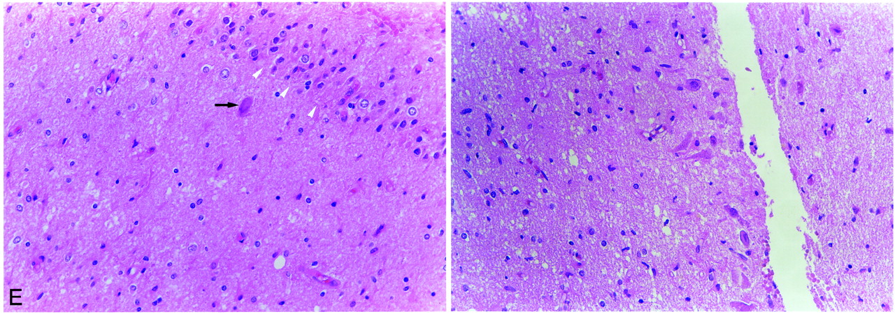
  • fig 4.
    • Download figure
    • Open in new tab
    • Download powerpoint
    fig 4.

    Continued.

    E, Light microscopic image of the left hippocampus obtained by amygdalohippocampectomy reveals extensive neuronal loss and gliosis in the CA4 area of the hippocampus (left: arrowheads, granule cell layer of dentate gyrus; arrow, remaining neuron in CA4) and neuronal loss and gliosis in the CA2 area of the hippocampus (right). More neurons are preserved in the CA2 area than in the other areas of the hippocampus (CA4, CA3, and CA1).

  • fig 5.
    • Download figure
    • Open in new tab
    • Download powerpoint
    fig 5.

    Images of a 51-year-old woman (patient 8) with generalized tonicoclonic status epilepticus.

    A, Initial FLAIR images show multiple increased signal intensity and swelling at the cortical gray matter and subcortical white matter of the right inferior frontal gyrus (left, arrowhead), right insular cortex (left, short arrow), left superior, middle, and inferior temporal gyri (right, open arrows), and bilateral hippocampi (long arrows).

    B, Initial diffusion-weighted images show increased signal intensity at the right inferior frontal gyrus (left, arrows), left temporal lobe (middle), and bilateral hippocampi (right, arrows).

    C, Follow-up diffusion-weighted images show resolution of the signal changes of the right frontal (left) and left temporal (middle) lobe. The increased signal intensity at the bilateral hippocampi did not resolve (right).

  • fig 6.
    • Download figure
    • Open in new tab
    • Download powerpoint
    fig 6.

    Images of a 3-year-old male patient (patient 1) with complex partial status epilepticus show the resolution process of the signal change.

    A, Initial T2-weighted image shows increased signal intensity in the cortical gray matter of bilateral cingulate gyri (arrows).

    B, Follow-up T2-weighted image shows partial resolution of the signal intensity 9 days after seizure onset.

    C, Follow-up T2-weighted image shows complete resolution of the signal intensity 30 days after seizure onset.

Tables

  • Figures
  • TABLE 1:
    • View popup
    • Download powerpoint
    TABLE 1:

    Patient profiles

  • TABLE 2:
    • View popup
    • Download powerpoint
    TABLE 2:

    Periictal MR signal changes and single-photon emission CT findings

  • TABLE 3:
    • View popup
    • Download powerpoint
    TABLE 3:

    Apparent diffusion coefficient of region of interest

PreviousNext
Back to top

In this issue

American Journal of Neuroradiology: 22 (6)
American Journal of Neuroradiology
Vol. 22, Issue 6
1 Jun 2001
  • Table of Contents
  • Index by author
Advertisement
Print
Download PDF
Email Article

Thank you for your interest in spreading the word on American Journal of Neuroradiology.

NOTE: We only request your email address so that the person you are recommending the page to knows that you wanted them to see it, and that it is not junk mail. We do not capture any email address.

Enter multiple addresses on separate lines or separate them with commas.
Transient MR Signal Changes in Patients with Generalized Tonicoclonic Seizure or Status Epilepticus: Periictal Diffusion-weighted Imaging
(Your Name) has sent you a message from American Journal of Neuroradiology
(Your Name) thought you would like to see the American Journal of Neuroradiology web site.
CAPTCHA
This question is for testing whether or not you are a human visitor and to prevent automated spam submissions.
Cite this article
Jeong-Ah Kim, Jin Il Chung, Pyeong Ho Yoon, Dong Ik Kim, Tae-Sub Chung, Eun-Ju Kim, Eun-Kee Jeong
Transient MR Signal Changes in Patients with Generalized Tonicoclonic Seizure or Status Epilepticus: Periictal Diffusion-weighted Imaging
American Journal of Neuroradiology Jun 2001, 22 (6) 1149-1160;

Citation Manager Formats

  • BibTeX
  • Bookends
  • EasyBib
  • EndNote (tagged)
  • EndNote 8 (xml)
  • Medlars
  • Mendeley
  • Papers
  • RefWorks Tagged
  • Ref Manager
  • RIS
  • Zotero
0 Responses
Respond to this article
Share
Bookmark this article
Transient MR Signal Changes in Patients with Generalized Tonicoclonic Seizure or Status Epilepticus: Periictal Diffusion-weighted Imaging
Jeong-Ah Kim, Jin Il Chung, Pyeong Ho Yoon, Dong Ik Kim, Tae-Sub Chung, Eun-Ju Kim, Eun-Kee Jeong
American Journal of Neuroradiology Jun 2001, 22 (6) 1149-1160;
del.icio.us logo Twitter logo Facebook logo Mendeley logo
  • Tweet Widget
  • Facebook Like
  • Google Plus One

Jump to section

  • Article
    • Abstract
    • Methods
    • Results
    • Discussion
    • Footnotes
    • References
  • Figures & Data
  • Info & Metrics
  • Responses
  • References
  • PDF

Related Articles

  • No related articles found.
  • PubMed
  • Google Scholar

Cited By...

  • Human Herpesvirus 6 in the CSF of a Woman With New-Onset Seizures: Encephalitis or Genomic Integration?
  • Diagnosing autoimmune limbic encephalitis
  • Magnetic resonance imaging findings in epileptic cats with a normal interictal neurological examination: 188 cases
  • Pearls & Oy-sters: Hemicrania epileptica: Unfolding the mystery of an unremitting migraine
  • Recognizing Autoimmune-Mediated Encephalitis in the Differential Diagnosis of Limbic Disorders
  • MRI Findings in Autoimmune Voltage-Gated Potassium Channel Complex Encephalitis with Seizures: One Potential Etiology for Mesial Temporal Sclerosis
  • Transient high-intensity signal of heterotopia on DWI in an epilepsy patient
  • Peri-ictal restricted diffusion in heterotopic gray matter assessed by MRI
  • Dr. Varaprasad and Dr. Agrawal reply
  • Neuronal surface antigen antibodies in limbic encephalitis: Clinical-immunologic associations
  • Practice parameter: diagnostic assessment of the child with status epilepticus (an evidence-based review): report of the Quality Standards Subcommittee of the American Academy of Neurology and the Practice Committee of the Child Neurology Society.
  • Acute symptomatic seizures and hippocampus damage: DWI and MRS findings
  • Cortical laminar necrosis related to prolonged focal status epilepticus
  • Transient lesion in the splenium of the corpus callosum and antiepileptic drug withdrawal
  • Evolution of MRI changes and development of bilateral hippocampal sclerosis during long lasting generalised status epilepticus
  • Diffusion-weighted MRI abnormalities as a clue to the diagnosis of herpes simplex encephalitis
  • Visualization of Evolving Status Epilepticus with Diffusion and Perfusion MR Imaging
  • Do Seizures Injure the Brain?
  • Crossref
  • Google Scholar

This article has not yet been cited by articles in journals that are participating in Crossref Cited-by Linking.

More in this TOC Section

  • Usefulness of Quantitative Susceptibility Mapping for the Diagnosis of Parkinson Disease
  • White Matter Alterations in the Brains of Patients with Active, Remitted, and Cured Cushing Syndrome: A DTI Study
  • Qualitative and Quantitative Analysis of MR Imaging Findings in Patients with Middle Cerebral Artery Stroke Implanted with Mesenchymal Stem Cells
Show more Brain

Similar Articles

Advertisement

Indexed Content

  • Current Issue
  • Accepted Manuscripts
  • Article Preview
  • Past Issues
  • Editorials
  • Editor's Choice
  • Fellows' Journal Club
  • Letters to the Editor
  • Video Articles

Cases

  • Case Collection
  • Archive - Case of the Week
  • Archive - Case of the Month
  • Archive - Classic Case

More from AJNR

  • Trainee Corner
  • Imaging Protocols
  • MRI Safety Corner
  • Book Reviews

Multimedia

  • AJNR Podcasts
  • AJNR Scantastics

Resources

  • Turnaround Time
  • Submit a Manuscript
  • Submit a Video Article
  • Submit an eLetter to the Editor/Response
  • Manuscript Submission Guidelines
  • Statistical Tips
  • Fast Publishing of Accepted Manuscripts
  • Graphical Abstract Preparation
  • Imaging Protocol Submission
  • Evidence-Based Medicine Level Guide
  • Publishing Checklists
  • Author Policies
  • Become a Reviewer/Academy of Reviewers
  • News and Updates

About Us

  • About AJNR
  • Editorial Board
  • Editorial Board Alumni
  • Alerts
  • Permissions
  • Not an AJNR Subscriber? Join Now
  • Advertise with Us
  • Librarian Resources
  • Feedback
  • Terms and Conditions
  • AJNR Editorial Board Alumni

American Society of Neuroradiology

  • Not an ASNR Member? Join Now

© 2025 by the American Society of Neuroradiology All rights, including for text and data mining, AI training, and similar technologies, are reserved.
Print ISSN: 0195-6108 Online ISSN: 1936-959X

Powered by HighWire