Advertisement

Main menu

  • Home
  • Content
    • Current Issue
    • Accepted Manuscripts
    • Article Preview
    • Past Issue Archive
    • Video Articles
    • AJNR Case Collection
    • Case of the Week Archive
    • Case of the Month Archive
    • Classic Case Archive
  • Special Collections
    • AJNR Awards
    • Low-Field MRI
    • Alzheimer Disease
    • ASNR Foundation Special Collection
    • Photon-Counting CT
    • View All
  • Multimedia
    • AJNR Podcasts
    • AJNR SCANtastic
    • Trainee Corner
    • MRI Safety Corner
    • Imaging Protocols
  • For Authors
    • Submit a Manuscript
    • Submit a Video Article
    • Submit an eLetter to the Editor/Response
    • Manuscript Submission Guidelines
    • Statistical Tips
    • Fast Publishing of Accepted Manuscripts
    • Graphical Abstract Preparation
    • Imaging Protocol Submission
    • Author Policies
  • About Us
    • About AJNR
    • Editorial Board
    • Editorial Board Alumni
  • More
    • Become a Reviewer/Academy of Reviewers
    • Subscribers
    • Permissions
    • Alerts
    • Feedback
    • Advertisers
    • ASNR Home

User menu

  • Alerts
  • Log in

Search

  • Advanced search
American Journal of Neuroradiology

American Journal of Neuroradiology

ASHNR American Society of Functional Neuroradiology ASHNR American Society of Pediatric Neuroradiology ASSR
  • Alerts
  • Log in

Advanced Search

  • Home
  • Content
    • Current Issue
    • Accepted Manuscripts
    • Article Preview
    • Past Issue Archive
    • Video Articles
    • AJNR Case Collection
    • Case of the Week Archive
    • Case of the Month Archive
    • Classic Case Archive
  • Special Collections
    • AJNR Awards
    • Low-Field MRI
    • Alzheimer Disease
    • ASNR Foundation Special Collection
    • Photon-Counting CT
    • View All
  • Multimedia
    • AJNR Podcasts
    • AJNR SCANtastic
    • Trainee Corner
    • MRI Safety Corner
    • Imaging Protocols
  • For Authors
    • Submit a Manuscript
    • Submit a Video Article
    • Submit an eLetter to the Editor/Response
    • Manuscript Submission Guidelines
    • Statistical Tips
    • Fast Publishing of Accepted Manuscripts
    • Graphical Abstract Preparation
    • Imaging Protocol Submission
    • Author Policies
  • About Us
    • About AJNR
    • Editorial Board
    • Editorial Board Alumni
  • More
    • Become a Reviewer/Academy of Reviewers
    • Subscribers
    • Permissions
    • Alerts
    • Feedback
    • Advertisers
    • ASNR Home
  • Follow AJNR on Twitter
  • Visit AJNR on Facebook
  • Follow AJNR on Instagram
  • Join AJNR on LinkedIn
  • RSS Feeds

AJNR Awards, New Junior Editors, and more. Read the latest AJNR updates

Research ArticleHead and Neck Imaging

The Accuracy of Sonography for Evaluation of Internal Derangement of the Temporomandibular Joint in Asymptomatic Elementary School Children: Comparison with MR and CT

Takafumi Hayashi, Jusuke Ito, Jun-ichi Koyama and Kazuhiro Yamada
American Journal of Neuroradiology April 2001, 22 (4) 728-734;
Takafumi Hayashi
  • Find this author on Google Scholar
  • Find this author on PubMed
  • Search for this author on this site
Jusuke Ito
  • Find this author on Google Scholar
  • Find this author on PubMed
  • Search for this author on this site
Jun-ichi Koyama
  • Find this author on Google Scholar
  • Find this author on PubMed
  • Search for this author on this site
Kazuhiro Yamada
  • Find this author on Google Scholar
  • Find this author on PubMed
  • Search for this author on this site
  • Article
  • Figures & Data
  • Info & Metrics
  • Responses
  • References
  • PDF
Loading

Article Figures & Data

Figures

  • Tables
  • fig 1.
    • Download figure
    • Open in new tab
    • Download powerpoint
    fig 1.

    A schematic drawing of the scanning plane of the transducer in a transverse direction running parallel to the Camper line intersecting the ala of the nose and the tragus of the ear
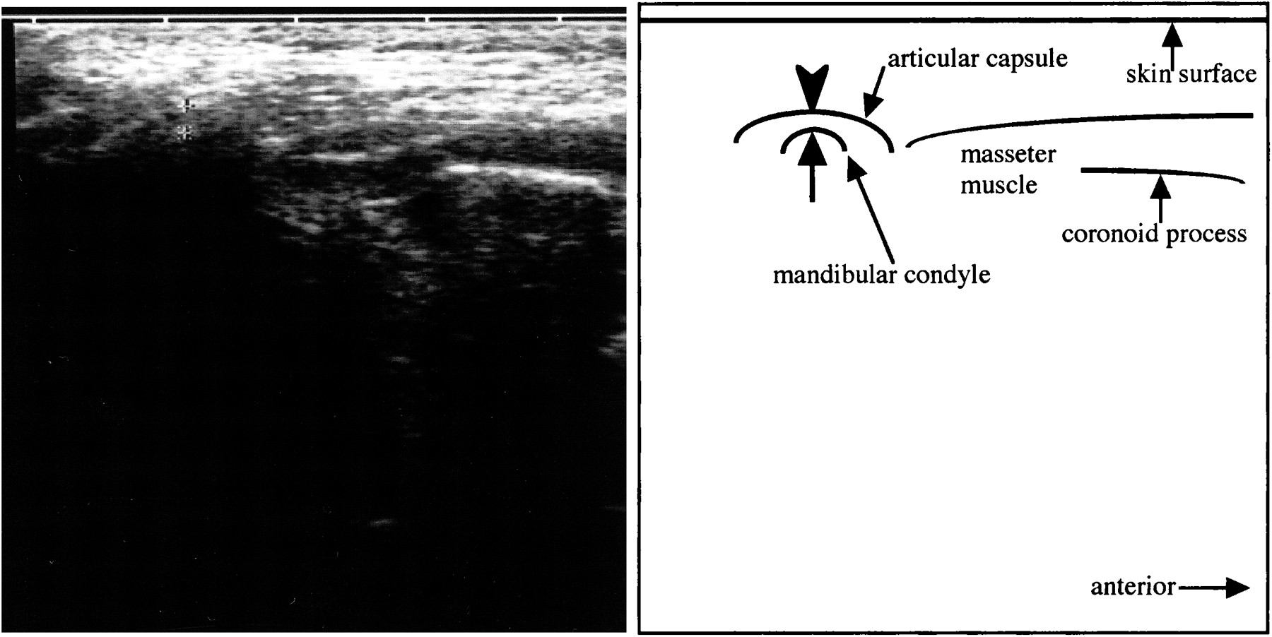
  • fig 2.
    • Download figure
    • Open in new tab
    • Download powerpoint
    fig 2.

    A transverse section of sonography of the normal right TMJ obtained while the patient was in the closed-mouth position. Note hyperechoic line (arrowhead) running lateral and parallel to the lateral surface (arrow) of the mandibular condyle, indicating the articular capsule. We measured the distance between the articular capsule and the lateral surface of the mandibular condyle (between the apexes of the arrow and arrowhead) on the display of the sonographic equipment

  • fig 3.
    • Download figure
    • Open in new tab
    • Download powerpoint
    fig 3.

    10-year-old girl.

    A, A transverse section of sonography of the right TMJ obtained while the patient was in the closed-mouth position. Note a hyperechoic line (arrowhead) running lateral and parallel to the lateral surface (arrow) of the mandibular condyle, indicating the articular capsule. The distance between the articular capsule and the lateral surface of the mandibular condyle is 4 mm.

    B, A sagittal proton density–weighted image of the right TMJ obtained while the patient was in the closed-mouth position. Note the posterior band of the articular disk located anterior to the mandibular condyle (anterior disk displacement). The evaluation of the disk displacement on A is a true-positive result.

    C, A transverse section of sonography of the left TMJ obtained while the patient was in the closed-mouth position. The distance between the articular capsule (arrowhead) and the lateral surface (arrow) of the mandibular condyle is 3 mm.

    D, A sagittal proton density–weighted image of the left TMJ obtained while the patient was in the closed-mouth position. Note the posterior band of the articular disk located superior to the mandibular condyle (normal superior position). The evaluation of the disk displacement on C is a true-negative result.

  • fig 4.
    • Download figure
    • Open in new tab
    • Download powerpoint
    fig 4.

    Same patient as shown in figure 3. A series of continuous axial T1-weighted images at the level of the TMJ obtained while the patient was in the closed-mouth position. Note a part of anterolaterally displaced disk (arrows) covering the lateral surface of the right mandibular condyle and a part of widened articular capsule (arrowhead) adjacent to the lateral surface of the condyle
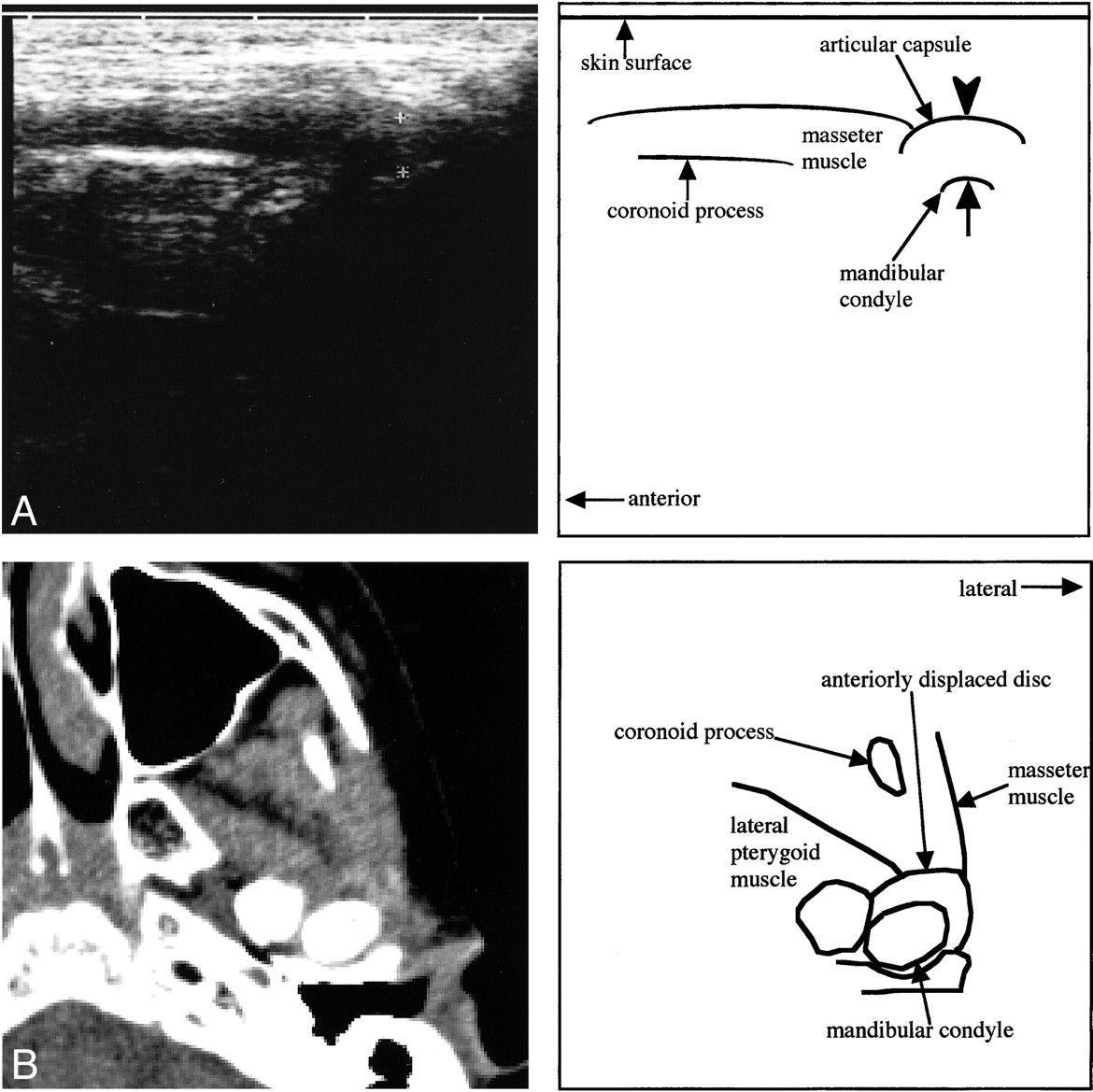
  • fig 5.
    • Download figure
    • Open in new tab
    • Download powerpoint
    fig 5.

    10-year-old boy.

    A, A transverse sonographic section of the left TMJ obtained while the patient was in the closed-mouth position. The distance between the articular capsule (arrowhead) and the lateral surface (arrow) of the mandibular condyle is 5 mm.

    B, A transverse CT section at the level of the mandibular condyle obtained while the patient was in a closed-mouth position. Note the hyperdense area in front of the left mandibular condyle indicating the anteriorly dislocated articular disk. Disk displacement on A is a true-positive result.

Tables

  • Figures
  • TABLE 1:
    • View popup
    • Download powerpoint
    TABLE 1:

    Clinical summary of the subjects

  • TABLE 2:
    • View popup
    • Download powerpoint
    TABLE 2:

    The relationship between the distance between the articular capsule and the lateral surface of the mandibular condyle and articular disk displacement of the TMJ detected on MR or helical CT images

  • TABLE 3:
    • View popup
    • Download powerpoint
    TABLE 3:

    The diagnostic reliability of the cut-off distance between the articular capsule and the lateral surface of the mandibular condyle on sonograms

PreviousNext
Back to top

In this issue

American Journal of Neuroradiology
Vol. 22, Issue 4
1 Apr 2001
  • Table of Contents
  • Index by author
Advertisement
Print
Download PDF
Email Article

Thank you for your interest in spreading the word on American Journal of Neuroradiology.

NOTE: We only request your email address so that the person you are recommending the page to knows that you wanted them to see it, and that it is not junk mail. We do not capture any email address.

Enter multiple addresses on separate lines or separate them with commas.
The Accuracy of Sonography for Evaluation of Internal Derangement of the Temporomandibular Joint in Asymptomatic Elementary School Children: Comparison with MR and CT
(Your Name) has sent you a message from American Journal of Neuroradiology
(Your Name) thought you would like to see the American Journal of Neuroradiology web site.
CAPTCHA
This question is for testing whether or not you are a human visitor and to prevent automated spam submissions.
Cite this article
Takafumi Hayashi, Jusuke Ito, Jun-ichi Koyama, Kazuhiro Yamada
The Accuracy of Sonography for Evaluation of Internal Derangement of the Temporomandibular Joint in Asymptomatic Elementary School Children: Comparison with MR and CT
American Journal of Neuroradiology Apr 2001, 22 (4) 728-734;

Citation Manager Formats

  • BibTeX
  • Bookends
  • EasyBib
  • EndNote (tagged)
  • EndNote 8 (xml)
  • Medlars
  • Mendeley
  • Papers
  • RefWorks Tagged
  • Ref Manager
  • RIS
  • Zotero
0 Responses
Respond to this article
Share
Bookmark this article
The Accuracy of Sonography for Evaluation of Internal Derangement of the Temporomandibular Joint in Asymptomatic Elementary School Children: Comparison with MR and CT
Takafumi Hayashi, Jusuke Ito, Jun-ichi Koyama, Kazuhiro Yamada
American Journal of Neuroradiology Apr 2001, 22 (4) 728-734;
del.icio.us logo Twitter logo Facebook logo Mendeley logo
  • Tweet Widget
  • Facebook Like
  • Google Plus One

Jump to section

  • Article
    • Abstract
    • Methods
    • Results
    • Discussion
    • Conclusion
    • Footnotes
    • References
  • Figures & Data
  • Info & Metrics
  • Responses
  • References
  • PDF

Related Articles

  • No related articles found.
  • PubMed
  • Google Scholar

Cited By...

  • Dynamic High-Resolution Sonography Compared to Magnetic Resonance Imaging for Diagnosis of Temporomandibular Joint Disk Displacement
  • Is high-resolution ultrasonography suitable for the detection of temporomandibular joint involvement in children with juvenile idiopathic arthritis?
  • Ultrasound and tomographic evaluation of temporomandibular joints in adolescents with and without signs and symptoms of temporomandibular disorders: a pilot study
  • Ultrasound assessment of increased capsular width as a predictor of temporomandibular joint effusion
  • Crossref
  • Google Scholar

This article has not yet been cited by articles in journals that are participating in Crossref Cited-by Linking.

More in this TOC Section

  • ASL Sensitivity for Head and Neck Paraganglioma
  • Post SRS Peritumoral Hyperintense Signal of VSs
  • Contrast Enhanced Pituitary CISS/FIESTA
Show more Head and Neck Imaging

Similar Articles

Advertisement

Indexed Content

  • Current Issue
  • Accepted Manuscripts
  • Article Preview
  • Past Issues
  • Editorials
  • Editor's Choice
  • Fellows' Journal Club
  • Letters to the Editor
  • Video Articles

Cases

  • Case Collection
  • Archive - Case of the Week
  • Archive - Case of the Month
  • Archive - Classic Case

More from AJNR

  • Trainee Corner
  • Imaging Protocols
  • MRI Safety Corner
  • Book Reviews

Multimedia

  • AJNR Podcasts
  • AJNR Scantastics

Resources

  • Turnaround Time
  • Submit a Manuscript
  • Submit a Video Article
  • Submit an eLetter to the Editor/Response
  • Manuscript Submission Guidelines
  • Statistical Tips
  • Fast Publishing of Accepted Manuscripts
  • Graphical Abstract Preparation
  • Imaging Protocol Submission
  • Evidence-Based Medicine Level Guide
  • Publishing Checklists
  • Author Policies
  • Become a Reviewer/Academy of Reviewers
  • News and Updates

About Us

  • About AJNR
  • Editorial Board
  • Editorial Board Alumni
  • Alerts
  • Permissions
  • Not an AJNR Subscriber? Join Now
  • Advertise with Us
  • Librarian Resources
  • Feedback
  • Terms and Conditions
  • AJNR Editorial Board Alumni

American Society of Neuroradiology

  • Not an ASNR Member? Join Now

© 2025 by the American Society of Neuroradiology All rights, including for text and data mining, AI training, and similar technologies, are reserved.
Print ISSN: 0195-6108 Online ISSN: 1936-959X

Powered by HighWire