Advertisement

Main menu

  • Home
  • Content
    • Current Issue
    • Accepted Manuscripts
    • Article Preview
    • Past Issue Archive
    • Video Articles
    • AJNR Case Collection
    • Case of the Week Archive
    • Case of the Month Archive
    • Classic Case Archive
  • Special Collections
    • AJNR Awards
    • Low-Field MRI
    • Alzheimer Disease
    • ASNR Foundation Special Collection
    • Photon-Counting CT
    • View All
  • Multimedia
    • AJNR Podcasts
    • AJNR SCANtastic
    • Trainee Corner
    • MRI Safety Corner
    • Imaging Protocols
  • For Authors
    • Submit a Manuscript
    • Submit a Video Article
    • Submit an eLetter to the Editor/Response
    • Manuscript Submission Guidelines
    • Statistical Tips
    • Fast Publishing of Accepted Manuscripts
    • Graphical Abstract Preparation
    • Imaging Protocol Submission
    • Author Policies
  • About Us
    • About AJNR
    • Editorial Board
    • Editorial Board Alumni
  • More
    • Become a Reviewer/Academy of Reviewers
    • Subscribers
    • Permissions
    • Alerts
    • Feedback
    • Advertisers
    • ASNR Home

User menu

  • Alerts
  • Log in

Search

  • Advanced search
American Journal of Neuroradiology

American Journal of Neuroradiology

ASHNR American Society of Functional Neuroradiology ASHNR American Society of Pediatric Neuroradiology ASSR
  • Alerts
  • Log in

Advanced Search

  • Home
  • Content
    • Current Issue
    • Accepted Manuscripts
    • Article Preview
    • Past Issue Archive
    • Video Articles
    • AJNR Case Collection
    • Case of the Week Archive
    • Case of the Month Archive
    • Classic Case Archive
  • Special Collections
    • AJNR Awards
    • Low-Field MRI
    • Alzheimer Disease
    • ASNR Foundation Special Collection
    • Photon-Counting CT
    • View All
  • Multimedia
    • AJNR Podcasts
    • AJNR SCANtastic
    • Trainee Corner
    • MRI Safety Corner
    • Imaging Protocols
  • For Authors
    • Submit a Manuscript
    • Submit a Video Article
    • Submit an eLetter to the Editor/Response
    • Manuscript Submission Guidelines
    • Statistical Tips
    • Fast Publishing of Accepted Manuscripts
    • Graphical Abstract Preparation
    • Imaging Protocol Submission
    • Author Policies
  • About Us
    • About AJNR
    • Editorial Board
    • Editorial Board Alumni
  • More
    • Become a Reviewer/Academy of Reviewers
    • Subscribers
    • Permissions
    • Alerts
    • Feedback
    • Advertisers
    • ASNR Home
  • Follow AJNR on Twitter
  • Visit AJNR on Facebook
  • Follow AJNR on Instagram
  • Join AJNR on LinkedIn
  • RSS Feeds

AJNR Awards, New Junior Editors, and more. Read the latest AJNR updates

Research ArticleBRAIN

Lateralizing Ability of Single-voxel Proton MR Spectroscopy in Hippocampal Sclerosis: Comparison with MR Imaging and Positron Emission Tomography

Sun-Won Park, Kee-Hyun Chang, Hong-Dae Kim, In Chan Song, Dong Soo Lee, Sang Kun Lee, Chun-Kee Chung, In Kyu Yu, Moon Hee Han and Yang Hee Park
American Journal of Neuroradiology April 2001, 22 (4) 625-631;
Sun-Won Park
  • Find this author on Google Scholar
  • Find this author on PubMed
  • Search for this author on this site
Kee-Hyun Chang
  • Find this author on Google Scholar
  • Find this author on PubMed
  • Search for this author on this site
Hong-Dae Kim
  • Find this author on Google Scholar
  • Find this author on PubMed
  • Search for this author on this site
In Chan Song
  • Find this author on Google Scholar
  • Find this author on PubMed
  • Search for this author on this site
Dong Soo Lee
  • Find this author on Google Scholar
  • Find this author on PubMed
  • Search for this author on this site
Sang Kun Lee
  • Find this author on Google Scholar
  • Find this author on PubMed
  • Search for this author on this site
Chun-Kee Chung
  • Find this author on Google Scholar
  • Find this author on PubMed
  • Search for this author on this site
In Kyu Yu
  • Find this author on Google Scholar
  • Find this author on PubMed
  • Search for this author on this site
Moon Hee Han
  • Find this author on Google Scholar
  • Find this author on PubMed
  • Search for this author on this site
Yang Hee Park
  • Find this author on Google Scholar
  • Find this author on PubMed
  • Search for this author on this site
  • Article
  • Figures & Data
  • Info & Metrics
  • Responses
  • References
  • PDF
Loading

Article Figures & Data

Figures

  • Tables
  • fig 1.
    • Download figure
    • Open in new tab
    • Download powerpoint
    fig 1.

    Concordance among MR imaging, MRS, and PET in right hippocampal sclerosis. Oblique coronal T2-weighted MR image (top row, middle) shows right hippocampal sclerosis (arrow), in concordance with PET scan (bottom row, middle), which shows decreased metabolism in right temporal lobe (arrow). The MR spectrum from the right hippocampus (Rt) shows marked decrease in NAA/Cho ratio (arrow). The MR spectrum from left hippocampus is normal (Lt)

  • fig 2.
    • Download figure
    • Open in new tab
    • Download powerpoint
    fig 2.

    Discordant MRS with MR imaging and PET in left hippocampal sclerosis. Oblique coronal T2-weighted MR image (top row, middle) shows left hippocampal atrophy (arrow), in concordance with PET scan (bottom row, middle), which shows decreased metabolism in left temporal lobe (arrow). However, the MR spectrum from the left hippocampus (Lt) appears within normal range (false-negative finding), based on the abnormal criteria of an NAA/Cho ratio of 0.8 or less or an NAA/Cr ratio of 1.0 or less. The MR spectrum from the left hippocampus (Lt) shows a decrease in NAA/Cho and NAA/Cr ratios (arrow) on right side (Rt) (false-positive finding)

  • fig 3.
    • Download figure
    • Open in new tab
    • Download powerpoint
    fig 3.

    Bilateral abnormality on MRS in left hippocampal sclerosis. Oblique coronal T2-weighted MR image (top row, middle) shows left hippocampal sclerosis with increased signal intensity (arrow), in concordance with PET scan (bottom row, middle), which shows decreased metabolism (arrow) in left temporal lobe. The MR spectra (Rt and Lt) show bilateral abnormalities (arrows), decreased NAA/Cho ratios bilaterally, and decreased NAA/Cr ratio on right side

Tables

  • Figures
  • Table1

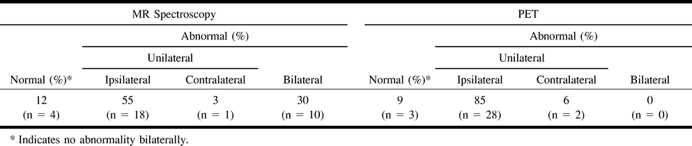
    Summary of MRS and PET results in lateralization of hippocampal sclerosis (n = 33)

PreviousNext
Back to top

In this issue

American Journal of Neuroradiology
Vol. 22, Issue 4
1 Apr 2001
  • Table of Contents
  • Index by author
Advertisement
Print
Download PDF
Email Article

Thank you for your interest in spreading the word on American Journal of Neuroradiology.

NOTE: We only request your email address so that the person you are recommending the page to knows that you wanted them to see it, and that it is not junk mail. We do not capture any email address.

Enter multiple addresses on separate lines or separate them with commas.
Lateralizing Ability of Single-voxel Proton MR Spectroscopy in Hippocampal Sclerosis: Comparison with MR Imaging and Positron Emission Tomography
(Your Name) has sent you a message from American Journal of Neuroradiology
(Your Name) thought you would like to see the American Journal of Neuroradiology web site.
CAPTCHA
This question is for testing whether or not you are a human visitor and to prevent automated spam submissions.
Cite this article
Sun-Won Park, Kee-Hyun Chang, Hong-Dae Kim, In Chan Song, Dong Soo Lee, Sang Kun Lee, Chun-Kee Chung, In Kyu Yu, Moon Hee Han, Yang Hee Park
Lateralizing Ability of Single-voxel Proton MR Spectroscopy in Hippocampal Sclerosis: Comparison with MR Imaging and Positron Emission Tomography
American Journal of Neuroradiology Apr 2001, 22 (4) 625-631;

Citation Manager Formats

  • BibTeX
  • Bookends
  • EasyBib
  • EndNote (tagged)
  • EndNote 8 (xml)
  • Medlars
  • Mendeley
  • Papers
  • RefWorks Tagged
  • Ref Manager
  • RIS
  • Zotero
0 Responses
Respond to this article
Share
Bookmark this article
Lateralizing Ability of Single-voxel Proton MR Spectroscopy in Hippocampal Sclerosis: Comparison with MR Imaging and Positron Emission Tomography
Sun-Won Park, Kee-Hyun Chang, Hong-Dae Kim, In Chan Song, Dong Soo Lee, Sang Kun Lee, Chun-Kee Chung, In Kyu Yu, Moon Hee Han, Yang Hee Park
American Journal of Neuroradiology Apr 2001, 22 (4) 625-631;
del.icio.us logo Twitter logo Facebook logo Mendeley logo
  • Tweet Widget
  • Facebook Like
  • Google Plus One

Jump to section

  • Article
    • Abstract
    • Methods
    • Results
    • Discussion
    • Conclusion
    • Footnotes
    • References
  • Figures & Data
  • Info & Metrics
  • Responses
  • References
  • PDF

Related Articles

  • No related articles found.
  • PubMed
  • Google Scholar

Cited By...

  • Differential Features of Metabolic Abnormalities Between Medial and Lateral Temporal Lobe Epilepsy: Quantitative Analysis of 18F-FDG PET Using SPM
  • Crossref
  • Google Scholar

This article has not yet been cited by articles in journals that are participating in Crossref Cited-by Linking.

More in this TOC Section

  • Statin Therapy Does Not Affect the Radiographic and Clinical Profile of Patients with TIA and Minor Stroke
  • Usefulness of Quantitative Susceptibility Mapping for the Diagnosis of Parkinson Disease
  • White Matter Alterations in the Brains of Patients with Active, Remitted, and Cured Cushing Syndrome: A DTI Study
Show more Brain

Similar Articles

Advertisement

Indexed Content

  • Current Issue
  • Accepted Manuscripts
  • Article Preview
  • Past Issues
  • Editorials
  • Editor's Choice
  • Fellows' Journal Club
  • Letters to the Editor
  • Video Articles

Cases

  • Case Collection
  • Archive - Case of the Week
  • Archive - Case of the Month
  • Archive - Classic Case

More from AJNR

  • Trainee Corner
  • Imaging Protocols
  • MRI Safety Corner
  • Book Reviews

Multimedia

  • AJNR Podcasts
  • AJNR Scantastics

Resources

  • Turnaround Time
  • Submit a Manuscript
  • Submit a Video Article
  • Submit an eLetter to the Editor/Response
  • Manuscript Submission Guidelines
  • Statistical Tips
  • Fast Publishing of Accepted Manuscripts
  • Graphical Abstract Preparation
  • Imaging Protocol Submission
  • Evidence-Based Medicine Level Guide
  • Publishing Checklists
  • Author Policies
  • Become a Reviewer/Academy of Reviewers
  • News and Updates

About Us

  • About AJNR
  • Editorial Board
  • Editorial Board Alumni
  • Alerts
  • Permissions
  • Not an AJNR Subscriber? Join Now
  • Advertise with Us
  • Librarian Resources
  • Feedback
  • Terms and Conditions
  • AJNR Editorial Board Alumni

American Society of Neuroradiology

  • Not an ASNR Member? Join Now

© 2025 by the American Society of Neuroradiology All rights, including for text and data mining, AI training, and similar technologies, are reserved.
Print ISSN: 0195-6108 Online ISSN: 1936-959X

Powered by HighWire