Advertisement

Main menu

  • Home
  • Content
    • Current Issue
    • Accepted Manuscripts
    • Article Preview
    • Past Issue Archive
    • Video Articles
    • AJNR Case Collection
    • Case of the Week Archive
    • Case of the Month Archive
    • Classic Case Archive
  • Special Collections
    • AJNR Awards
    • Low-Field MRI
    • Alzheimer Disease
    • ASNR Foundation Special Collection
    • Photon-Counting CT
    • View All
  • Multimedia
    • AJNR Podcasts
    • AJNR SCANtastic
    • Trainee Corner
    • MRI Safety Corner
    • Imaging Protocols
  • For Authors
    • Submit a Manuscript
    • Submit a Video Article
    • Submit an eLetter to the Editor/Response
    • Manuscript Submission Guidelines
    • Statistical Tips
    • Fast Publishing of Accepted Manuscripts
    • Graphical Abstract Preparation
    • Imaging Protocol Submission
    • Author Policies
  • About Us
    • About AJNR
    • Editorial Board
    • Editorial Board Alumni
  • More
    • Become a Reviewer/Academy of Reviewers
    • Subscribers
    • Permissions
    • Alerts
    • Feedback
    • Advertisers
    • ASNR Home

User menu

  • Alerts
  • Log in

Search

  • Advanced search
American Journal of Neuroradiology

American Journal of Neuroradiology

ASHNR American Society of Functional Neuroradiology ASHNR American Society of Pediatric Neuroradiology ASSR
  • Alerts
  • Log in

Advanced Search

  • Home
  • Content
    • Current Issue
    • Accepted Manuscripts
    • Article Preview
    • Past Issue Archive
    • Video Articles
    • AJNR Case Collection
    • Case of the Week Archive
    • Case of the Month Archive
    • Classic Case Archive
  • Special Collections
    • AJNR Awards
    • Low-Field MRI
    • Alzheimer Disease
    • ASNR Foundation Special Collection
    • Photon-Counting CT
    • View All
  • Multimedia
    • AJNR Podcasts
    • AJNR SCANtastic
    • Trainee Corner
    • MRI Safety Corner
    • Imaging Protocols
  • For Authors
    • Submit a Manuscript
    • Submit a Video Article
    • Submit an eLetter to the Editor/Response
    • Manuscript Submission Guidelines
    • Statistical Tips
    • Fast Publishing of Accepted Manuscripts
    • Graphical Abstract Preparation
    • Imaging Protocol Submission
    • Author Policies
  • About Us
    • About AJNR
    • Editorial Board
    • Editorial Board Alumni
  • More
    • Become a Reviewer/Academy of Reviewers
    • Subscribers
    • Permissions
    • Alerts
    • Feedback
    • Advertisers
    • ASNR Home
  • Follow AJNR on Twitter
  • Visit AJNR on Facebook
  • Follow AJNR on Instagram
  • Join AJNR on LinkedIn
  • RSS Feeds

AJNR Awards, New Junior Editors, and more. Read the latest AJNR updates

Research ArticleSPINE

Spinal Cord Involvement in Primary Angiitis of the Central Nervous System: A Report of Two Cases

Adriana Campi, G. Benndorf, V. Martinelli, M. R. Terreni and G. Scotti
American Journal of Neuroradiology March 2001, 22 (3) 577-582;
Adriana Campi
  • Find this author on Google Scholar
  • Find this author on PubMed
  • Search for this author on this site
G. Benndorf
  • Find this author on Google Scholar
  • Find this author on PubMed
  • Search for this author on this site
V. Martinelli
  • Find this author on Google Scholar
  • Find this author on PubMed
  • Search for this author on this site
M. R. Terreni
  • Find this author on Google Scholar
  • Find this author on PubMed
  • Search for this author on this site
G. Scotti
  • Find this author on Google Scholar
  • Find this author on PubMed
  • Search for this author on this site
  • Article
  • Figures & Data
  • Info & Metrics
  • Responses
  • References
  • PDF
Loading

Article Figures & Data

Figures

  • fig 1.
    • Download figure
    • Open in new tab
    • Download powerpoint
    fig 1.

    Patient 1: 29-year-old woman with primary angiitis of the CNS whose progressive paraparesis started 1 month after the onset of cerebral symptoms.

    A–F, First MR study. Sagittal FSE T2-weighted (4700/112) images (A–C) show diffuse increased signal intensity within the cervical cord (arrows, A). Skip areas of slightly increased signal intensity are detectable within the upper and lower thoracic cord (arrows, B and C). Sagittal contrast-enhanced SE T1-weighted (500/12) images (D and E) show multiple small homogeneously enhancing areas of the cervical and thoracic spinal cord (arrows), primarily posterior in location, and pial enhancement of the conus. Axial contrast-enhanced SE T1-weighted (500/15) image (F) shows two posteriorly located punctate areas of homogeneous enhancement (arrows).

  • fig 1.
    • Download figure
    • Open in new tab
    • Download powerpoint
    fig 1.

    fig. 1. Continued.

    G–I, Follow-up MR study 12 months later. Sagittal FSE T2-weighted (4700/112) images (G and H) show regression of the increased signal intensity within the entire cord. Sagittal contrast-enhanced SE T1-weighted (500/12) image (I) shows no evidence of contrast enhancement of the cervico-thoracic cord.

    J and K, Microscopic sections show a small cortical vessel with its wall infiltrated by a proliferation of mononuclear cells in rarefied gliotic cerebral cortex (J) and fibrinoid necrosis with nuclear and cytoplasmic debris (arrows, K) and loss of normal structure of the white matter (K) (hematoxylin-eosin, original magnification ×275).
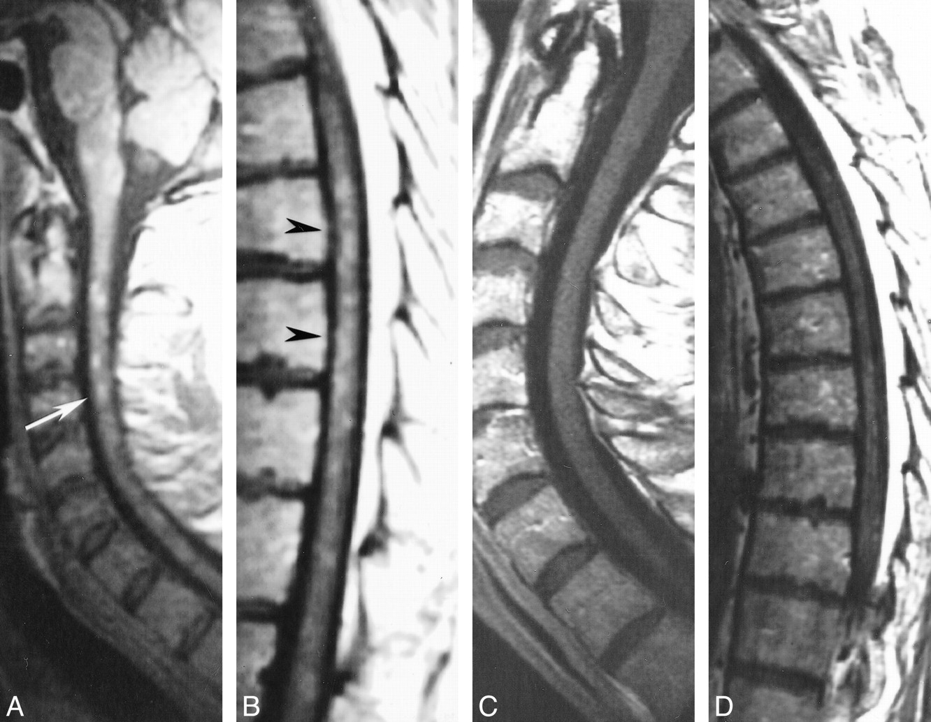
  • fig 2.
    • Download figure
    • Open in new tab
    • Download powerpoint
    fig 2.

    Patient 2: 50-year-old man with primary angiitis of the CNS who presented with progressive paraparesis.

    A and B, First MR study, obtained at 0.5 T. Sagittal contrast-enhanced SE T1-weighted images show numerous small homogeneously enhancing lesions located in the cervical and thoracic cord (arrow, A; arrowheads, B). Enhancing lesions can also be seen in the medulla, both dorsally and ventrally.

    C and D, Follow-up MR study 36 months after the onset of disease. Sagittal contrast-enhanced SE T1-weighted (500/12) images show no evidence of contrast enhancement of the cord.

PreviousNext
Back to top

In this issue

American Journal of Neuroradiology
Vol. 22, Issue 3
1 Mar 2001
  • Table of Contents
  • Index by author
Advertisement
Print
Download PDF
Email Article

Thank you for your interest in spreading the word on American Journal of Neuroradiology.

NOTE: We only request your email address so that the person you are recommending the page to knows that you wanted them to see it, and that it is not junk mail. We do not capture any email address.

Enter multiple addresses on separate lines or separate them with commas.
Spinal Cord Involvement in Primary Angiitis of the Central Nervous System: A Report of Two Cases
(Your Name) has sent you a message from American Journal of Neuroradiology
(Your Name) thought you would like to see the American Journal of Neuroradiology web site.
CAPTCHA
This question is for testing whether or not you are a human visitor and to prevent automated spam submissions.
Cite this article
Adriana Campi, G. Benndorf, V. Martinelli, M. R. Terreni, G. Scotti
Spinal Cord Involvement in Primary Angiitis of the Central Nervous System: A Report of Two Cases
American Journal of Neuroradiology Mar 2001, 22 (3) 577-582;

Citation Manager Formats

  • BibTeX
  • Bookends
  • EasyBib
  • EndNote (tagged)
  • EndNote 8 (xml)
  • Medlars
  • Mendeley
  • Papers
  • RefWorks Tagged
  • Ref Manager
  • RIS
  • Zotero
0 Responses
Respond to this article
Share
Bookmark this article
Spinal Cord Involvement in Primary Angiitis of the Central Nervous System: A Report of Two Cases
Adriana Campi, G. Benndorf, V. Martinelli, M. R. Terreni, G. Scotti
American Journal of Neuroradiology Mar 2001, 22 (3) 577-582;
del.icio.us logo Twitter logo Facebook logo Mendeley logo
  • Tweet Widget
  • Facebook Like
  • Google Plus One

Jump to section

  • Article
    • Abstract
    • Case Reports
    • Discussion
    • Conclusion
    • Footnotes
    • References
  • Figures & Data
  • Info & Metrics
  • Responses
  • References
  • PDF

Related Articles

  • No related articles found.
  • PubMed
  • Google Scholar

Cited By...

  • Primary CNS vasculitis with spinal cord involvement
  • Crossref
  • Google Scholar

This article has not yet been cited by articles in journals that are participating in Crossref Cited-by Linking.

More in this TOC Section

  • Bern Score Validity for SIH
  • MP2RAGE 7T in MS Lesions of the Cervical Spine
  • Resisted Inspiration for CSF-Venous Fistula
Show more Spine

Similar Articles

Advertisement

Indexed Content

  • Current Issue
  • Accepted Manuscripts
  • Article Preview
  • Past Issues
  • Editorials
  • Editor's Choice
  • Fellows' Journal Club
  • Letters to the Editor
  • Video Articles

Cases

  • Case Collection
  • Archive - Case of the Week
  • Archive - Case of the Month
  • Archive - Classic Case

More from AJNR

  • Trainee Corner
  • Imaging Protocols
  • MRI Safety Corner
  • Book Reviews

Multimedia

  • AJNR Podcasts
  • AJNR Scantastics

Resources

  • Turnaround Time
  • Submit a Manuscript
  • Submit a Video Article
  • Submit an eLetter to the Editor/Response
  • Manuscript Submission Guidelines
  • Statistical Tips
  • Fast Publishing of Accepted Manuscripts
  • Graphical Abstract Preparation
  • Imaging Protocol Submission
  • Evidence-Based Medicine Level Guide
  • Publishing Checklists
  • Author Policies
  • Become a Reviewer/Academy of Reviewers
  • News and Updates

About Us

  • About AJNR
  • Editorial Board
  • Editorial Board Alumni
  • Alerts
  • Permissions
  • Not an AJNR Subscriber? Join Now
  • Advertise with Us
  • Librarian Resources
  • Feedback
  • Terms and Conditions
  • AJNR Editorial Board Alumni

American Society of Neuroradiology

  • Not an ASNR Member? Join Now

© 2025 by the American Society of Neuroradiology All rights, including for text and data mining, AI training, and similar technologies, are reserved.
Print ISSN: 0195-6108 Online ISSN: 1936-959X

Powered by HighWire