
Article Figures & Data
Figures
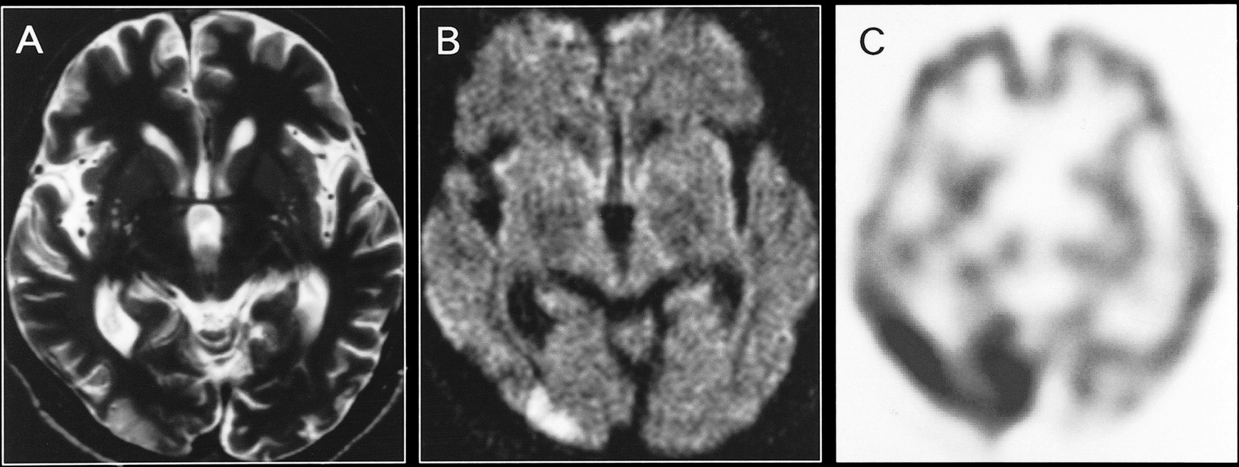
- fig 1.
A, Axial T2-weighted image obtained at the time of initial hospitalization (8 days after the onset of symptoms).
B, Contrast-enhanced T1-weighted image reveals small hyperintense lesions with patchy and punctate enhancement in the bilateral occipital cortices.
C and D, Follow-up images obtained 2 months later show resolution of the lesions.
- fig 2.
A, Axial T2-weighted image obtained at the time of the second hospitalization (2 days after the onset of symptoms).
B, Diffusion-weighted image shows an extensive hyperintense lesion involving the cortex and subcortical white matter in the left occipital lobe.
C, Corresponding ADC map reveals no obvious abnormalities.
D, 99mTc-HMPAO SPECT scan obtained on day 3 shows a remarkable increase in tracer accumulation in the left occipital lobe. Follow-up images obtained 2 weeks later (not shown) indicated the resolution of the left occipital abnormality with residual localized atrophy.
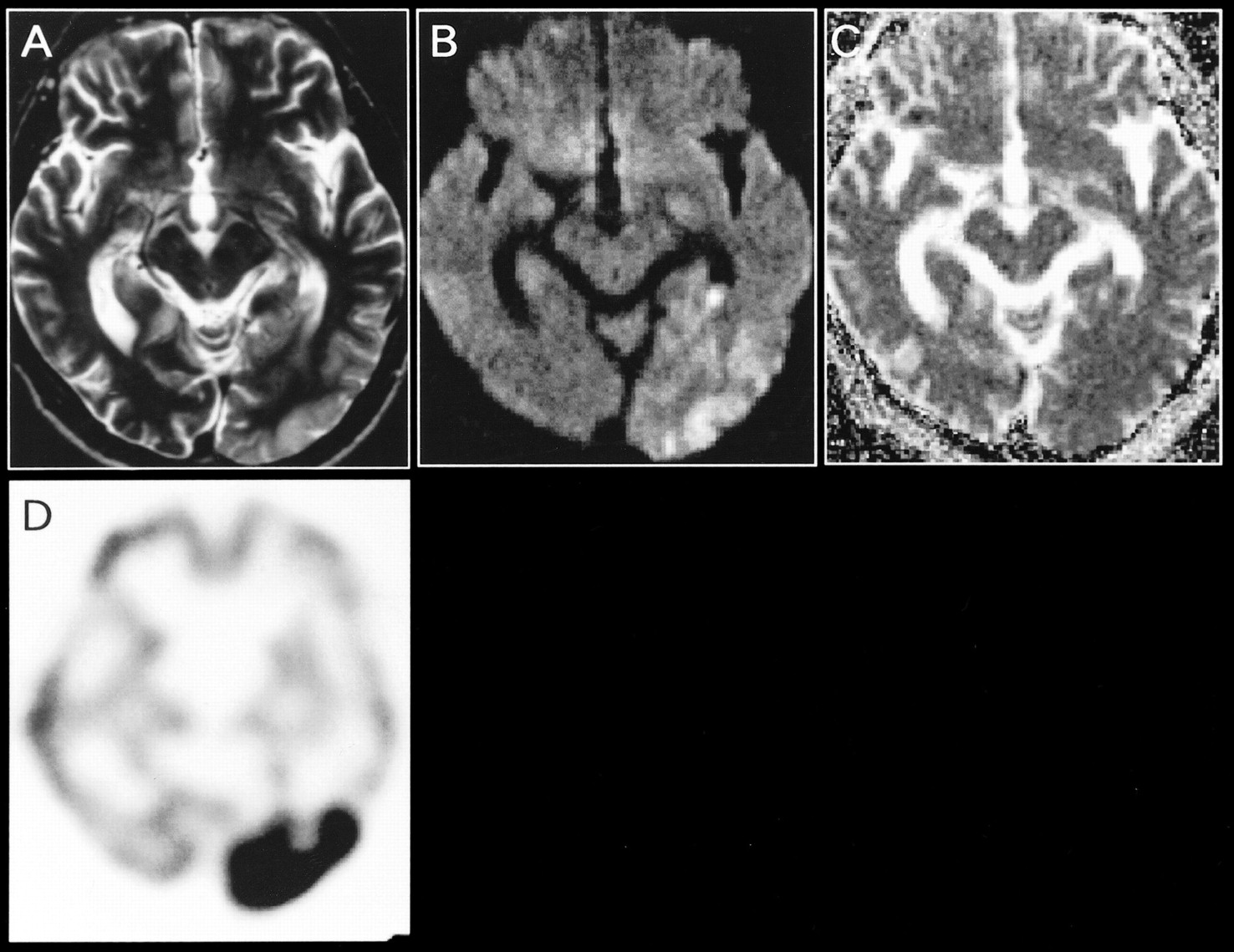
- fig 3.
A, Axial T2-weighted image obtained at the time of the third hospitalization (2 days after the onset of symptoms).
B, Diffusion-weighted image reveals a cortical hyperintense lesion in the right occipital lobe.
C, 99mTc-HMPAO SPECT scan obtained on the same day shows a remarkable increase in tracer accumulation in the right occipital cortex. Follow-up MR images obtained 2 weeks later (not shown) revealed resolution of the right occipital abnormality, and SPECT scans obtained 16 days after the onset of symptoms (not shown) showed a normal accumulation in the right occipital cortex.


