Advertisement

Main menu

  • Home
  • Content
    • Current Issue
    • Accepted Manuscripts
    • Article Preview
    • Past Issue Archive
    • Video Articles
    • AJNR Case Collection
    • Case of the Week Archive
    • Case of the Month Archive
    • Classic Case Archive
  • Special Collections
    • AJNR Awards
    • Low-Field MRI
    • Alzheimer Disease
    • ASNR Foundation Special Collection
    • Photon-Counting CT
    • View All
  • Multimedia
    • AJNR Podcasts
    • AJNR SCANtastic
    • Trainee Corner
    • MRI Safety Corner
    • Imaging Protocols
  • For Authors
    • Submit a Manuscript
    • Submit a Video Article
    • Submit an eLetter to the Editor/Response
    • Manuscript Submission Guidelines
    • Statistical Tips
    • Fast Publishing of Accepted Manuscripts
    • Graphical Abstract Preparation
    • Imaging Protocol Submission
    • Author Policies
  • About Us
    • About AJNR
    • Editorial Board
    • Editorial Board Alumni
  • More
    • Become a Reviewer/Academy of Reviewers
    • Subscribers
    • Permissions
    • Alerts
    • Feedback
    • Advertisers
    • ASNR Home

User menu

  • Alerts
  • Log in

Search

  • Advanced search
American Journal of Neuroradiology

American Journal of Neuroradiology

ASHNR American Society of Functional Neuroradiology ASHNR American Society of Pediatric Neuroradiology ASSR
  • Alerts
  • Log in

Advanced Search

  • Home
  • Content
    • Current Issue
    • Accepted Manuscripts
    • Article Preview
    • Past Issue Archive
    • Video Articles
    • AJNR Case Collection
    • Case of the Week Archive
    • Case of the Month Archive
    • Classic Case Archive
  • Special Collections
    • AJNR Awards
    • Low-Field MRI
    • Alzheimer Disease
    • ASNR Foundation Special Collection
    • Photon-Counting CT
    • View All
  • Multimedia
    • AJNR Podcasts
    • AJNR SCANtastic
    • Trainee Corner
    • MRI Safety Corner
    • Imaging Protocols
  • For Authors
    • Submit a Manuscript
    • Submit a Video Article
    • Submit an eLetter to the Editor/Response
    • Manuscript Submission Guidelines
    • Statistical Tips
    • Fast Publishing of Accepted Manuscripts
    • Graphical Abstract Preparation
    • Imaging Protocol Submission
    • Author Policies
  • About Us
    • About AJNR
    • Editorial Board
    • Editorial Board Alumni
  • More
    • Become a Reviewer/Academy of Reviewers
    • Subscribers
    • Permissions
    • Alerts
    • Feedback
    • Advertisers
    • ASNR Home
  • Follow AJNR on Twitter
  • Visit AJNR on Facebook
  • Follow AJNR on Instagram
  • Join AJNR on LinkedIn
  • RSS Feeds

AJNR Awards, New Junior Editors, and more. Read the latest AJNR updates

Research ArticleBRAIN

Diffusion-weighted Imaging Patterns of Brain Damage Associated with Cerebral Venous Thrombosis

Denis Ducreux, Catherine Oppenheim, Xavier Vandamme, Didier Dormont, Yves Samson, Gérald Rancurel, Guy Cosnard and Claude Marsault
American Journal of Neuroradiology February 2001, 22 (2) 261-268;
Denis Ducreux
  • Find this author on Google Scholar
  • Find this author on PubMed
  • Search for this author on this site
Catherine Oppenheim
  • Find this author on Google Scholar
  • Find this author on PubMed
  • Search for this author on this site
Xavier Vandamme
  • Find this author on Google Scholar
  • Find this author on PubMed
  • Search for this author on this site
Didier Dormont
  • Find this author on Google Scholar
  • Find this author on PubMed
  • Search for this author on this site
Yves Samson
  • Find this author on Google Scholar
  • Find this author on PubMed
  • Search for this author on this site
Gérald Rancurel
  • Find this author on Google Scholar
  • Find this author on PubMed
  • Search for this author on this site
Guy Cosnard
  • Find this author on Google Scholar
  • Find this author on PubMed
  • Search for this author on this site
Claude Marsault
  • Find this author on Google Scholar
  • Find this author on PubMed
  • Search for this author on this site
  • Article
  • Figures & Data
  • Info & Metrics
  • Responses
  • References
  • PDF
Loading

Article Figures & Data

Figures

  • Tables
  • fig 1.
    • Download figure
    • Open in new tab
    • Download powerpoint
    fig 1.

    Patient 9, a 34-year-old woman with headaches, cognitive function loss, and sudden-onset right hemiparesis.

    A, MR angiogram (axial 2D time-of-flight method, maximum intensity pixel reconstruction, lateral view), obtained 1 day after the onset of neurologic symptoms, shows an occlusion of the deep and superficial venous systems (vein of Galen, straight sinus, internal cerebral veins, anterior part of the superior sagittal sinus).

    B, Initial MR image obtained 1 day after the onset of neurologic symptoms. Axial spin-echo T1-weighted image shows bilateral areas of hypointensity at the level of the capsulae. Initial fast-FLAIR (10002/148/2200 [TR/TE/TI]) and diffusion-weighted (4000/120 [TR/TE], b = 1000 s/mm2) images show widespread bilateral areas of hyperintensity in putamen, caudate nuclei, and the capsulae. ADC maps show markedly decreased ADC values (0.33–0.42 10−3 mm2/s) (ie, z scores <−3 when compared with normal-appearing brain).

    C, Follow-up MR image obtained 3 months after onset shows normal-appearing brain at the level of the gray nuclei, indicating that most lesions with initially decreased ADC values were reversible. Only focal slight areas of hyperintensity were visible in the centrum semiovale (not shown). Symptoms regressed completely within a few weeks.

  • fig 2.
    • Download figure
    • Open in new tab
    • Download powerpoint
    fig 2.

    Patient 2, a 41-year-old woman with vomiting, headaches, right hemianopsia, and right sensorimotor deficit of sudden onset.

    A, MR angiogram (coronal 2D time-of-flight method, maximum intensity pixel reconstruction, lateral view), obtained 4 days after the onset of neurologic symptoms, shows an occlusion of the deep venous system (internal cerebral veins, straight sinus, vein of Galen). The right transverse sinus was also occluded (not shown).

    B, Initial MR imaging and CT were performed 4 days after the onset of neurologic symptoms. CT scan shows a left capsulothalamic hematoma surrounded by edema. Axial FLAIR (10002/148/2200 [TR/TE/TI]) and diffusion-weighted (4000/120 [TR/TE], b = 1000 s/mm2) images show central thalamic hypoisointensity surrounded by areas of hyperintensity. ADC values (0.59 10−3 mm2/s) measured in the hemorrhagic thalamic lesion are at the lower limit of the normal range (z score of −1.91) and increased at the periphery (1.3 10−3 mm2/s, z score = 2.8).

    C, Three months later, right hemiparesis persists along with areas of hyperintensity on T1- and T2-weighted images, suggesting the presence of hemorrhagic sequelae.

  • fig 3.
    • Download figure
    • Open in new tab
    • Download powerpoint
    fig 3.

    Patient 5, a 20-year-old woman with seizures and left motor deficit of sudden onset.

    A, CT angiogram, obtained 1 day after onset, shows an occluded superior sagittal sinus. The deep venous system was intact (not shown).

    B, Initial MR imaging and CT were performed 1 day after onset. CT scan shows a large area of hypodensity with focal areas of hyperdensity corresponding to a recent hematoma. Axial FLAIR (10002/148/2200 [TR/TE/TI]) and diffusion-weighted (4000/120 [TR/TE], b = 1000 s/mm2) images show a heterogeneous area with predominant areas of hyperintensity and hypointensity at the site of the hematoma seen on the CT scan. ADC map shows mixed areas of increased ADC values (1.66 10−3 mm2/s) (ie, z score >3), at the lower limit of normal values (0.59 10−3 mm2/s) (ie, z score = −1.9), or slightly decreased ADC values (0.58 10−3 mm2/s) (ie, z score <−2).

    C, Follow-up MR image obtained 6 months after onset shows left frontal sequelae in areas with initially decreased or normal ADC values (2D time-of-flight source image used to illustrate MR sequelae). The patient had no clinical deficit.

Tables

  • Figures
  • Table1

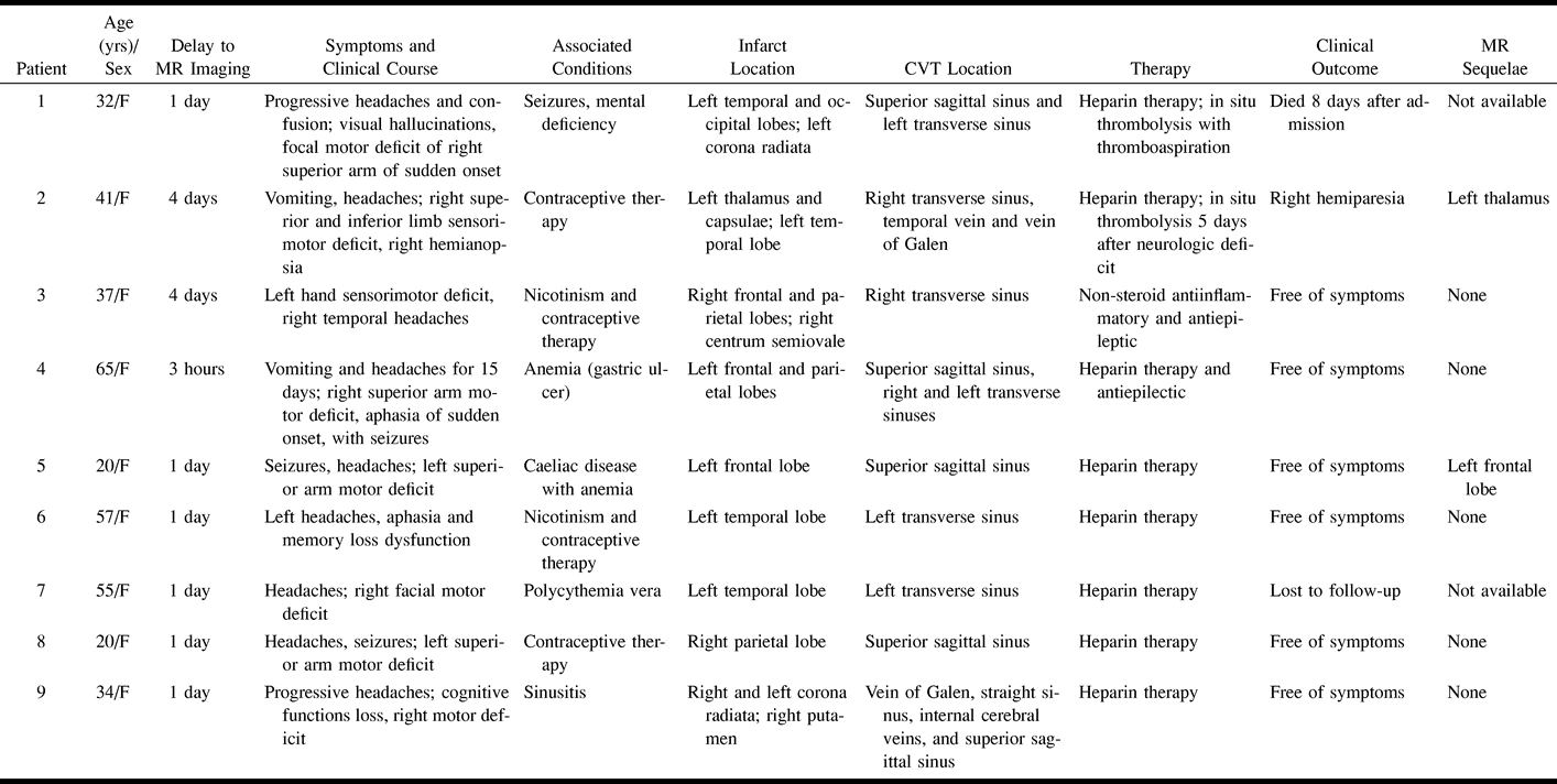
    Synopsis of clinical and imaging findings and follow-up in nine patients with venous stroke

PreviousNext
Back to top

In this issue

American Journal of Neuroradiology
Vol. 22, Issue 2
1 Feb 2001
  • Table of Contents
  • Index by author
Advertisement
Print
Download PDF
Email Article

Thank you for your interest in spreading the word on American Journal of Neuroradiology.

NOTE: We only request your email address so that the person you are recommending the page to knows that you wanted them to see it, and that it is not junk mail. We do not capture any email address.

Enter multiple addresses on separate lines or separate them with commas.
Diffusion-weighted Imaging Patterns of Brain Damage Associated with Cerebral Venous Thrombosis
(Your Name) has sent you a message from American Journal of Neuroradiology
(Your Name) thought you would like to see the American Journal of Neuroradiology web site.
CAPTCHA
This question is for testing whether or not you are a human visitor and to prevent automated spam submissions.
Cite this article
Denis Ducreux, Catherine Oppenheim, Xavier Vandamme, Didier Dormont, Yves Samson, Gérald Rancurel, Guy Cosnard, Claude Marsault
Diffusion-weighted Imaging Patterns of Brain Damage Associated with Cerebral Venous Thrombosis
American Journal of Neuroradiology Feb 2001, 22 (2) 261-268;

Citation Manager Formats

  • BibTeX
  • Bookends
  • EasyBib
  • EndNote (tagged)
  • EndNote 8 (xml)
  • Medlars
  • Mendeley
  • Papers
  • RefWorks Tagged
  • Ref Manager
  • RIS
  • Zotero
0 Responses
Respond to this article
Share
Bookmark this article
Diffusion-weighted Imaging Patterns of Brain Damage Associated with Cerebral Venous Thrombosis
Denis Ducreux, Catherine Oppenheim, Xavier Vandamme, Didier Dormont, Yves Samson, Gérald Rancurel, Guy Cosnard, Claude Marsault
American Journal of Neuroradiology Feb 2001, 22 (2) 261-268;
del.icio.us logo Twitter logo Facebook logo Mendeley logo
  • Tweet Widget
  • Facebook Like
  • Google Plus One

Jump to section

  • Article
    • Abstract
    • Methods
    • Results
    • Discussion
    • Conclusion
    • Footnotes
    • References
  • Figures & Data
  • Info & Metrics
  • Responses
  • References
  • PDF

Related Articles

  • No related articles found.
  • PubMed
  • Google Scholar

Cited By...

  • Current endovascular strategies for cerebral venous thrombosis: report of the SNIS Standards and Guidelines Committee
  • Imaging Characteristics of Venous Parenchymal Abnormalities
  • Diagnosis and Management of Cerebral Venous Thrombosis: A Statement for Healthcare Professionals From the American Heart Association/American Stroke Association
  • Compromise of Brain Tissue Caused by Cortical Venous Reflux of Intracranial Dural Arteriovenous Fistulas: Assessment With Diffusion-Weighted Magnetic Resonance Imaging
  • Role of imaging in the diagnosis of acute bacterial meningitis and its complications
  • Should Decompressive Surgery Be Performed in Malignant Cerebral Venous Thrombosis?: A Series of 12 Patients * Supplemental Material
  • Roles of Inflammation and the Activated Protein C Pathway in the Brain Edema Associated With Cerebral Venous Sinus Thrombosis
  • MR Imaging Features of Isolated Cortical Vein Thrombosis: Diagnosis and Follow-Up
  • Is Heparin Treatment the Optimal Management for Cerebral Venous Thrombosis?: Effect of Abciximab, Recombinant Tissue Plasminogen Activator, and Enoxaparin in Experimentally Induced Superior Sagittal Sinus Thrombosis
  • Reversible parkinsonism and MRI diffusion abnormalities in cortical venous thrombosis
  • Diffusion-Weighted MR Imaging of an Acute Venous Stroke: Case Report
  • Crossref
  • Google Scholar

This article has not yet been cited by articles in journals that are participating in Crossref Cited-by Linking.

More in this TOC Section

  • Usefulness of Quantitative Susceptibility Mapping for the Diagnosis of Parkinson Disease
  • White Matter Alterations in the Brains of Patients with Active, Remitted, and Cured Cushing Syndrome: A DTI Study
  • Qualitative and Quantitative Analysis of MR Imaging Findings in Patients with Middle Cerebral Artery Stroke Implanted with Mesenchymal Stem Cells
Show more BRAIN

Similar Articles

Advertisement

Indexed Content

  • Current Issue
  • Accepted Manuscripts
  • Article Preview
  • Past Issues
  • Editorials
  • Editor's Choice
  • Fellows' Journal Club
  • Letters to the Editor
  • Video Articles

Cases

  • Case Collection
  • Archive - Case of the Week
  • Archive - Case of the Month
  • Archive - Classic Case

More from AJNR

  • Trainee Corner
  • Imaging Protocols
  • MRI Safety Corner
  • Book Reviews

Multimedia

  • AJNR Podcasts
  • AJNR Scantastics

Resources

  • Turnaround Time
  • Submit a Manuscript
  • Submit a Video Article
  • Submit an eLetter to the Editor/Response
  • Manuscript Submission Guidelines
  • Statistical Tips
  • Fast Publishing of Accepted Manuscripts
  • Graphical Abstract Preparation
  • Imaging Protocol Submission
  • Evidence-Based Medicine Level Guide
  • Publishing Checklists
  • Author Policies
  • Become a Reviewer/Academy of Reviewers
  • News and Updates

About Us

  • About AJNR
  • Editorial Board
  • Editorial Board Alumni
  • Alerts
  • Permissions
  • Not an AJNR Subscriber? Join Now
  • Advertise with Us
  • Librarian Resources
  • Feedback
  • Terms and Conditions
  • AJNR Editorial Board Alumni

American Society of Neuroradiology

  • Not an ASNR Member? Join Now

© 2025 by the American Society of Neuroradiology All rights, including for text and data mining, AI training, and similar technologies, are reserved.
Print ISSN: 0195-6108 Online ISSN: 1936-959X

Powered by HighWire