
Article Figures & Data
Figures
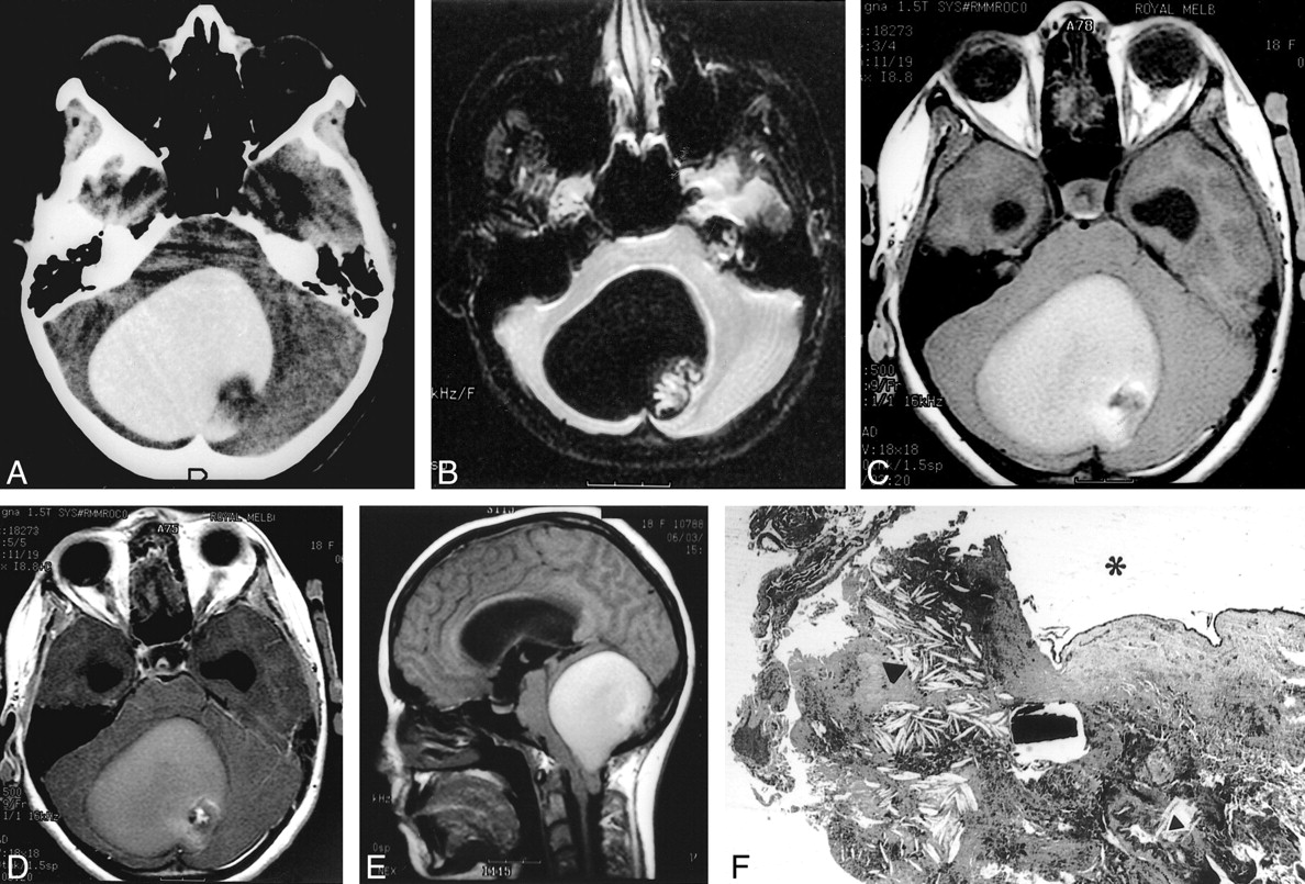
- fig 1.
18-year-old girl with occipital headaches of 8 weeks' duration.
A, Unenhanced CT scan shows a hyperattenuating posterior fossa lesion with posterior mural nodule.
B, Gradient-echo MR image shows marked hypointensity with a frondlike mural nodule.
C and D, Pre- (C) and postcontrast (D) T1-weighted MR images show hyperintensity of the cystic contents and clear evidence of contrast enhancement of the mural nodule.
E, Sagittal unenhanced T1-weighted MR image shows marked cerebellar tonsillar herniation through the foramen magnum and obliteration of the fourth ventricle.
F, Section of cyst wall (lumen indicated by asterisk) shows a lozenge-shaped deposit of calcium with a clear space centrally, numerous cholesterol deposits (large black arrowhead), and a leash of vessels (small black arrowhead) (hematoxylin-eosin, original magnification ×25).

In this issue


Jump to section
Related Articles
Cited By...
- No citing articles found.