Advertisement

Main menu

  • Home
  • Content
    • Current Issue
    • Accepted Manuscripts
    • Article Preview
    • Past Issue Archive
    • Video Articles
    • AJNR Case Collection
    • Case of the Week Archive
    • Case of the Month Archive
    • Classic Case Archive
  • Special Collections
    • AJNR Awards
    • Low-Field MRI
    • Alzheimer Disease
    • ASNR Foundation Special Collection
    • Photon-Counting CT
    • View All
  • Multimedia
    • AJNR Podcasts
    • AJNR SCANtastic
    • Trainee Corner
    • MRI Safety Corner
    • Imaging Protocols
  • For Authors
    • Submit a Manuscript
    • Submit a Video Article
    • Submit an eLetter to the Editor/Response
    • Manuscript Submission Guidelines
    • Statistical Tips
    • Fast Publishing of Accepted Manuscripts
    • Graphical Abstract Preparation
    • Imaging Protocol Submission
    • Author Policies
  • About Us
    • About AJNR
    • Editorial Board
    • Editorial Board Alumni
  • More
    • Become a Reviewer/Academy of Reviewers
    • Subscribers
    • Permissions
    • Alerts
    • Feedback
    • Advertisers
    • ASNR Home

User menu

  • Alerts
  • Log in

Search

  • Advanced search
American Journal of Neuroradiology

American Journal of Neuroradiology

ASHNR American Society of Functional Neuroradiology ASHNR American Society of Pediatric Neuroradiology ASSR
  • Alerts
  • Log in

Advanced Search

  • Home
  • Content
    • Current Issue
    • Accepted Manuscripts
    • Article Preview
    • Past Issue Archive
    • Video Articles
    • AJNR Case Collection
    • Case of the Week Archive
    • Case of the Month Archive
    • Classic Case Archive
  • Special Collections
    • AJNR Awards
    • Low-Field MRI
    • Alzheimer Disease
    • ASNR Foundation Special Collection
    • Photon-Counting CT
    • View All
  • Multimedia
    • AJNR Podcasts
    • AJNR SCANtastic
    • Trainee Corner
    • MRI Safety Corner
    • Imaging Protocols
  • For Authors
    • Submit a Manuscript
    • Submit a Video Article
    • Submit an eLetter to the Editor/Response
    • Manuscript Submission Guidelines
    • Statistical Tips
    • Fast Publishing of Accepted Manuscripts
    • Graphical Abstract Preparation
    • Imaging Protocol Submission
    • Author Policies
  • About Us
    • About AJNR
    • Editorial Board
    • Editorial Board Alumni
  • More
    • Become a Reviewer/Academy of Reviewers
    • Subscribers
    • Permissions
    • Alerts
    • Feedback
    • Advertisers
    • ASNR Home
  • Follow AJNR on Twitter
  • Visit AJNR on Facebook
  • Follow AJNR on Instagram
  • Join AJNR on LinkedIn
  • RSS Feeds

AJNR Awards, New Junior Editors, and more. Read the latest AJNR updates

Research ArticleBRAIN

Congenital Absence of the Internal Carotid Artery: Case Reports and Review of the Collateral Circulation

Curtis A. Given II, Frank Huang-Hellinger, Michael D. Baker, Neeraj B. Chepuri and P. Pearse Morris
American Journal of Neuroradiology November 2001, 22 (10) 1953-1959;
Curtis A. Given II
  • Find this author on Google Scholar
  • Find this author on PubMed
  • Search for this author on this site
Frank Huang-Hellinger
  • Find this author on Google Scholar
  • Find this author on PubMed
  • Search for this author on this site
Michael D. Baker
  • Find this author on Google Scholar
  • Find this author on PubMed
  • Search for this author on this site
Neeraj B. Chepuri
  • Find this author on Google Scholar
  • Find this author on PubMed
  • Search for this author on this site
P. Pearse Morris
  • Find this author on Google Scholar
  • Find this author on PubMed
  • Search for this author on this site
  • Article
  • Figures & Data
  • Info & Metrics
  • Responses
  • References
  • PDF
Loading

Article Figures & Data

Figures

  • fig 1.
    • Download figure
    • Open in new tab
    • Download powerpoint
    fig 1.

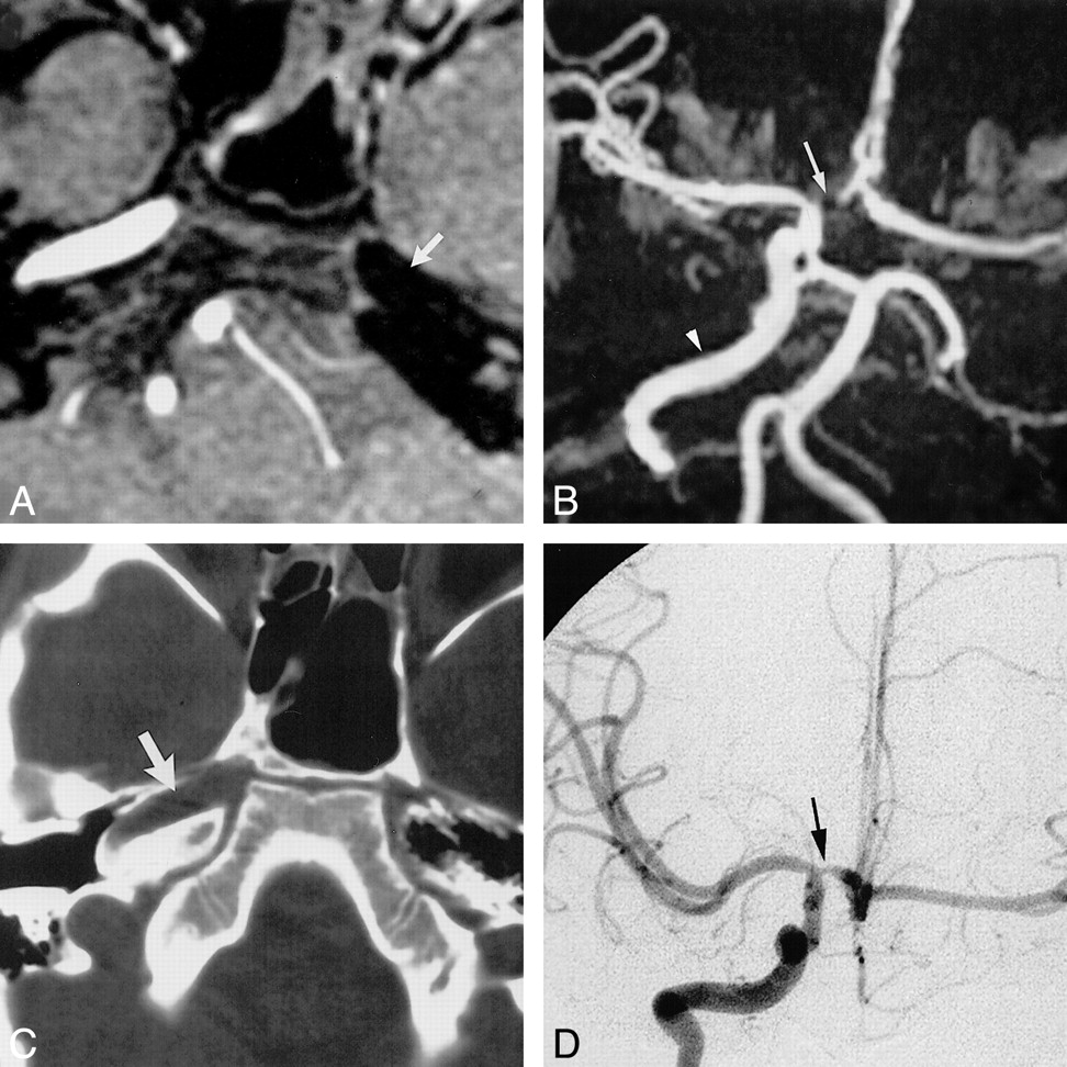
    Case 1: Hypoplasia of the left ICA.

    A, Source image from a 3D time-of-flight MR angiogram shows diminished flow-related signal intensity within the petrous portions of the left ICA (arrow).

    B, Compressed image from the MR angiogram shows a tortuous, enlarged PCOM (arrow) extending forward to supply the left MCA. The left ACA is supplied via a patent ACOM (arrowhead). There is no perceivable flow-related signal intensity within the supraclinoid left ICA on the compressed image.

    C, Axial image from a CT angiogram at the level of the petrous ICA shows hypoplasia of the left carotid canal (arrowhead).

  • fig 2.
    • Download figure
    • Open in new tab
    • Download powerpoint
    fig 2.

    Case 2: Agenesis of the left ICA.

    A, Source image from a 3D time-of-flight MR angiogram reveals absence of flow-related signal within the left petrous ICA (arrow).

    B, Compressed view from the MR angiogram displays absence of flow-related signal intensity within the left ICA with collateral supply to the left hemisphere through a patent ACOM. Normal flow is present within the right ICA (arrowhead). Focal loss of flow-related signal intensity within the A1 segment of the right ACA (arrow) represents a clinically insignificant stenosis.

    C, Axial CT scan of the skull base shows absence of the left carotid canal and a normally developed right carotid canal (arrow).

    D, Collateral supply to the left cerebral hemisphere is provided through a patent ACOM, as demonstrated on the frontal view from the right carotid arteriogram. Stenosis of the A1 segment of the right ACA is revealed again (arrow).

  • fig 3.
    • Download figure
    • Open in new tab
    • Download powerpoint
    fig 3.

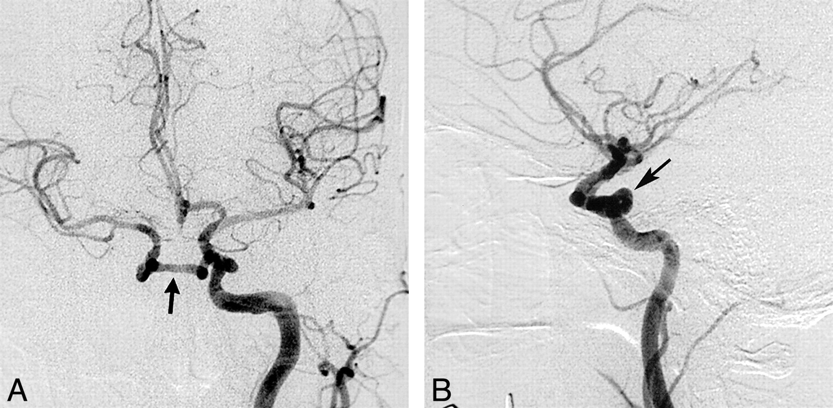
    Case 3: Absence of the right ICA.

    A and B, Frontal (A) and lateral (B) projections from a left CCA arteriogram show an anomalous communication between the cavernous portions of the ICAs (arrow). This anomalous communication courses through the sella turcica. The right ACA is supplied via a patent ACOM, with the right A1 segment either being aplastic or extremely hypoplastic. The right MCA is a continuation of the right supraclinoid ICA.

  • fig 4.
    • Download figure
    • Open in new tab
    • Download powerpoint
    fig 4.

    Case 4: Aplasia of the left ICA.

    A, Frontal view from a right CCA arteriogram shows collateral flow to the left ACA across a patent ACOM (arrow).

    B, Frontal view from a left vertebral arteriogram displays collateral flow to the left MCA (arrow) via forward flow through a patent PCOM (arrowhead).

    C, Axial CT scan through the skull base reveals a diminutive left carotid canal (arrow).

  • fig 5.
    • Download figure
    • Open in new tab
    • Download powerpoint
    fig 5.

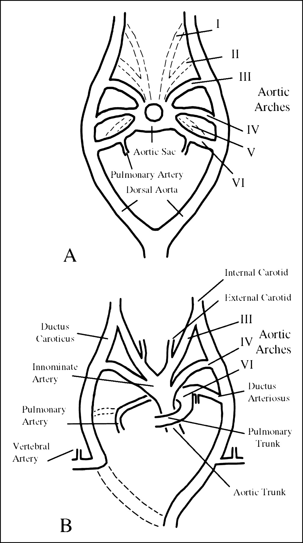
    A and B, Illustrations of the developing embryo at 6 mm (A) and 11 mm (B). After Congdon, as reproduced in (5); see text

  • fig 6.
    • Download figure
    • Open in new tab
    • Download powerpoint
    fig 6.

    Absence of the ICA: pathways of collateral blood flow. After Lie (5) and Quint et al (6); see text

PreviousNext
Back to top

In this issue

American Journal of Neuroradiology: 22 (10)
American Journal of Neuroradiology
Vol. 22, Issue 10
1 Nov 2001
  • Table of Contents
  • Index by author
Advertisement
Print
Download PDF
Email Article

Thank you for your interest in spreading the word on American Journal of Neuroradiology.

NOTE: We only request your email address so that the person you are recommending the page to knows that you wanted them to see it, and that it is not junk mail. We do not capture any email address.

Enter multiple addresses on separate lines or separate them with commas.
Congenital Absence of the Internal Carotid Artery: Case Reports and Review of the Collateral Circulation
(Your Name) has sent you a message from American Journal of Neuroradiology
(Your Name) thought you would like to see the American Journal of Neuroradiology web site.
CAPTCHA
This question is for testing whether or not you are a human visitor and to prevent automated spam submissions.
Cite this article
Curtis A. Given II, Frank Huang-Hellinger, Michael D. Baker, Neeraj B. Chepuri, P. Pearse Morris
Congenital Absence of the Internal Carotid Artery: Case Reports and Review of the Collateral Circulation
American Journal of Neuroradiology Nov 2001, 22 (10) 1953-1959;

Citation Manager Formats

  • BibTeX
  • Bookends
  • EasyBib
  • EndNote (tagged)
  • EndNote 8 (xml)
  • Medlars
  • Mendeley
  • Papers
  • RefWorks Tagged
  • Ref Manager
  • RIS
  • Zotero
0 Responses
Respond to this article
Share
Bookmark this article
Congenital Absence of the Internal Carotid Artery: Case Reports and Review of the Collateral Circulation
Curtis A. Given II, Frank Huang-Hellinger, Michael D. Baker, Neeraj B. Chepuri, P. Pearse Morris
American Journal of Neuroradiology Nov 2001, 22 (10) 1953-1959;
del.icio.us logo Twitter logo Facebook logo Mendeley logo
  • Tweet Widget
  • Facebook Like
  • Google Plus One

Jump to section

  • Article
    • Abstract
    • Case Reports
    • Discussion
    • Conclusion
    • Footnotes
    • References
  • Figures & Data
  • Info & Metrics
  • Responses
  • References
  • PDF

Related Articles

  • No related articles found.
  • PubMed
  • Google Scholar

Cited By...

  • Cerebral neurovascular embryology, anatomic variations, and congenital brain arteriovenous lesions
  • Republished: Rete mirabile associated with pial arteriovenous fistula: imaging features with literature review
  • Rete mirabile associated with pial arteriovenous fistula: imaging features with literature review
  • Congenital absence of internal carotid artery: an unsuspected diagnosis
  • Congenital absence of bilateral ICA: an unusual incidental finding in an adult male
  • Internal Carotid Artery Hypoplasia: Role of Color-Coded Carotid Duplex Sonography
  • Non-visualization of the internal carotid artery with a normal ipsilateral common carotid artery Doppler waveform: a finding suggesting congenital absence of the ICA on colour Doppler ultrasound.
  • Bilateral absence of the internal carotid artery: MR angiography and ultrasound findings
  • Crossref
  • Google Scholar

This article has not yet been cited by articles in journals that are participating in Crossref Cited-by Linking.

More in this TOC Section

  • Usefulness of Quantitative Susceptibility Mapping for the Diagnosis of Parkinson Disease
  • White Matter Alterations in the Brains of Patients with Active, Remitted, and Cured Cushing Syndrome: A DTI Study
  • Qualitative and Quantitative Analysis of MR Imaging Findings in Patients with Middle Cerebral Artery Stroke Implanted with Mesenchymal Stem Cells
Show more BRAIN

Similar Articles

Advertisement

Indexed Content

  • Current Issue
  • Accepted Manuscripts
  • Article Preview
  • Past Issues
  • Editorials
  • Editor's Choice
  • Fellows' Journal Club
  • Letters to the Editor
  • Video Articles

Cases

  • Case Collection
  • Archive - Case of the Week
  • Archive - Case of the Month
  • Archive - Classic Case

More from AJNR

  • Trainee Corner
  • Imaging Protocols
  • MRI Safety Corner
  • Book Reviews

Multimedia

  • AJNR Podcasts
  • AJNR Scantastics

Resources

  • Turnaround Time
  • Submit a Manuscript
  • Submit a Video Article
  • Submit an eLetter to the Editor/Response
  • Manuscript Submission Guidelines
  • Statistical Tips
  • Fast Publishing of Accepted Manuscripts
  • Graphical Abstract Preparation
  • Imaging Protocol Submission
  • Evidence-Based Medicine Level Guide
  • Publishing Checklists
  • Author Policies
  • Become a Reviewer/Academy of Reviewers
  • News and Updates

About Us

  • About AJNR
  • Editorial Board
  • Editorial Board Alumni
  • Alerts
  • Permissions
  • Not an AJNR Subscriber? Join Now
  • Advertise with Us
  • Librarian Resources
  • Feedback
  • Terms and Conditions
  • AJNR Editorial Board Alumni

American Society of Neuroradiology

  • Not an ASNR Member? Join Now

© 2025 by the American Society of Neuroradiology All rights, including for text and data mining, AI training, and similar technologies, are reserved.
Print ISSN: 0195-6108 Online ISSN: 1936-959X

Powered by HighWire