
Article Figures & Data
Figures
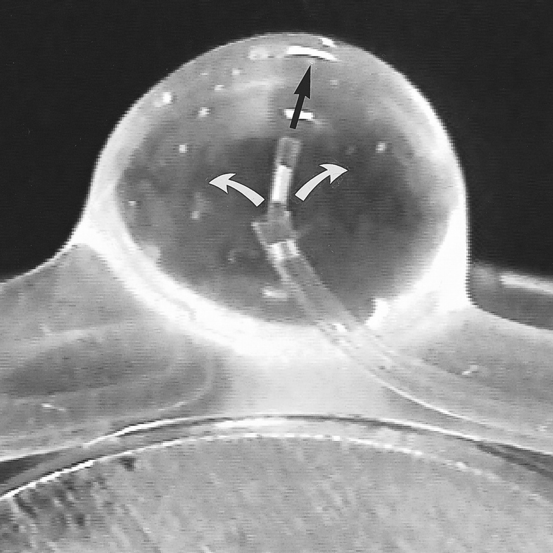
- fig 1.
Double-lumen microcatheter placed in the silicone-made aneurysm. A small 1.7F microcatheter system was used for the injection of Onyx (black arrow indicates direction of flow), and a large 2.7F microcatheter system was used for the injection of saline to flush the stagnant DMSO in the aneurysmal sac (white arrows indicate direction of flow)
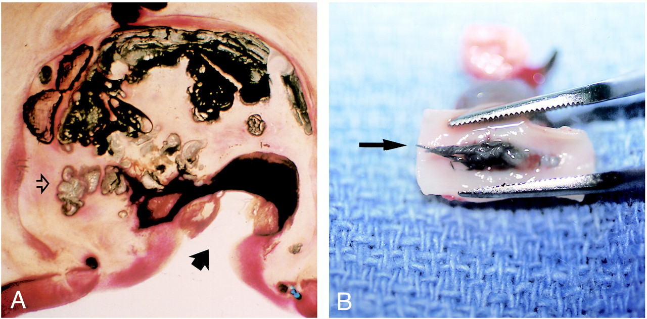
- fig 2.
A, Sagittal view of aneurysm embolized with Onyx. Note incomplete packing of the aneurysm, multiple fragmentation of the embolic mass (open arrow), and recanalization of the aneurysm inflow zone (solid arrow) (hematoxylin-eosin, ×4).
B, Macroscopic appearance of the neck of the aneurysm with Onyx migration into the parent artery (arrow). No significant thrombus formation was observed.
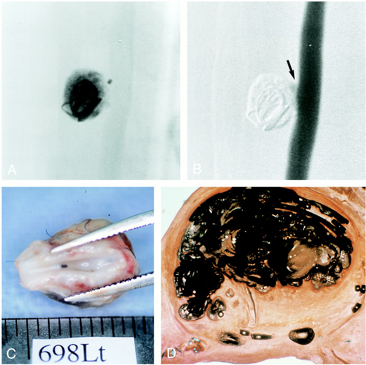
- fig 3.
A, Nonsubtraction angiogram shows dense packing of the aneurysmal sac with the combination of a GDC and Onyx. The coil was placed in the aneurysm.
B, Digital subtraction view shows small neck remnant at the inflow zone (arrow).
C, Macroscopic appearance of the neck of the aneurysm with complete neoendothelial lining.
D, Sagittal view of aneurysm embolized with Onyx and a GDC. Note denser packing with Onyx and endothelial lining across the neck of the aneurysm.
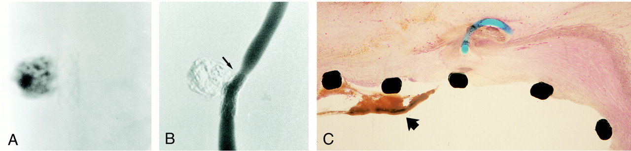
- fig 4.
A, Plain radiograph shows immediate poststenting and Onyx embolization of the aneurysm.
B, Follow-up angiogram at 14 days shows moderate degree of stenosis of the parent artery due to intimal hyperplasia (note stent deformity, arrow). The aneurysm was completely occluded with Onyx.
C, Light microscopic findings of aneurysmal neck 14 days after embolization with Onyx and microstent show thrombus deposition on stent wire (arrow) (hematoxylin-eosin, ×10).
- fig 5.
A, Graph shows IAP change during balloon inflation. Note significant increase of IAP with complete balloon inflation with or without saline flush.
B, Angiogram of experimental aneurysm created on the common carotid artery of a swine.
C, Approximately 80% occlusion of the aneurysm was achieved with combination of Onyx and a microballoon.
D, Embolization of the remnant at the inflow zone using nearly complete occlusion technique. Note small space between balloon and distal portion of the aneurysmal neck (arrow).
E, Example of successful occlusion of the aneurysm with Onyx and balloon technique. Note small concave shape of the Onyx along with balloon shape.
- fig 6.
A, Light microscopic findings in region of aneurysmal dome 14 day after embolization with Onyx (hematoxylin-eosin, ×25). Dense connective tissue containing moderate number of mixed inflammatory cells is observed along with polymer surfaces. No angionecrosis is present.
B, Light microscopic findings in region of aneurysmal dome 14 days after embolization with GDCs (hematoxylin-eosin, ×25). Minimum to mild inflammatory reaction is observed along with coils.
C, Light microscopic findings of aneurysmal neck 14 days after embolization with Onyx. Note tonguelike migration of Onyx into the parent artery and complete coverage with neointima (hematoxylin-eosin, ×10).
D, Light microscopic findings of aneurysmal neck 14 days after embolization with Onyx and microstent. Note significant intimal hyperplasia (wide arrow) and stent deformity due to tissue reaction (thin arrow) (hematoxylin-eosin, ×5).


Tables

Angiographic findings in 20 swine with 40 experimental lateral-wall aneurysms


