Advertisement

Main menu

  • Home
  • Content
    • Current Issue
    • Accepted Manuscripts
    • Article Preview
    • Past Issue Archive
    • Video Articles
    • AJNR Case Collection
    • Case of the Week Archive
    • Case of the Month Archive
    • Classic Case Archive
  • Special Collections
    • AJNR Awards
    • Low-Field MRI
    • Alzheimer Disease
    • ASNR Foundation Special Collection
    • Photon-Counting CT
    • View All
  • Multimedia
    • AJNR Podcasts
    • AJNR SCANtastic
    • Trainee Corner
    • MRI Safety Corner
    • Imaging Protocols
  • For Authors
    • Submit a Manuscript
    • Submit a Video Article
    • Submit an eLetter to the Editor/Response
    • Manuscript Submission Guidelines
    • Statistical Tips
    • Fast Publishing of Accepted Manuscripts
    • Graphical Abstract Preparation
    • Imaging Protocol Submission
    • Author Policies
  • About Us
    • About AJNR
    • Editorial Board
    • Editorial Board Alumni
  • More
    • Become a Reviewer/Academy of Reviewers
    • Subscribers
    • Permissions
    • Alerts
    • Feedback
    • Advertisers
    • ASNR Home

User menu

  • Alerts
  • Log in

Search

  • Advanced search
American Journal of Neuroradiology

American Journal of Neuroradiology

ASHNR American Society of Functional Neuroradiology ASHNR American Society of Pediatric Neuroradiology ASSR
  • Alerts
  • Log in

Advanced Search

  • Home
  • Content
    • Current Issue
    • Accepted Manuscripts
    • Article Preview
    • Past Issue Archive
    • Video Articles
    • AJNR Case Collection
    • Case of the Week Archive
    • Case of the Month Archive
    • Classic Case Archive
  • Special Collections
    • AJNR Awards
    • Low-Field MRI
    • Alzheimer Disease
    • ASNR Foundation Special Collection
    • Photon-Counting CT
    • View All
  • Multimedia
    • AJNR Podcasts
    • AJNR SCANtastic
    • Trainee Corner
    • MRI Safety Corner
    • Imaging Protocols
  • For Authors
    • Submit a Manuscript
    • Submit a Video Article
    • Submit an eLetter to the Editor/Response
    • Manuscript Submission Guidelines
    • Statistical Tips
    • Fast Publishing of Accepted Manuscripts
    • Graphical Abstract Preparation
    • Imaging Protocol Submission
    • Author Policies
  • About Us
    • About AJNR
    • Editorial Board
    • Editorial Board Alumni
  • More
    • Become a Reviewer/Academy of Reviewers
    • Subscribers
    • Permissions
    • Alerts
    • Feedback
    • Advertisers
    • ASNR Home
  • Follow AJNR on Twitter
  • Visit AJNR on Facebook
  • Follow AJNR on Instagram
  • Join AJNR on LinkedIn
  • RSS Feeds

AJNR Awards, New Junior Editors, and more. Read the latest AJNR updates

Research ArticleHead and Neck Imaging

MR Imaging for Predicting Neoplastic Invasion of the Cervical Esophagus

Sudipta Roychowdhury, Laurie A. Loevner, David M. Yousem, Ara Chalian and Kathleen T. Montone
American Journal of Neuroradiology October 2000, 21 (9) 1681-1687;
Sudipta Roychowdhury
  • Find this author on Google Scholar
  • Find this author on PubMed
  • Search for this author on this site
Laurie A. Loevner
  • Find this author on Google Scholar
  • Find this author on PubMed
  • Search for this author on this site
David M. Yousem
  • Find this author on Google Scholar
  • Find this author on PubMed
  • Search for this author on this site
Ara Chalian
  • Find this author on Google Scholar
  • Find this author on PubMed
  • Search for this author on this site
Kathleen T. Montone
  • Find this author on Google Scholar
  • Find this author on PubMed
  • Search for this author on this site
  • Article
  • Figures & Data
  • Info & Metrics
  • Responses
  • References
  • PDF
Loading

Article Figures & Data

Figures

  • Tables
  • fig 1.
    • Download figure
    • Open in new tab
    • Download powerpoint
    fig 1.

    This 68-year-old woman with a benign, left, thyroidal multinodular goiter demonstrates the normal MR imaging appearance of the adjacent esophagus.

    A, Axial T1-weighted image (500/16 [TR/TE]) shows an intact fat plane (arrow) between the esophagus and the mass. The thyroid mass is adjacent to the esophagus, and the esophageal lumen is collapsed.

    B, Axial T2-weighted image (3500/84) shows normal hypointense signal of the wall musculature of the esophagus (arrow).

    C, Axial enhanced 3D FMPSPGR image (220/27; flip angle, 90o) shows mild enhancement of the mucosal lining of the esophageal wall (arrow).
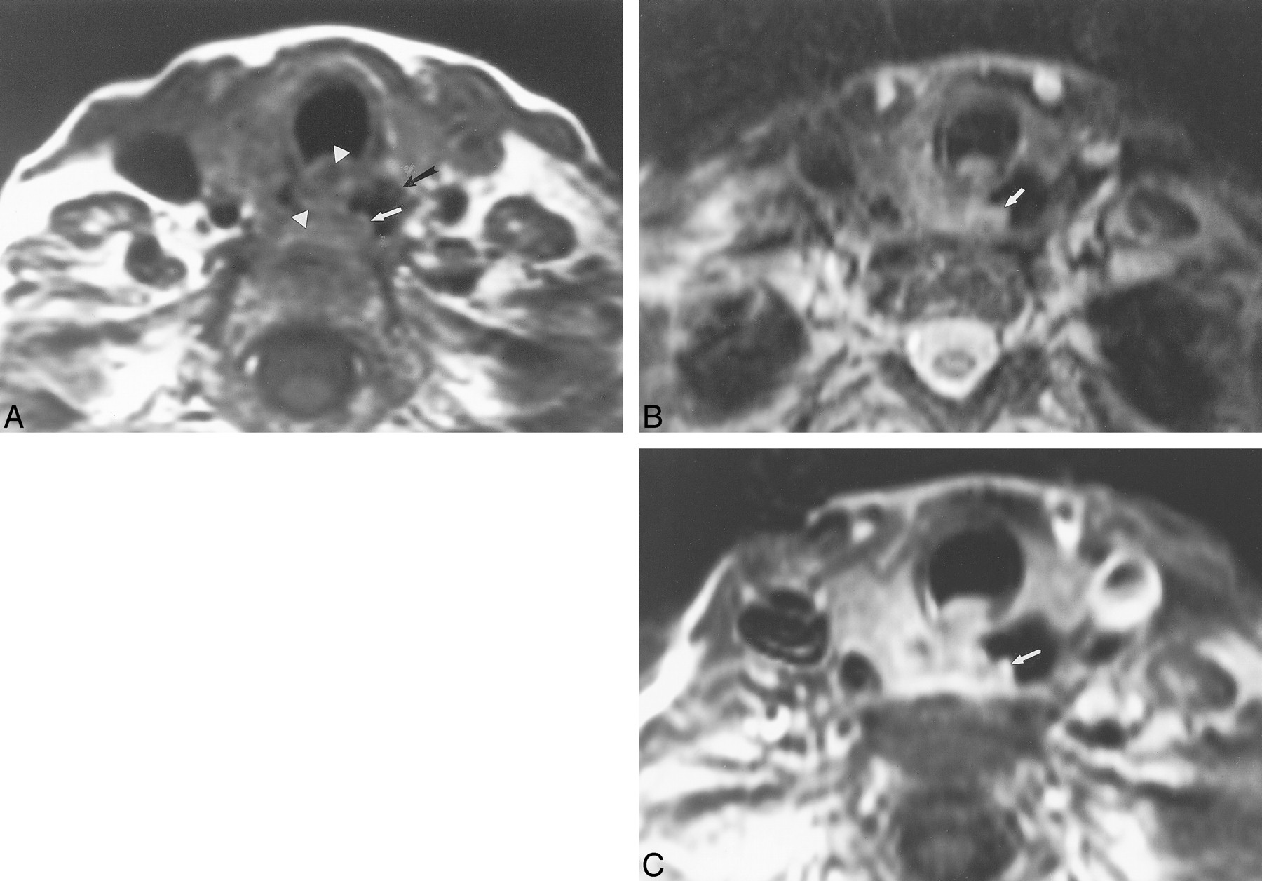
  • fig 2.
    • Download figure
    • Open in new tab
    • Download powerpoint
    fig 2.

    This 62-year-old man with metastatic laryngeal carcinoma to cervical lymph nodes had surgically proven esophageal invasion.

    A, Axial T1-weighted image (700/17) shows obliteration of the fat plane between the mass (arrowheads) and the right lateral esophageal wall. There is focal wall thickening of the esophagus (white arrow). The esophageal lumen is distended with air (black arrow). The mass abuts the esophageal wall for less than 180o.

    B, Axial T2-weighted image (4000/80) demonstrates increased esophageal wall signal (arrow) adjacent to the mass.

    C, Axial enhanced 3D FMPSPGR image (235/21; flip angle, 90o) shows increased enhancement of the esophageal wall (arrow) as well as diffuse enhancement of the adjacent mass.

  • fig 3.
    • Download figure
    • Open in new tab
    • Download powerpoint
    fig 3.

    This 66-year-old man had direct EI by laryngeal carcinoma, resulting in a tracheo-esophageal fistula.

    A, Axial T1-weighted image (600/17) shows a mass that surrounds the esophagus (arrows) by approximately 270o. The fat plane between the mass and the esophagus is obliterated. The esophageal lumen contains air.

    B, Axial T2-weighted image (4000/80) shows focal increased signal (arrow) in the esophageal wall adjacent to the mass.

  • fig 4.
    • Download figure
    • Open in new tab
    • Download powerpoint
    fig 4.

    This 72-year-old woman had follicular carcinoma of the left lobe of the thyroid. This case represents a false-positive interpretation of esophageal invasion. There was no evidence of esophageal invasion by surgical or pathologic evaluation.

    A, Axial T1-weighted image (700/17) shows effacement of the fat plane between the esophagus and the left thyroid (arrow).

    B, Axial T2-weighted image (3000/78) shows focal increased signal within the esophageal wall adjacent to the thyroid mass (arrowheads) compared with the normal right lateral wall of the esophagus (arrow).
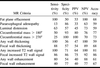
Tables

  • Figures
  • TABLE 1:
    • View popup
    • Download powerpoint
    TABLE 1:

    Findings revealed by MR imaging

  • TABLE 2:
    • View popup
    • Download powerpoint
    TABLE 2:

    Esophageal invasion. Comparison of MR imaging criteria

PreviousNext
Back to top

In this issue

American Journal of Neuroradiology
Vol. 21, Issue 9
1 Oct 2000
  • Table of Contents
  • Index by author
Advertisement
Print
Download PDF
Email Article

Thank you for your interest in spreading the word on American Journal of Neuroradiology.

NOTE: We only request your email address so that the person you are recommending the page to knows that you wanted them to see it, and that it is not junk mail. We do not capture any email address.

Enter multiple addresses on separate lines or separate them with commas.
MR Imaging for Predicting Neoplastic Invasion of the Cervical Esophagus
(Your Name) has sent you a message from American Journal of Neuroradiology
(Your Name) thought you would like to see the American Journal of Neuroradiology web site.
CAPTCHA
This question is for testing whether or not you are a human visitor and to prevent automated spam submissions.
Cite this article
Sudipta Roychowdhury, Laurie A. Loevner, David M. Yousem, Ara Chalian, Kathleen T. Montone
MR Imaging for Predicting Neoplastic Invasion of the Cervical Esophagus
American Journal of Neuroradiology Oct 2000, 21 (9) 1681-1687;

Citation Manager Formats

  • BibTeX
  • Bookends
  • EasyBib
  • EndNote (tagged)
  • EndNote 8 (xml)
  • Medlars
  • Mendeley
  • Papers
  • RefWorks Tagged
  • Ref Manager
  • RIS
  • Zotero
0 Responses
Respond to this article
Share
Bookmark this article
MR Imaging for Predicting Neoplastic Invasion of the Cervical Esophagus
Sudipta Roychowdhury, Laurie A. Loevner, David M. Yousem, Ara Chalian, Kathleen T. Montone
American Journal of Neuroradiology Oct 2000, 21 (9) 1681-1687;
del.icio.us logo Twitter logo Facebook logo Mendeley logo
  • Tweet Widget
  • Facebook Like
  • Google Plus One

Jump to section

  • Article
    • Abstract
    • Methods
    • Results
    • Discussion
    • Conclusion
    • Footnotes
    • References
  • Figures & Data
  • Info & Metrics
  • Responses
  • References
  • PDF

Related Articles

  • No related articles found.
  • PubMed
  • Google Scholar

Cited By...

  • No citing articles found.
  • Crossref
  • Google Scholar

This article has not yet been cited by articles in journals that are participating in Crossref Cited-by Linking.

More in this TOC Section

  • Hydrops Herniation into the Semicircular Canals
  • ASL Sensitivity for Head and Neck Paraganglioma
  • Post SRS Peritumoral Hyperintense Signal of VSs
Show more Head and Neck Imaging

Similar Articles

Advertisement

Indexed Content

  • Current Issue
  • Accepted Manuscripts
  • Article Preview
  • Past Issues
  • Editorials
  • Editor's Choice
  • Fellows' Journal Club
  • Letters to the Editor
  • Video Articles

Cases

  • Case Collection
  • Archive - Case of the Week
  • Archive - Case of the Month
  • Archive - Classic Case

More from AJNR

  • Trainee Corner
  • Imaging Protocols
  • MRI Safety Corner
  • Book Reviews

Multimedia

  • AJNR Podcasts
  • AJNR Scantastics

Resources

  • Turnaround Time
  • Submit a Manuscript
  • Submit a Video Article
  • Submit an eLetter to the Editor/Response
  • Manuscript Submission Guidelines
  • Statistical Tips
  • Fast Publishing of Accepted Manuscripts
  • Graphical Abstract Preparation
  • Imaging Protocol Submission
  • Evidence-Based Medicine Level Guide
  • Publishing Checklists
  • Author Policies
  • Become a Reviewer/Academy of Reviewers
  • News and Updates

About Us

  • About AJNR
  • Editorial Board
  • Editorial Board Alumni
  • Alerts
  • Permissions
  • Not an AJNR Subscriber? Join Now
  • Advertise with Us
  • Librarian Resources
  • Feedback
  • Terms and Conditions
  • AJNR Editorial Board Alumni

American Society of Neuroradiology

  • Not an ASNR Member? Join Now

© 2025 by the American Society of Neuroradiology All rights, including for text and data mining, AI training, and similar technologies, are reserved.
Print ISSN: 0195-6108 Online ISSN: 1936-959X

Powered by HighWire