Advertisement

Main menu

  • Home
  • Content
    • Current Issue
    • Accepted Manuscripts
    • Article Preview
    • Past Issue Archive
    • Video Articles
    • AJNR Case Collection
    • Case of the Week Archive
    • Case of the Month Archive
    • Classic Case Archive
  • Special Collections
    • AJNR Awards
    • Low-Field MRI
    • Alzheimer Disease
    • ASNR Foundation Special Collection
    • Photon-Counting CT
    • View All
  • Multimedia
    • AJNR Podcasts
    • AJNR SCANtastic
    • Trainee Corner
    • MRI Safety Corner
    • Imaging Protocols
  • For Authors
    • Submit a Manuscript
    • Submit a Video Article
    • Submit an eLetter to the Editor/Response
    • Manuscript Submission Guidelines
    • Statistical Tips
    • Fast Publishing of Accepted Manuscripts
    • Graphical Abstract Preparation
    • Imaging Protocol Submission
    • Author Policies
  • About Us
    • About AJNR
    • Editorial Board
    • Editorial Board Alumni
  • More
    • Become a Reviewer/Academy of Reviewers
    • Subscribers
    • Permissions
    • Alerts
    • Feedback
    • Advertisers
    • ASNR Home

User menu

  • Alerts
  • Log in

Search

  • Advanced search
American Journal of Neuroradiology

American Journal of Neuroradiology

ASHNR American Society of Functional Neuroradiology ASHNR American Society of Pediatric Neuroradiology ASSR
  • Alerts
  • Log in

Advanced Search

  • Home
  • Content
    • Current Issue
    • Accepted Manuscripts
    • Article Preview
    • Past Issue Archive
    • Video Articles
    • AJNR Case Collection
    • Case of the Week Archive
    • Case of the Month Archive
    • Classic Case Archive
  • Special Collections
    • AJNR Awards
    • Low-Field MRI
    • Alzheimer Disease
    • ASNR Foundation Special Collection
    • Photon-Counting CT
    • View All
  • Multimedia
    • AJNR Podcasts
    • AJNR SCANtastic
    • Trainee Corner
    • MRI Safety Corner
    • Imaging Protocols
  • For Authors
    • Submit a Manuscript
    • Submit a Video Article
    • Submit an eLetter to the Editor/Response
    • Manuscript Submission Guidelines
    • Statistical Tips
    • Fast Publishing of Accepted Manuscripts
    • Graphical Abstract Preparation
    • Imaging Protocol Submission
    • Author Policies
  • About Us
    • About AJNR
    • Editorial Board
    • Editorial Board Alumni
  • More
    • Become a Reviewer/Academy of Reviewers
    • Subscribers
    • Permissions
    • Alerts
    • Feedback
    • Advertisers
    • ASNR Home
  • Follow AJNR on Twitter
  • Visit AJNR on Facebook
  • Follow AJNR on Instagram
  • Join AJNR on LinkedIn
  • RSS Feeds

AJNR Awards, New Junior Editors, and more. Read the latest AJNR updates

Research ArticleHead and Neck Imaging

Traumatic Neuroma after Neck Dissection: CT Characteristics in Four Cases

Lynn F. Huang, Jane L. Weissman and Chun-yung Fan
American Journal of Neuroradiology October 2000, 21 (9) 1676-1680;
Lynn F. Huang
  • Find this author on Google Scholar
  • Find this author on PubMed
  • Search for this author on this site
Jane L. Weissman
  • Find this author on Google Scholar
  • Find this author on PubMed
  • Search for this author on this site
Chun-yung Fan
  • Find this author on Google Scholar
  • Find this author on PubMed
  • Search for this author on this site
  • Article
  • Figures & Data
  • Info & Metrics
  • Responses
  • References
  • PDF
Loading

Article Figures & Data

Figures

  • fig 1.
    • Download figure
    • Open in new tab
    • Download powerpoint
    fig 1.

    59-year-old man with squamous cell carcinoma of the larynx after left-sided modified radical neck dissection (type 1) with sparing of the spinal accessory nerve.

    A, Axial contrast-enhanced CT scan of the neck shows a nodule (large arrow) with a radiolucent center, peripherally dense rim, and an intact layer of overlying fat (small arrow). The nodule lies posterolateral and close to the left CCA (arrowhead). The left sternocleidomastoid muscle and left internal jugular vein are absent.

    B, Axial contrast-enhanced CT scan of the neck obtained 1 year after A shows the nodule (arrow) to be minimally increased in size, which is highly atypical for tumor.
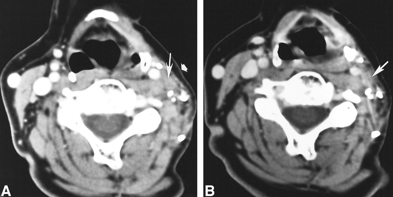
  • fig 2.
    • Download figure
    • Open in new tab
    • Download powerpoint
    fig 2.

    45-year-old man after left-sided modified radical neck dissection (type 1) with sparing of the spinal accessory nerve 1 year after a left partial glossectomy and selective neck dissection (types 1 and 2) for squamous cell carcinoma of the tongue.

    A, Axial contrast-enhanced CT scan shows a nodule (large straight arrow) with a radiolucent center, peripherally dense rim, and an intact layer of overlying subcutaneous fat (small arrow). This nodule is posterolateral and close to the left CCA (arrowhead). Left-sided hypoglossal denervation (curved arrow) is also present.

    B, Axial contrast-enhanced CT scan 1 year after A shows this nodule (large arrow) and its intact layer of overlying fat (small arrow) remain unchanged.
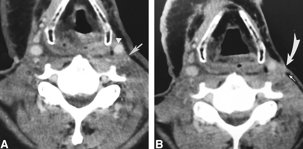
  • fig 3.
    • Download figure
    • Open in new tab
    • Download powerpoint
    fig 3.

    54-year-old woman 4 years after left-sided modified radical neck dissection with sparing of the spinal accessory nerve for parotid adenocarcinoma now with a trigger point.

    A, Axial contrast-enhanced CT scan shows a nodule (long arrow) with faint central radiolucency, a peripherally dense rim, and an intact layer of overlying fat (short arrow). This nodule lies posterolateral and close to the left ICA.

    B, Axial contrast-enhanced CT obtained 2 years after A shows this nodule (arrow) to be stable.

  • fig 4.
    • Download figure
    • Open in new tab
    • Download powerpoint
    fig 4.

    64-year-old man with a right-sided neck mass 7 months after right-sided modified radical neck dissection and knife excision of zones I through V for squamous cell carcinoma of the larynx.

    A, Axial contrast-enhanced CT scan reveals a nodule (arrow) posterolateral to but separate from the right ICA (arrowhead). The nodule lacks a radiolucent center and peripherally dense rim.

    B, Microscopic section of the nodule reveals tangled and interwoven neurofibrils (arrowheads) mixed with connective tissue, typical of a traumatic neuroma (hematoxylin-eosin, ×400).

    C, Another microscopic section of the nodule also reveals other areas containing abnormally mitotic nuclei (arrows), typical of carcinoma mixed with areas typical of traumatic neuroma (hematoxylin-eosin, ×400).

  • fig 5.
    • Download figure
    • Open in new tab
    • Download powerpoint
    fig 5.

    52-year-old man with a possible neck mass after prior bilateral radical neck dissections for squamous cell carcinoma of the tongue.

    A, Axial contrast-enhanced CT scan shows a heterogeneous nodule (arrow), posterolateral to the left ICA (arrowhead), which lacks a radiolucent center and peripherally dense rim.

    B, Axial contrast-enhanced CT scan 3 months after A shows the nodule (curved arrow) to be enlarged and invading the overlying subcutaneous fat (straight arrow). This nodule was pathologically proved to be recurrent squamous cell carcinoma.

PreviousNext
Back to top

In this issue

American Journal of Neuroradiology
Vol. 21, Issue 9
1 Oct 2000
  • Table of Contents
  • Index by author
Advertisement
Print
Download PDF
Email Article

Thank you for your interest in spreading the word on American Journal of Neuroradiology.

NOTE: We only request your email address so that the person you are recommending the page to knows that you wanted them to see it, and that it is not junk mail. We do not capture any email address.

Enter multiple addresses on separate lines or separate them with commas.
Traumatic Neuroma after Neck Dissection: CT Characteristics in Four Cases
(Your Name) has sent you a message from American Journal of Neuroradiology
(Your Name) thought you would like to see the American Journal of Neuroradiology web site.
CAPTCHA
This question is for testing whether or not you are a human visitor and to prevent automated spam submissions.
Cite this article
Lynn F. Huang, Jane L. Weissman, Chun-yung Fan
Traumatic Neuroma after Neck Dissection: CT Characteristics in Four Cases
American Journal of Neuroradiology Oct 2000, 21 (9) 1676-1680;

Citation Manager Formats

  • BibTeX
  • Bookends
  • EasyBib
  • EndNote (tagged)
  • EndNote 8 (xml)
  • Medlars
  • Mendeley
  • Papers
  • RefWorks Tagged
  • Ref Manager
  • RIS
  • Zotero
0 Responses
Respond to this article
Share
Bookmark this article
Traumatic Neuroma after Neck Dissection: CT Characteristics in Four Cases
Lynn F. Huang, Jane L. Weissman, Chun-yung Fan
American Journal of Neuroradiology Oct 2000, 21 (9) 1676-1680;
del.icio.us logo Twitter logo Facebook logo Mendeley logo
  • Tweet Widget
  • Facebook Like
  • Google Plus One

Jump to section

  • Article
    • Abstract
    • Methods
    • Results
    • Discussion
    • Conclusion
    • Footnotes
    • References
  • Figures & Data
  • Info & Metrics
  • Responses
  • References
  • PDF

Related Articles

  • No related articles found.
  • PubMed
  • Google Scholar

Cited By...

  • Chronic daily headaches secondary to greater auricular and lesser occipital neuromas following endolymphatic shunt surgery
  • Traumatic Neuromas in Breast Cancer Patients after Mastectomy
  • Non-neoplastic process after neck dissection demonstrated on enhanced CT in patients with head and neck cancer
  • Crossref
  • Google Scholar

This article has not yet been cited by articles in journals that are participating in Crossref Cited-by Linking.

More in this TOC Section

  • ASL Sensitivity for Head and Neck Paraganglioma
  • Post SRS Peritumoral Hyperintense Signal of VSs
  • Normal Facial Nerve Enhancement on VIBE Sequence
Show more Head and Neck Imaging

Similar Articles

Advertisement

Indexed Content

  • Current Issue
  • Accepted Manuscripts
  • Article Preview
  • Past Issues
  • Editorials
  • Editor's Choice
  • Fellows' Journal Club
  • Letters to the Editor
  • Video Articles

Cases

  • Case Collection
  • Archive - Case of the Week
  • Archive - Case of the Month
  • Archive - Classic Case

More from AJNR

  • Trainee Corner
  • Imaging Protocols
  • MRI Safety Corner
  • Book Reviews

Multimedia

  • AJNR Podcasts
  • AJNR Scantastics

Resources

  • Turnaround Time
  • Submit a Manuscript
  • Submit a Video Article
  • Submit an eLetter to the Editor/Response
  • Manuscript Submission Guidelines
  • Statistical Tips
  • Fast Publishing of Accepted Manuscripts
  • Graphical Abstract Preparation
  • Imaging Protocol Submission
  • Evidence-Based Medicine Level Guide
  • Publishing Checklists
  • Author Policies
  • Become a Reviewer/Academy of Reviewers
  • News and Updates

About Us

  • About AJNR
  • Editorial Board
  • Editorial Board Alumni
  • Alerts
  • Permissions
  • Not an AJNR Subscriber? Join Now
  • Advertise with Us
  • Librarian Resources
  • Feedback
  • Terms and Conditions
  • AJNR Editorial Board Alumni

American Society of Neuroradiology

  • Not an ASNR Member? Join Now

© 2025 by the American Society of Neuroradiology All rights, including for text and data mining, AI training, and similar technologies, are reserved.
Print ISSN: 0195-6108 Online ISSN: 1936-959X

Powered by HighWire