Advertisement

Main menu

  • Home
  • Content
    • Current Issue
    • Accepted Manuscripts
    • Article Preview
    • Past Issue Archive
    • Video Articles
    • AJNR Case Collection
    • Case of the Week Archive
    • Case of the Month Archive
    • Classic Case Archive
  • Special Collections
    • AJNR Awards
    • Low-Field MRI
    • Alzheimer Disease
    • ASNR Foundation Special Collection
    • Photon-Counting CT
    • View All
  • Multimedia
    • AJNR Podcasts
    • AJNR SCANtastic
    • Trainee Corner
    • MRI Safety Corner
    • Imaging Protocols
  • For Authors
    • Submit a Manuscript
    • Submit a Video Article
    • Submit an eLetter to the Editor/Response
    • Manuscript Submission Guidelines
    • Statistical Tips
    • Fast Publishing of Accepted Manuscripts
    • Graphical Abstract Preparation
    • Imaging Protocol Submission
    • Author Policies
  • About Us
    • About AJNR
    • Editorial Board
    • Editorial Board Alumni
  • More
    • Become a Reviewer/Academy of Reviewers
    • Subscribers
    • Permissions
    • Alerts
    • Feedback
    • Advertisers
    • ASNR Home

User menu

  • Alerts
  • Log in

Search

  • Advanced search
American Journal of Neuroradiology

American Journal of Neuroradiology

ASHNR American Society of Functional Neuroradiology ASHNR American Society of Pediatric Neuroradiology ASSR
  • Alerts
  • Log in

Advanced Search

  • Home
  • Content
    • Current Issue
    • Accepted Manuscripts
    • Article Preview
    • Past Issue Archive
    • Video Articles
    • AJNR Case Collection
    • Case of the Week Archive
    • Case of the Month Archive
    • Classic Case Archive
  • Special Collections
    • AJNR Awards
    • Low-Field MRI
    • Alzheimer Disease
    • ASNR Foundation Special Collection
    • Photon-Counting CT
    • View All
  • Multimedia
    • AJNR Podcasts
    • AJNR SCANtastic
    • Trainee Corner
    • MRI Safety Corner
    • Imaging Protocols
  • For Authors
    • Submit a Manuscript
    • Submit a Video Article
    • Submit an eLetter to the Editor/Response
    • Manuscript Submission Guidelines
    • Statistical Tips
    • Fast Publishing of Accepted Manuscripts
    • Graphical Abstract Preparation
    • Imaging Protocol Submission
    • Author Policies
  • About Us
    • About AJNR
    • Editorial Board
    • Editorial Board Alumni
  • More
    • Become a Reviewer/Academy of Reviewers
    • Subscribers
    • Permissions
    • Alerts
    • Feedback
    • Advertisers
    • ASNR Home
  • Follow AJNR on Twitter
  • Visit AJNR on Facebook
  • Follow AJNR on Instagram
  • Join AJNR on LinkedIn
  • RSS Feeds

AJNR Awards, New Junior Editors, and more. Read the latest AJNR updates

Research ArticleBRAIN

Optic Tract Edema in a Meningioma of the Tuberculum Sellae

Evelyn M. L. Sklar, Norman J. Schatz, Joel S. Glaser, Linda Sternau and Firas Seffo
American Journal of Neuroradiology October 2000, 21 (9) 1661-1663;
Evelyn M. L. Sklar
  • Find this author on Google Scholar
  • Find this author on PubMed
  • Search for this author on this site
Norman J. Schatz
  • Find this author on Google Scholar
  • Find this author on PubMed
  • Search for this author on this site
Joel S. Glaser
  • Find this author on Google Scholar
  • Find this author on PubMed
  • Search for this author on this site
Linda Sternau
  • Find this author on Google Scholar
  • Find this author on PubMed
  • Search for this author on this site
Firas Seffo
  • Find this author on Google Scholar
  • Find this author on PubMed
  • Search for this author on this site
  • Article
  • Figures & Data
  • Info & Metrics
  • Responses
  • References
  • PDF
Loading

Abstract

Summary: We report a case of tuberculum sellae meningioma with optic tract edema. Contrary to a prior report on this topic, edema along the optic tract is not only seen in craniopharyngiomas but may be seen (although rarely) in other common parasellar tumors, as in our case of a tuberculum sellae meningioma. The pathogenesis of this edema in meningioma is controversial.

Tuberculum sellae meningiomas represent 3% to 10% of all meningiomas (1). They are more common in women between the ages of 30 and 60 years. Visual prognosis is variable, with most series reporting an improvement in visual function in 40% to 60% of cases after surgery (1). Several factors appear to influence the visual prognosis in patients who undergo surgery for removal of tuberculum sellae meningiomas, including duration of visual symptoms, tumor size, location, and preoperative visual function. The role of edema of the visual pathways in the visual prognosis is unknown.

Case Report

A 40-year-old woman inadvertently discovered diminished vision in her right eye during evaluation for laser refractive surgery. Vision was 20/15 in each eye with no afferent pupillary defect. Confrontational visual fields suggested a temporal defect in the right eye. She had noted increased thirst and frequency of urination for some period of time. Two weeks later, a neuroophthalmologic examination revealed a right afferent pupillary defect. The Humphrey automated visual fields showed an inferior temporal hemianopic scotoma in the right eye only.

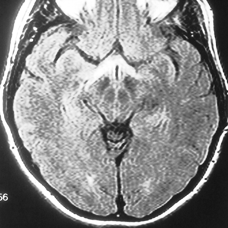
MR imaging revealed a 1.5-cm-diameter homogeneously enhancing suprasellar lesion with extension anteriorly as a “dural tail” on the planum sphenoidale, displacing the chiasm (Figs 1 and 2). There was increased signal in the optic tracts bilaterally both on fast spin-echo T2-weighted and fluid-attenuated inversion recovery (FLAIR) images (Fig 3).

fig 1.
  • Download figure
  • Open in new tab
  • Download powerpoint
fig 1.

Sagittal contrast-enhanced T1-weighted image (649/20/1) shows a homogeneously enhancing suprasellar lesion with extension anteriorly as a dural tail on the planum sphenoidale.fig 2. A and B, Coronal T1-weighted images (656/16/2) before (A) and after (B) administration of contrast material show the suprasellar lesion extending superiorly and displacing the chiasm

fig 3.
  • Download figure
  • Open in new tab
  • Download powerpoint
fig 3.

Increased signal in the optic tracts bilaterally is noted on this axial FLAIR image (6000/128/1, TI = 2000)

Extensive thickening of the arachnoid was discovered during surgery. The tumor spread forward along the olfactory nerves, encompassed the right optic nerve adjacent to the optic chiasm, infiltrated the surface of the brain, and was sheathed posteriorly along the chiasm. Complete removal was not attempted. The patient underwent postoperative radiation therapy.

Discussion

Meningioma is a benign tumor arising in the cranial cavity or in the spine. Grossly, meningioma is a well-circumscribed, firm, tan or grayish lesion arising from the meninges. Microscopically, a benign meningioma usually has a bland whorled appearance with little anaplasia or mitotic activity. Malignant varieties may be identified on the basis of clinical behavior (rapid growth, recurrence, or invasiveness), microscopic features of malignancy (cellular or nuclear anaplasia, mitotic figures), or certain histologic variants.

On MR images, meningiomas tend to be isointense with gray matter, increasing in visibility with contrast enhancement. In our patient, contrast enhancement revealed a homogeneously enhancing mass, although inhomogeneity may have been due to calcification, cystic areas, blood vessels, or histologic variables within the tumor. Enhancement and thickening of the dura adjacent to the neoplasm, the dural tail, may have been due to tumor infiltration or to reactive proliferation of loose connective tissue in the dura (2, 3).

Edema in association with meningioma is quite variable. Some very large meningiomas occur without any edema, whereas even small tumors may be accompanied by pronounced edema. Brain edema is apparently a reaction of the brain tissue to the presence of the neoplasm, even though the tumor is extracerebral; it is not related to the histologic characteristics of the neoplasm (4). Invasion of the brain with disruption of the blood-brain barrier is an unproved hypothesis.

The source of this peritumoral edema has long been a subject of discussion. Domingo et al (5) used dynamic perfusion scanning and proton spectroscopy to study perfusion and metabolism in the peritumoral edema surrounding eight meningiomas preoperatively. These investigators found extremely limited passage of contrast material and low cerebral blood volume in the 2-cm peritumoral region, as well as elevated lactate, suggesting that oligemia and altered metabolism may be part of the pathophysiology in peritumoral edema.

Vascular endothelial growth factor (VEGF) and vascular permeability factor (VPF) may also play a role in peritumoral edema (6, 7). It has been shown that activation of receptors for VEGF/VPF increases permeability of endothelial cells. Meningiomas with a large amount of peritumoral edema have elevated expression of VEGF/VPF. Other researchers have found that platelet activating factor (PAF), which may arise from infiltrating leukocytes, is important to the development of peritumoral edema in patients with meningioma (8). A significant positive correlation was found between peritumoral edema and PAF concentration. It has also been hypothesized that prostaglandin may play a role in the formation of edema (9). Levels of 6-ketoprostaglandin F1α, the stable metabolite of prostacyclin, were found to correlate well with the extent of edema.

The role of macrophages and leukotrines in association with peritumoral edema has also been described (10). Correlation between macrophage infiltration and peritumoral edema was good: in many cases of peritumoral edema, leukotrines were detected, whereas in most cases without peritumoral edema, leukotrines were not detected.

In a recent article, Nagahata et al (11) reported that five of eight patients with craniopharyngioma had optic tract edema, whereas 15 patients with large pituitary adenomas compressing the optic chiasm and six patients with tuberculum sellae meningiomas did not have such edema. The authors concluded that edema caused by craniopharyngioma tends to spread along the optic tracts and that this may be a useful MR finding in distinguishing craniopharyngioma from other common parasellar tumors. However, optic tract edema may be seen in tuberculum sellae meningioma (as in the present case) in pituitary metastases, and it has recently been reported in a case of muslinoma (12).

Conclusion

Optic tract edema, although rare, may be seen in conjunction with suprasellar meningiomas. The pathogenesis of this edema has been a source of discussion, and many hypotheses have been suggested. The role of edema in the visual prognosis is unknown. We think that our case shows that, although rare, optic tract edema may be found in association with suprasellar meningiomas, contrary to prior reports, and that meningioma cannot be excluded on the basis of the presence of edema.

Footnotes

  • ↵1 Address reprint requests to Evelyn M. L. Sklar, MD, Department of Radiology (R-308), University of Miami School of Medicine, 1115 NW 14th St., Miami, FL 33136.

References

  1. ↵
    Miller NR. Walsh and Hoyt's Clinical Neuro-Ophthalmology.. Baltimore: Williams & Wilkins 1991;
  2. ↵
    Goldsher D, Litt AW, Pinto RS, et al. Dural “tail” associated with meningiomas on Gd-DTPA-enhanced MR images: characteristics, differential diagnostic value, and possible implications of treatment. Radiology 1990;176:447-450
    PubMed
  3. Tobumaru A, O'uchi T, Eguchi T, et al. Prominent meningeal enhancement adjacent to meningioma on Gd-DTPA-enhanced MR images: histopathologic correlation. Radiology 1990;175:431-433
    PubMed
  4. ↵
    Chen TC, Zee CS, Miller CA, et al. Magnetic resonance imaging and pathological correlates of meningiomas. Neurosurgery 1992;31:1015-1021
    PubMed
  5. ↵
    Domingo Z, Rowe G, Blamire AM, Cadoux-Hudson TA. Role of ischemia in the genesis of oedema surrounding meningiomas assessed using magnetic resonance imaging and spectroscopy. Br J Neurosurg 1998;12:414-418
    CrossRefPubMed
  6. ↵
    Provias J, Claffey K, del Anguila L, Lau N, Feldkamp M, Guha A. Meningiomas: role of vascular endothelial growth factor/vascular permeability factor in angiogenesis and peritumoral edema. Neurosurgery 1997;40:1016-1026
    CrossRefPubMed
  7. Kalkanis SN, Carroll RS, Zhang J, Zamani AA, Black PM. Correlation of vascular endothelial growth factor messenger RNA expression with peritumoral vasogenic cerebral edema in meningiomas. J Neurosurg 1996;85:1095-1101
    PubMed
  8. ↵
    Hirashima Y, Hayashi N, Fukuda O, Ito H, Endo S, Takaku A. Platelet-activating factor and edema surrounding meningiomas. J Neurosurg 1998;88:304-307
    PubMed
  9. ↵
    Costantini S, Tamir J, Gamori MJ, Shohami E. Tumor prostaglandin levels correlate with edema around supratentorial meningiomas. Neurosurgery 1993;33:204-211
    PubMed
  10. ↵
    Shinonaga M, Chang CC, Kuwabara T. Relation between macrophage infiltrates and peritumoral edema. Adv Neurol 1990;52:475-481
    PubMed
  11. ↵
    Nagahata M, Hosoya T, Kayana T, Yamaguchi K. Edema along the optic tract: a useful finding for the diagnosis of craniopharyngiomas. AJNR Am J Neuroradiol 1998;19:1753-1757
    Abstract
  12. ↵
    Bhatti MT, Holder Ca, Newman NJ, Hudgins PA. MR characteristics of muslin-induced optic neuropathy: report of two cases and review of the literature. AJNR Am J Neuroradiol 2000;21:346-352
    Abstract/FREE Full Text
  • Received February 3, 2000.
  • Copyright © American Society of Neuroradiology
View Abstract
PreviousNext
Back to top

In this issue

American Journal of Neuroradiology
Vol. 21, Issue 9
1 Oct 2000
  • Table of Contents
  • Index by author
Advertisement
Print
Download PDF
Email Article

Thank you for your interest in spreading the word on American Journal of Neuroradiology.

NOTE: We only request your email address so that the person you are recommending the page to knows that you wanted them to see it, and that it is not junk mail. We do not capture any email address.

Enter multiple addresses on separate lines or separate them with commas.
Optic Tract Edema in a Meningioma of the Tuberculum Sellae
(Your Name) has sent you a message from American Journal of Neuroradiology
(Your Name) thought you would like to see the American Journal of Neuroradiology web site.
CAPTCHA
This question is for testing whether or not you are a human visitor and to prevent automated spam submissions.
Cite this article
Evelyn M. L. Sklar, Norman J. Schatz, Joel S. Glaser, Linda Sternau, Firas Seffo
Optic Tract Edema in a Meningioma of the Tuberculum Sellae
American Journal of Neuroradiology Oct 2000, 21 (9) 1661-1663;

Citation Manager Formats

  • BibTeX
  • Bookends
  • EasyBib
  • EndNote (tagged)
  • EndNote 8 (xml)
  • Medlars
  • Mendeley
  • Papers
  • RefWorks Tagged
  • Ref Manager
  • RIS
  • Zotero
0 Responses
Respond to this article
Share
Bookmark this article
Optic Tract Edema in a Meningioma of the Tuberculum Sellae
Evelyn M. L. Sklar, Norman J. Schatz, Joel S. Glaser, Linda Sternau, Firas Seffo
American Journal of Neuroradiology Oct 2000, 21 (9) 1661-1663;
del.icio.us logo Twitter logo Facebook logo Mendeley logo
  • Tweet Widget
  • Facebook Like
  • Google Plus One

Jump to section

  • Article
    • Abstract
    • Case Report
    • Discussion
    • Conclusion
    • Footnotes
    • References
  • Figures & Data
  • Info & Metrics
  • Responses
  • References
  • PDF

Related Articles

  • No related articles found.
  • PubMed
  • Google Scholar

Cited By...

  • Primary hypothalamic third ventriclular Burkitt's lymphoma: a case report with emphasis on differential diagnosis
  • Optic chiasm enhancement associated with giant aneurysm and yttrium treated pituitary adenoma
  • Crossref
  • Google Scholar

This article has not yet been cited by articles in journals that are participating in Crossref Cited-by Linking.

More in this TOC Section

  • Evaluating the Effects of White Matter Multiple Sclerosis Lesions on the Volume Estimation of 6 Brain Tissue Segmentation Methods
  • Quiet PROPELLER MRI Techniques Match the Quality of Conventional PROPELLER Brain Imaging Techniques
  • Predictors of Reperfusion in Patients with Acute Ischemic Stroke
Show more BRAIN

Similar Articles

Advertisement

Indexed Content

  • Current Issue
  • Accepted Manuscripts
  • Article Preview
  • Past Issues
  • Editorials
  • Editor's Choice
  • Fellows' Journal Club
  • Letters to the Editor
  • Video Articles

Cases

  • Case Collection
  • Archive - Case of the Week
  • Archive - Case of the Month
  • Archive - Classic Case

More from AJNR

  • Trainee Corner
  • Imaging Protocols
  • MRI Safety Corner

Multimedia

  • AJNR Podcasts
  • AJNR Scantastics

Resources

  • Turnaround Time
  • Submit a Manuscript
  • Submit a Video Article
  • Submit an eLetter to the Editor/Response
  • Manuscript Submission Guidelines
  • Statistical Tips
  • Fast Publishing of Accepted Manuscripts
  • Graphical Abstract Preparation
  • Imaging Protocol Submission
  • Evidence-Based Medicine Level Guide
  • Publishing Checklists
  • Author Policies
  • Become a Reviewer/Academy of Reviewers
  • News and Updates

About Us

  • About AJNR
  • Editorial Board
  • Editorial Board Alumni
  • Alerts
  • Permissions
  • Not an AJNR Subscriber? Join Now
  • Advertise with Us
  • Librarian Resources
  • Feedback
  • Terms and Conditions
  • AJNR Editorial Board Alumni

American Society of Neuroradiology

  • Not an ASNR Member? Join Now

© 2025 by the American Society of Neuroradiology All rights, including for text and data mining, AI training, and similar technologies, are reserved.
Print ISSN: 0195-6108 Online ISSN: 1936-959X

Powered by HighWire