
Article Figures & Data
Figures
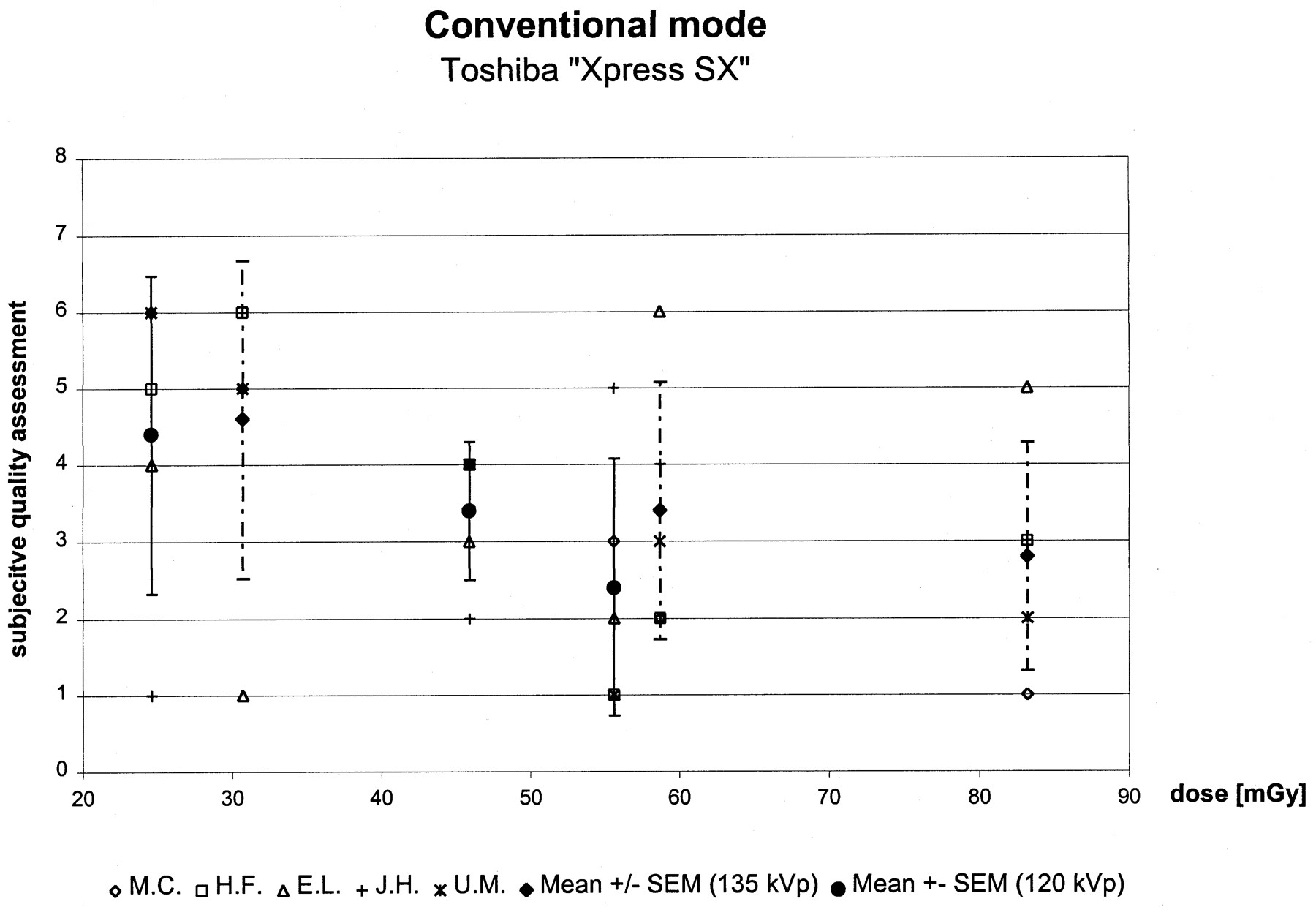
- fig 1.
Subjective quality assessment (grade 1–6) plotted against surface dose (mGy) used in conventional scanning with the Toshiba Xpress SX, showing an inverse linear relationship. Dotted lines represent the standard error of the mean in scans with 135 kVp, full lines in scans with 120 kVp
- fig 2.
Subjective quality assessment (grade 1–6) plotted against surface dose (mGy) used in conventional scanning with the Siemens Somatom AR.SP, showing an inverse linear relationship. Dotted lines represent the standard error of the mean in scans with 130 kVp, full lines in scans with 110 kVp
- fig 3.
Subjective quality assessment (grade 1–7) plotted against surface dose (mGy) used in helical scanning with the Toshiba Xpress SX. Dotted lines represent standard error of the mean of scans with 120 kVp, full lines of scans with 100 kVp. The 80 kVp scan was unanimously rated as a 7
- fig 4.
Subjective quality assessment (grade 1–7) plotted against surface dose (mGy) used in helical scanning with the Siemens Somatom AR.SP. Dotted lines represent the standard error of the mean of scans with 130 kVp, full lines of scans with 110 kVp


Tables

- TABLE 1:
TABLE 2A: Scan parameter, skin entrance dose, and image noise of cerebellar and supratentorial parenchyma (conventional mode, Xpress SX)
- TABLE 1:
TABLE 2B: Scan parameter, skin entrance dose, and image noise of cerebellar and supratentorial parenchyma (conventional mode, somatom AR.SP)
- TABLE 1:
TABLE 3A: Scan parameter, skin entrance dose, and image noise of cerebellar and supratentorial parenchyma (helical mode, Xpress SX)
- TABLE 1:
TABLE 3B: Scan parameter, skin entrance dose, and image noise of cerebellar and supratentorial parenchyma (Helical mode, Somatom AR.SP)

In this issue


Jump to section
Related Articles
Cited By...
- Effect of CTA Tube Current on Spot Sign Detection and Accuracy for Prediction of Intracerebral Hemorrhage Expansion
- Whole-Brain Adaptive 70-kVp Perfusion Imaging with Variable and Extended Sampling Improves Quality and Consistency While Reducing Dose
- CT Radiation Dose and Image Quality Optimization Using a Porcine Model
- Dose reduction and image quality in CT angiography for cerebralaneurysm with various tube potentials and current settings
- Effect of Tube Voltage on Image Quality in 64-Section Multidetector 3D CT Angiography: Evaluation with a Vascular Phantom with Superimposed Bone Skull Structures
- Comparison of Image Quality and Radiation Dose between Fixed Tube Current and Combined Automatic Tube Current Modulation in Craniocervical CT Angiography
- Accuracy of three-dimensional (3D) craniofacial cephalometric landmarks on a low-dose 3D computed tomograph