Advertisement
  • Main menu
  • User menu
  • Search
  • English ▼
    • English
    • Afrikaans
    • Albanian
    • Amharic
    • Arabic
    • Armenian
    • Azerbaijani
    • Basque
    • Belarusian
    • Bengali
    • Bosnian
    • Bulgarian
    • Catalan
    • Cebuano
    • Chichewa
    • Chinese (Simplified)
    • Chinese (Traditional)
    • Corsican
    • Croatian
    • Czech
    • Danish
    • Dutch
    • Esperanto
    • Estonian
    • Filipino
    • Finnish
    • French
    • Frisian
    • Galician
    • Georgian
    • German
    • Greek
    • Gujarati
    • Haitian Creole
    • Hausa
    • Hawaiian
    • Hebrew
    • Hindi
    • Hmong
    • Hungarian
    • Icelandic
    • Igbo
    • Indonesian
    • Irish
    • Italian
    • Japanese
    • Javanese
    • Kannada
    • Kazakh
    • Khmer
    • Korean
    • Kurdish (Kurmanji)
    • Kyrgyz
    • Lao
    • Latin
    • Latvian
    • Lithuanian
    • Luxembourgish
    • Macedonian
    • Malagasy
    • Malay
    • Malayalam
    • Maltese
    • Maori
    • Marathi
    • Mongolian
    • Myanmar (Burmese)
    • Nepali
    • Norwegian
    • Pashto
    • Persian
    • Polish
    • Portuguese
    • Punjabi
    • Romanian
    • Russian
    • Samoan
    • Scottish Gaelic
    • Serbian
    • Sesotho
    • Shona
    • Sindhi
    • Sinhala
    • Slovak
    • Slovenian
    • Somali
    • Spanish
    • Sudanese
    • Swahili
    • Swedish
    • Tajik
    • Tamil
    • Telugu
    • Thai
    • Turkish
    • Ukrainian
    • Urdu
    • Uzbek
    • Vietnamese
    • Welsh
    • Xhosa
    • Yiddish
    • Yoruba
    • Zulu

Main menu

  • Home
  • Content
    • Current Issue
    • Accepted Manuscripts
    • Article Preview
    • Past Issue Archive
    • Video Articles
    • AJNR Case Collection
    • Case of the Week Archive
    • Case of the Month Archive
    • Classic Case Archive
  • Special Collections
    • Advancing NeuroMRI with High-Relaxivity Contrast Agents
    • AJNR Awards
    • Low-Field MRI
    • Alzheimer Disease
    • ASNR Foundation Special Collection
    • Photon-Counting CT
    • View All
  • Multimedia
    • AJNR Podcasts
    • AJNR SCANtastic
    • Trainee Corner
    • MRI Safety Corner
    • Imaging Protocols
  • For Authors
    • Submit a Manuscript
    • Submit a Video Article
    • Submit an eLetter to the Editor/Response
    • Manuscript Submission Guidelines
    • Statistical Tips
    • Fast Publishing of Accepted Manuscripts
    • Graphical Abstract Preparation
    • Imaging Protocol Submission
    • Author Policies
  • About Us
    • About AJNR
    • Editorial Board
    • Editorial Board Alumni
  • More
    • Become a Reviewer/Academy of Reviewers
    • Subscribers
    • Permissions
    • Alerts
    • Feedback
    • Advertisers
    • ASNR Home

User menu

  • Alerts
  • Log in

Search

  • Advanced search
American Journal of Neuroradiology

American Journal of Neuroradiology

ASHNR American Society of Functional Neuroradiology ASHNR American Society of Pediatric Neuroradiology ASSR
  • Alerts
  • Log in

Advanced Search

English ▼
  • English
  • Afrikaans
  • Albanian
  • Amharic
  • Arabic
  • Armenian
  • Azerbaijani
  • Basque
  • Belarusian
  • Bengali
  • Bosnian
  • Bulgarian
  • Catalan
  • Cebuano
  • Chichewa
  • Chinese (Simplified)
  • Chinese (Traditional)
  • Corsican
  • Croatian
  • Czech
  • Danish
  • Dutch
  • Esperanto
  • Estonian
  • Filipino
  • Finnish
  • French
  • Frisian
  • Galician
  • Georgian
  • German
  • Greek
  • Gujarati
  • Haitian Creole
  • Hausa
  • Hawaiian
  • Hebrew
  • Hindi
  • Hmong
  • Hungarian
  • Icelandic
  • Igbo
  • Indonesian
  • Irish
  • Italian
  • Japanese
  • Javanese
  • Kannada
  • Kazakh
  • Khmer
  • Korean
  • Kurdish (Kurmanji)
  • Kyrgyz
  • Lao
  • Latin
  • Latvian
  • Lithuanian
  • Luxembourgish
  • Macedonian
  • Malagasy
  • Malay
  • Malayalam
  • Maltese
  • Maori
  • Marathi
  • Mongolian
  • Myanmar (Burmese)
  • Nepali
  • Norwegian
  • Pashto
  • Persian
  • Polish
  • Portuguese
  • Punjabi
  • Romanian
  • Russian
  • Samoan
  • Scottish Gaelic
  • Serbian
  • Sesotho
  • Shona
  • Sindhi
  • Sinhala
  • Slovak
  • Slovenian
  • Somali
  • Spanish
  • Sudanese
  • Swahili
  • Swedish
  • Tajik
  • Tamil
  • Telugu
  • Thai
  • Turkish
  • Ukrainian
  • Urdu
  • Uzbek
  • Vietnamese
  • Welsh
  • Xhosa
  • Yiddish
  • Yoruba
  • Zulu
  • Home
  • Content
    • Current Issue
    • Accepted Manuscripts
    • Article Preview
    • Past Issue Archive
    • Video Articles
    • AJNR Case Collection
    • Case of the Week Archive
    • Case of the Month Archive
    • Classic Case Archive
  • Special Collections
    • Advancing NeuroMRI with High-Relaxivity Contrast Agents
    • AJNR Awards
    • Low-Field MRI
    • Alzheimer Disease
    • ASNR Foundation Special Collection
    • Photon-Counting CT
    • View All
  • Multimedia
    • AJNR Podcasts
    • AJNR SCANtastic
    • Trainee Corner
    • MRI Safety Corner
    • Imaging Protocols
  • For Authors
    • Submit a Manuscript
    • Submit a Video Article
    • Submit an eLetter to the Editor/Response
    • Manuscript Submission Guidelines
    • Statistical Tips
    • Fast Publishing of Accepted Manuscripts
    • Graphical Abstract Preparation
    • Imaging Protocol Submission
    • Author Policies
  • About Us
    • About AJNR
    • Editorial Board
    • Editorial Board Alumni
  • More
    • Become a Reviewer/Academy of Reviewers
    • Subscribers
    • Permissions
    • Alerts
    • Feedback
    • Advertisers
    • ASNR Home
  • Follow AJNR on Twitter
  • Visit AJNR on Facebook
  • Follow AJNR on Instagram
  • Join AJNR on LinkedIn
  • RSS Feeds

AJNR Awards, New Junior Editors, and more. Read the latest AJNR updates


Improved Turnaround Times | Median time to first decision: 12 days

Research ArticleBrain

Functional MR Imaging Activation after Finger Tapping Has a Shorter Duration in the Basal Ganglia Than in the Sensorimotor Cortex

Chad H. Moritz, M. Elizabeth Meyerand, Dietmar Cordes and Victor M. Haughton
American Journal of Neuroradiology August 2000, 21 (7) 1228-1234;
Chad H. Moritz
aFrom the Departments of Radiology (C.H.M., V.M.H.) and Medical Physics (M.E.M., D.C.), University of Wisconsin, Madison.
  • Find this author on Google Scholar
  • Find this author on PubMed
  • Search for this author on this site
M. Elizabeth Meyerand
aFrom the Departments of Radiology (C.H.M., V.M.H.) and Medical Physics (M.E.M., D.C.), University of Wisconsin, Madison.
  • Find this author on Google Scholar
  • Find this author on PubMed
  • Search for this author on this site
Dietmar Cordes
aFrom the Departments of Radiology (C.H.M., V.M.H.) and Medical Physics (M.E.M., D.C.), University of Wisconsin, Madison.
  • Find this author on Google Scholar
  • Find this author on PubMed
  • Search for this author on this site
Victor M. Haughton
aFrom the Departments of Radiology (C.H.M., V.M.H.) and Medical Physics (M.E.M., D.C.), University of Wisconsin, Madison.
  • Find this author on Google Scholar
  • Find this author on PubMed
  • Search for this author on this site
  • Article
  • Figures & Data
  • Info & Metrics
  • Responses
  • References
  • PDF
Loading

Article Figures & Data

Figures

  • Tables
  • fig 1.
    • Download figure
    • Open in new tab
    • Download powerpoint
    fig 1.

    Reference functions and task timing

  • fig 2.
    • Download figure
    • Open in new tab
    • Download powerpoint
    fig 2.

    A–F, Functional MR images comparing activation detected by reference function 1 (right-handed task) (A–C) and reference function 2 (left-handed task) (D–F). Activation is detected in the SMC, thalamus, putamen, and cerebellum; activation is contralateral to the fingers that were active in the SMC and the thalamus, bilateral in the putamen, and ipsilateral in the cerebellum. (Note that all images are conventional radiologic display format, with the subject's right side appearing on the viewer's left.)

  • fig 3.
    • Download figure
    • Open in new tab
    • Download powerpoint
    fig 3.

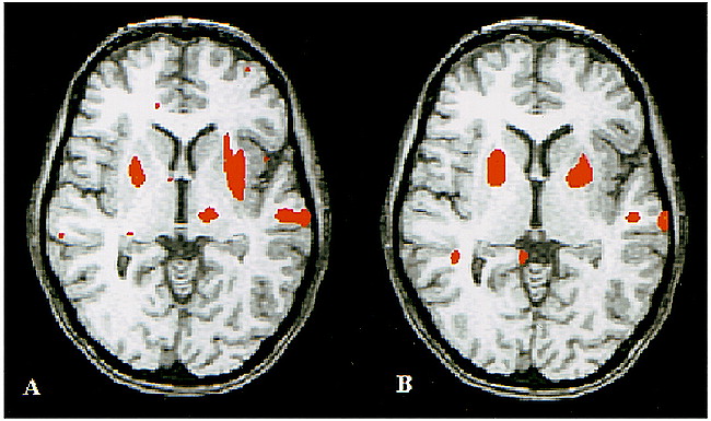
    A and B, Functional MR images showing subcortical activation for the same subject as in figure 2 detected by reference function 3 (right-handed initiation) (A) and reference function 4 (left-handed initiation) (B). Activation is detected in the putamen, thalamus, and transverse temporal gyrus. As compared with reference functions 1 and 2 (see fig 2B and E), more activation is detected in the putamen

  • fig 4.
    • Download figure
    • Open in new tab
    • Download powerpoint
    fig 4.

    Functional MR images showing activation detected by reference function 5. Activation is detected in the SMC, SMA, thalamus, and cerebellum. As compared with the other reference functions (figs 2 and 3), activation is detected bilaterally in the SMC, thalamus, and cerebellum, but with decreased specificity for the SMC

  • fig 5.
    • Download figure
    • Open in new tab
    • Download powerpoint
    fig 5.

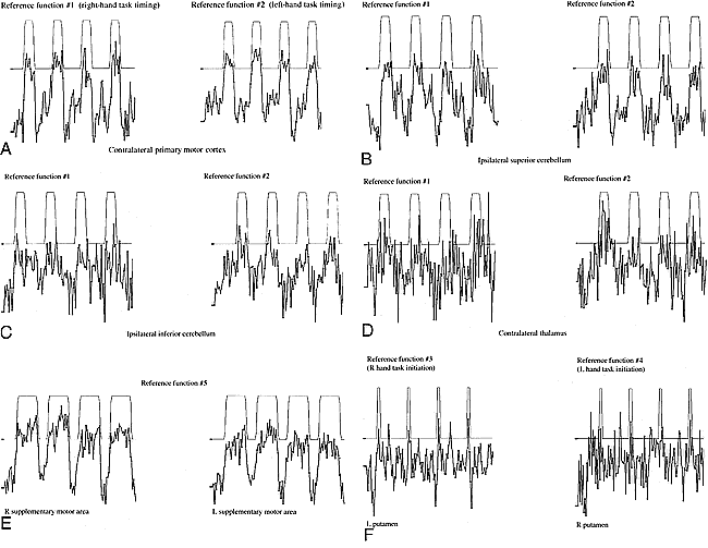
    Representative time courses from selected ROIs.

    A, Right and left primary motor cortex.

    B, Right and left superior cerebellum.

    C, Right and left inferior cerebellum.

    D, Right and left thalamus.

    E, Right and left SMA.

    F, Right and left putamen.

  • fig 6.
    • Download figure
    • Open in new tab
    • Download powerpoint
    fig 6.

    A and B, Time courses are averaged across the four epochs from representative pixels in the right and left putamen (A) and left and right primary motor cortex (B). Vertical gray lines indicate timing intervals for the initiation of each 20-second cycle of right- and left-handed activation and rest

Tables

  • Figures
  • Table 1:
    • View popup
    • Download powerpoint
    Table 1:

    Number of subjects who showed activation in specific regions for reference functions 1 and 2

  • Table 2:
    • View popup
    • Download powerpoint
    Table 2:

    Number of subjects who showed activation in the basal ganglia for reference functions 1–4

PreviousNext
Back to top

In this issue

American Journal of Neuroradiology
Vol. 21, Issue 7
1 Aug 2000
  • Table of Contents
  • Index by author
Advertisement
Print
Download PDF
Email Article
Cite this article
0 Responses
Respond to this article
Share
Bookmark this article
Functional MR Imaging Activation after Finger Tapping Has a Shorter Duration in the Basal Ganglia Than in the Sensorimotor Cortex
Chad H. Moritz, M. Elizabeth Meyerand, Dietmar Cordes, Victor M. Haughton
American Journal of Neuroradiology Aug 2000, 21 (7) 1228-1234;
del.icio.us logo Twitter logo Facebook logo Mendeley logo
  • Tweet Widget

Jump to section

  • Article
    • Abstract
    • Methods
    • Results
    • Discussion
    • Conclusion
    • Footnotes
    • References
  • Figures & Data
  • Info & Metrics
  • Responses
  • References
  • PDF

Related Articles

  • No related articles found.
  • PubMed
  • Google Scholar

Cited By...

  • Improving fMRI in Parkinsons Disease by Accounting for Brain Region-Specific Activity Patterns
  • Crossref
  • Google Scholar

This article has not yet been cited by articles in journals that are participating in Crossref Cited-by Linking.

More in this TOC Section

  • Usefulness of Quantitative Susceptibility Mapping for the Diagnosis of Parkinson Disease
  • White Matter Alterations in the Brains of Patients with Active, Remitted, and Cured Cushing Syndrome: A DTI Study
  • Qualitative and Quantitative Analysis of MR Imaging Findings in Patients with Middle Cerebral Artery Stroke Implanted with Mesenchymal Stem Cells
Show more Brain

Similar Articles

Advertisement

Indexed Content

  • Current Issue
  • Accepted Manuscripts
  • Article Preview
  • Past Issues
  • Editorials
  • Editor's Choice
  • Fellows' Journal Club
  • Letters to the Editor
  • Video Articles

Cases

  • Case Collection
  • Archive - Case of the Week
  • Archive - Case of the Month
  • Archive - Classic Case

More from AJNR

  • Trainee Corner
  • Imaging Protocols
  • MRI Safety Corner
  • Book Reviews

Multimedia

  • AJNR Podcasts
  • AJNR Scantastics

Resources

  • Turnaround Time
  • Submit a Manuscript
  • Submit a Video Article
  • Submit an eLetter to the Editor/Response
  • Manuscript Submission Guidelines
  • Statistical Tips
  • Fast Publishing of Accepted Manuscripts
  • Graphical Abstract Preparation
  • Imaging Protocol Submission
  • Evidence-Based Medicine Level Guide
  • Publishing Checklists
  • Author Policies
  • Become a Reviewer/Academy of Reviewers
  • News and Updates

About Us

  • About AJNR
  • Editorial Board
  • Editorial Board Alumni
  • Alerts
  • Permissions
  • Not an AJNR Subscriber? Join Now
  • Advertise with Us
  • Librarian Resources
  • Feedback
  • Terms and Conditions
  • AJNR Editorial Board Alumni

American Society of Neuroradiology

  • Not an ASNR Member? Join Now

© 2025 by the American Society of Neuroradiology All rights, including for text and data mining, AI training, and similar technologies, are reserved.
Print ISSN: 0195-6108 Online ISSN: 1936-959X

Powered by HighWire
Email this Article

Thank you for your interest in spreading the word on American Journal of Neuroradiology.

NOTE: We only request your email address so that the person you are recommending the page to knows that you wanted them to see it, and that it is not junk mail. We do not capture any email address.

Enter multiple addresses on separate lines or separate them with commas.
Functional MR Imaging Activation after Finger Tapping Has a Shorter Duration in the Basal Ganglia Than in the Sensorimotor Cortex
(Your Name) has sent you a message from American Journal of Neuroradiology
(Your Name) thought you would like to see the American Journal of Neuroradiology web site.
CAPTCHA
This question is for testing whether or not you are a human visitor and to prevent automated spam submissions.
Cite this article
Chad H. Moritz, M. Elizabeth Meyerand, Dietmar Cordes, Victor M. Haughton
Functional MR Imaging Activation after Finger Tapping Has a Shorter Duration in the Basal Ganglia Than in the Sensorimotor Cortex
American Journal of Neuroradiology Aug 2000, 21 (7) 1228-1234;

Citation Manager Formats

  • BibTeX
  • Bookends
  • EasyBib
  • EndNote (tagged)
  • EndNote 8 (xml)
  • Medlars
  • Mendeley
  • Papers
  • RefWorks Tagged
  • Ref Manager
  • RIS
  • Zotero

We use cookies on this site to enhance your user experience. By clicking any link on this page you are giving your consent for us to set cookies.