
Article Figures & Data
Figures
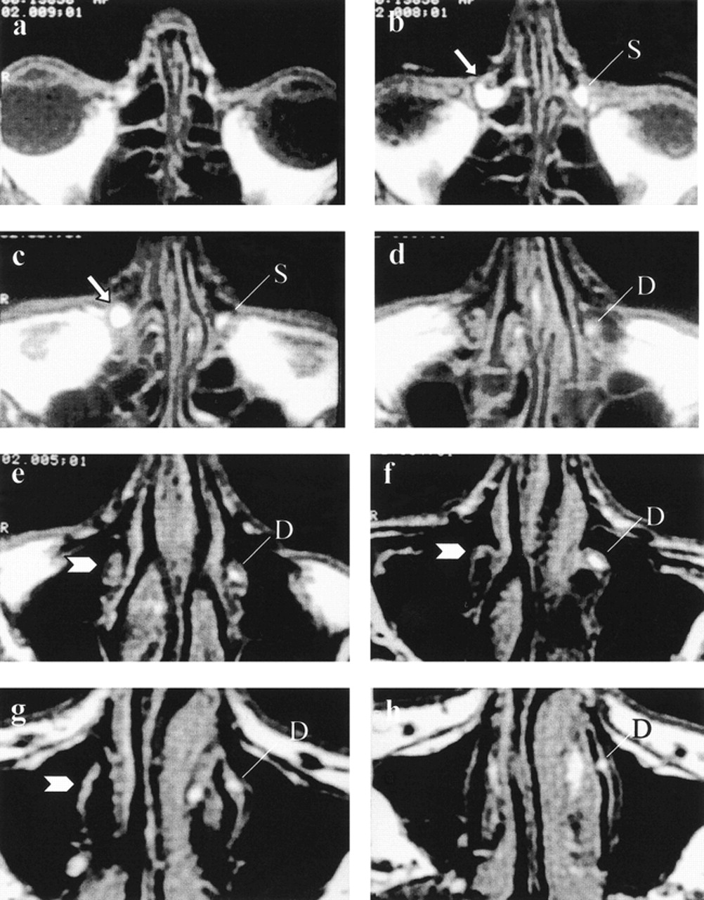
- fig 1.
MRD images of a patient with right postlacrimal sac stenosis and healthy left lacrimal system. Axial T1-weighted (600/30/1 [TR/TE/excitations]) images obtained after the topical administration of diluted gadopentetate solution from the nasolacrimal duct to the orbital level. On the left side, healthy drainage of contrast-enhanced tear can be appreciated inside the lacrimal sac (L) and the nasolacrimal duct (N). On the right side, postsaccular stenosis causes dilation of the right lacrimal sac (arrows), and no contrast media is appreciated down into the ipsilateral nasolacrimal duct (arrowheads).
a, Nasolacrimal duct, most inferior section.
b through g, Progression of sections from inferior to superior.
h, Orbital level, superior section.
- fig 2.
Right postlacrimal sac stenosis and healthy left lacrimal system.
a, Axial CTD scan obtained at the level of the lacrimal sac. Dilation of the right lacrimal sac (white arrow) and absence of contrast medium inside the right nasolacrimal duct (arrowhead) can be appreciated. For comparison, see the healthy drainage of the contrast medium on the left side (thin arrows).
b, Axial CTD scan obtained at the level of the nasolacrimal duct.
c, Right postlacrimal sac stenosis and healthy left lacrimal system. Axial MRDt images (600/30/1) obtained after the injection of iodinate and gadopentetate contrast media at the level of the lacrimal sac. Dilation of the lacrimal sac (white arrow) and absence of contrast media below the lacrimal sac level on the right side (arrowhead) are equally depicted on MRDt images. The healthy contrast media drainage on the left side is also appreciated (thin arrows).
d, Axial MRDt images (600/30/1) obtained after the injection of iodinate and gadopentetate contrast media at the level of the nasolacrimal duct.
- fig 3.
Bilateral dacryocystophlegmon.
a, CTD scan obtained at the level of the lacrimal sac. Dilation of the lacrimal sac, showing dense material inside, with a hyperdense foreign body inside the left one (arrowhead) is visible.
b through d, Four-in-one image with caudocranial contiguous axial T1-weighted MRDc images (600/30/1) using lacrimal canaliculus cannulation. After the injection of a diluted gadopentetate solution, only partial inflow of the contrast medium is appreciated inside the lacrimal sacs (white arrows), with intraluminal exudate being isointense to the cortex. See the small hypointense foreign body on the right lacrimal sac (arrowhead).
- fig 4.
Right post-lacrimal sac stenosis and healthy left lacrimal system.
a, Axial MRDt image (600/30/1) obtained before fat saturation. Although no additional information is detected on the fat saturation image, higher contrast between the gadopentetate dimeglumine solution (arrowheads) and perilacrimal soft tissue is shown.
b, Axial MRDt image (600/30/1) obtained after fat saturation.
- fig 5.
Schematic drawing of the lacrimal system (modified from Sobotta/Becher Atlas, USES 1976). C, lacrimal canaliculi; S, lacrimal sac; R, Rosenmüller's valve; H, Hasner's valve


Tables

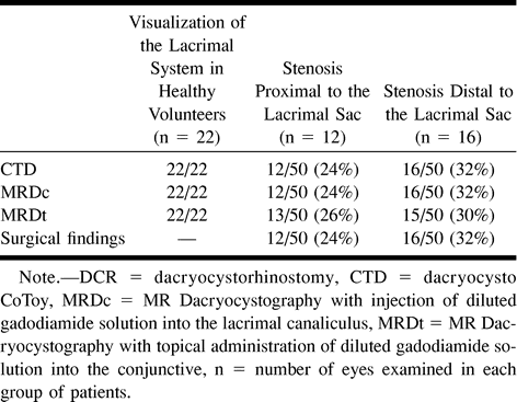
Results of MR and CT dacryocystography in 11 healthy volunteers (22 eyes) and 25 patients with epiphora (50 eyes)


