
Article Figures & Data
Figures
- fig 1.
A–C, Graphs comparing peak height (A), peak area (B), and width at half maximum (C) for NAA, Cho, and Cr in 10 patients with high-grade brain astrocytomas. There are no significant differences in the studies obtained before and after contrast administration
- fig 2.
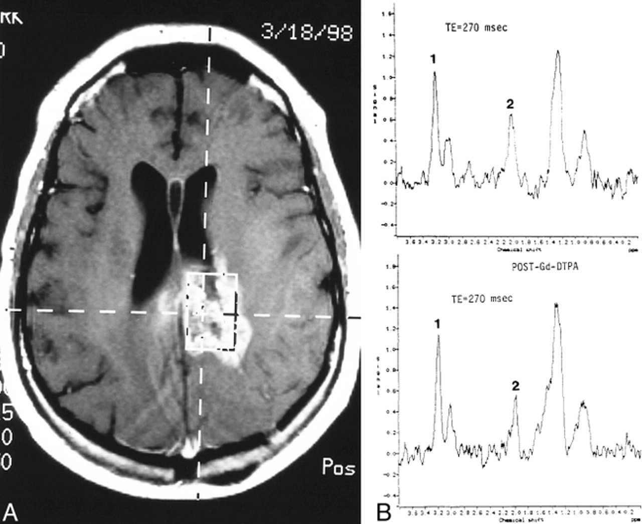
Recurrent glioblastoma multiforme.
A, Axial contrast-enhanced T1-weighted image (560/15/1) shows placement of VOI encompassing mostly enhancing tumor.
B, Pre- (top panel) and postcontrast (bottom panel) MR spectroscopy studies show no significant differences. There is significant elevation of Cho (1) and reduction of NAA (2) on both examinations.
- fig 3.
Recurrent anaplastic astrocytoma.
A, Axial contrast-enhanced T1-weighted image (560/15/1) shows placement of VOI containing mostly enhancing tumor.
B, Pre- (top panel) and postcontrast (bottom panel) MR spectroscopy studies show no significant differences. The Cho level is high and the NAA level is low on both examinations.
- fig 4.
Recurrent glioblastoma multiforme.
A, Axial contrast-enhanced T1-weighted image (560/15/1) shows placement of VOI containing mostly enhancing tumor.
B, Pre- (top panel) and postcontrast (bottom panel) MR spectroscopy studies show reduction of the Cho (1) resonance from 1.0 before to 0.7 after contrast administration. Despite this reduction, the Cho peak remains markedly elevated in relation to the NAA peak (2), indicating an increase in number and/or activity of tumor cells.
- fig 5.
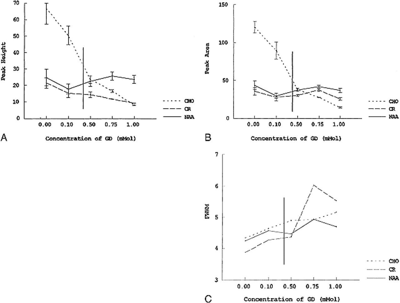
A–C, Graphs depicting the effect of variable concentrations of contrast material on peak height (A), peak area (B), and full width at half maximum (FWHM) (C) in a phantom containing a 10-mmol concentration of NAA, Cho, and Cr in phosphate-buffered saline. As the concentration of contrast material increases from 0.1 to 1.0 mmol, there is a significant decline in peak height and area for Cho. The NAA and Cr peaks show no significant alterations. Some peak broadening occurs for all the metabolites with increasing contrast material concentration. Vertical line indicates estimated extracellular space concentration of contrast material in an average patient, based on dose and volume of distribution.


Tables

Table of peak height, peak area, and width at half maximum for NAA, Cho, and Cr peaks in 10 patients with high-grade brain astrocytomas


