
Article Figures & Data
Figures
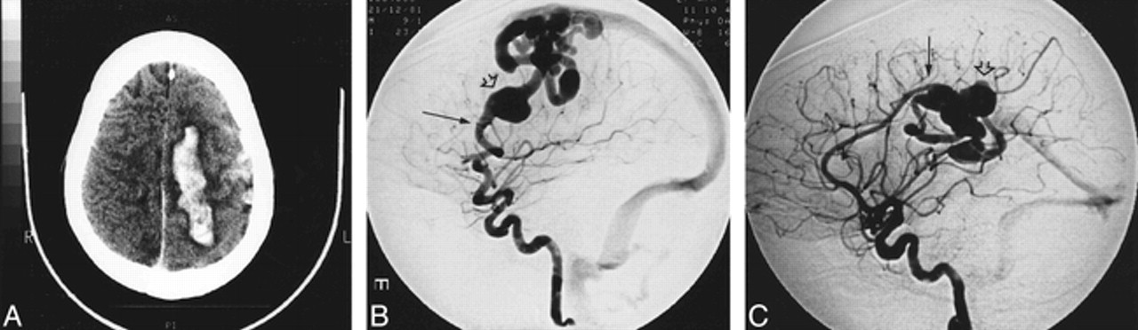
- fig 1.
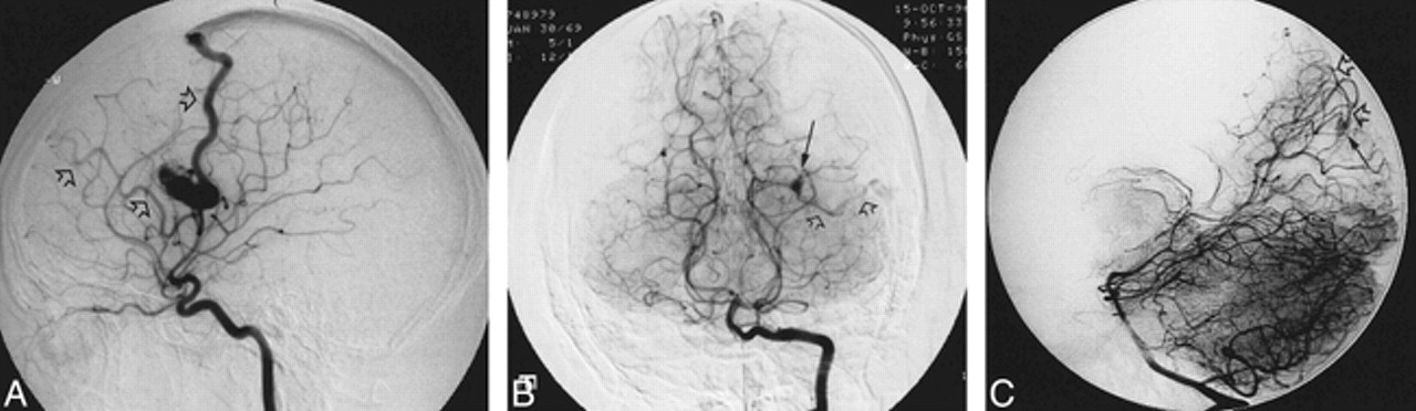
Angiograms of a 21-year-old man who presented with a grand mal seizure, a history of epistaxis, lower-lip telangiectasia, and a family history of HHT.
A, Lateral-view angiogram of the right internal carotid artery shows the small AVM involving right parietal lobe cortex filled by the two branches of the right middle cerebral artery with two draining veins (open arrows).
B, Anteroposterior-view angiogram of the vertebral artery shows a second AVM (micro AVM) (arrow), involving the left occipital cortex, fed by a branch of the posterior cerebral artery and draining into a single vein (open arrows).
C, Lateral-view angiogram shows another micro AVM (arrow) involving the right occipital lobe cortex supplied by a parieto-occipital artery with a single draining vein (open arrows).
- fig 2.
Images from the case of a 15-year-old woman who presented with right hemiplegia, a history of recurrent epistaxis, and a family history of HHT. She was also subsequently diagnosed with a pulmonary AVM.
A, Unenhanced CT scan shows an intracerebral hemorrhage and subarachnoid hemorrhage.
B, Lateral-view angiogram of the left internal carotid artery shows an AVF (arrow) involving the left frontal lobe cortex, arising from a middle cerebral artery branch, with cortical drainage and venous pouch (open arrow).
C, Lateral-view angiogram of the right internal carotid artery shows another AVF (arrow) involving the right parietal cortex supplied by the right pericallosal artery with deep drainage and venous pouch (open arrow).


Tables
 TABLE 1:
Relationship between initial presentation and cerebral AVM type in HHT (n = 28)
In this issue


Jump to section
Related Articles
Cited By...
- Arteriovenous malformation from a patient with JP-HHT harbours two second-hit somatic DNA alterations in SMAD4
- Natural history of brain capillary vascular malformations in hereditary hemorrhagic telangiectasia patients
- Neurovascular Manifestations in Hereditary Hemorrhagic Telangiectasia: Imaging Features and Genotype-Phenotype Correlations
- Brain Arteriovenous Malformation Multiplicity Predicts the Diagnosis of Hereditary Hemorrhagic Telangiectasia: Quantitative Assessment
- International guidelines for the diagnosis and management of hereditary haemorrhagic telangiectasia
- Screening for pulmonary and cerebral arteriovenous malformations in children with hereditary haemorrhagic telangiectasia
- Multiple Symptomatic Cerebral Arteriovenous Malformations in a Patient with HIV
- De Novo Cerebral Arteriovenous Malformation: Case Report and Literature Review
- Management of Stroke in Infants and Children: A Scientific Statement From a Special Writing Group of the American Heart Association Stroke Council and the Council on Cardiovascular Disease in the Young
- Endothelial Notch4 signaling induces hallmarks of brain arteriovenous malformations in mice
- Polymorphisms in Transforming Growth Factor-{beta}-Related Genes ALK1 and ENG Are Associated With Sporadic Brain Arteriovenous Malformations
- Are There Genetic Influences on Sporadic Brain Arteriovenous Malformations?
- Cerebral Vascular Abnormalities in a Murine Model of Hereditary Hemorrhagic Telangiectasia
- Compelling Reasons to Screen Brain in HHT Response