
Article Figures & Data
Figures
- fig 1.
Voxel location in a 60-year-old man with definite ALS and clinically severe UMN signs.
A, Precentral gyrus.
B, Cuneus gyrus.
- fig 2.
Proton spectra from same subject and voxel locations as shown in figure 1. The dark line represents the LCModel fit of the spectrum. The assignments of the principal peaks (in ppm) in the in vivo spectrum are as follows: NA = NAA (2.01) + NAAG (2.05), Cr (3.03), Ins (3.56). The complex multiplets of Glu and Gln, whose sum is Glx, are located at approximately 2.1 to 2.5 ppm and 3.7 to 3.8 ppm (see [41]).
A, Precentral gyrus.
B, Cuneus gyrus.
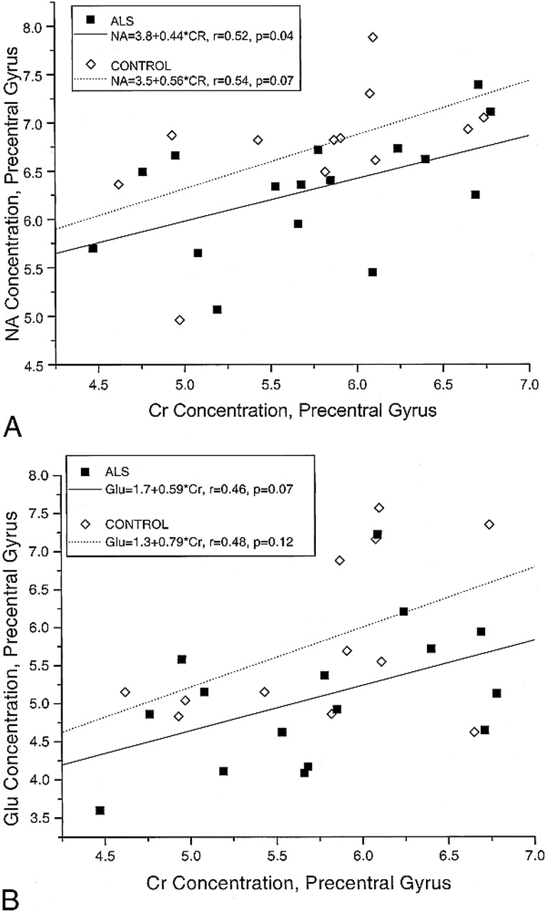
- fig 3.
Correlation between metabolite concentrations (mmol/L). The formulas for the regression lines are given in the box in the upper left-hand corner. Asterisk indicates multiplication; r, Pearson correlation coefficient for each group.
A, NA and Cr.
B, Glu and Cr.
- fig 4.
Precentral gyrus metabolite concentrations (mmol/L) as a function of the severity of UMN clinical signs. The length of the line extending above each rectangle indicates the standard error of the mean (SEM).
A, NA.
B, Cho.
C, Glu.
D, Ins.
- fig 5.
Signal intensity changes in the precentral gyri of a 50-year-old woman with definite ALS (clinically moderate UMN signs).
A–D, Adjacent T2-weighted FSE images (A and B) and corresponding FLAIR images (C and D) show a curvilinear hypointensity (closed arrows) in the region of the motor cortex of the precentral gyrus. In the subcortical white matter of the precentral gyrus, and extending toward the superior frontal gyrus, faint hyperintensity (open arrows) is detected on the FLAIR images but not on the FSE images.


Tables
 TABLE 1:
Metabolite concentrations (mean ± SEM, mmol/L) from STEAM (TE = 20) spectra
- TABLE 2:
Difference in precentral gyrus metabolite concentrations (mean ± SEM, mmol/L) between first and second studies of ALS patients
- TABLE 4:
Change in mean metabolite concentration and MR image signal intensity in the precentral gyrus of ALS patients


