Advertisement

Main menu

  • Home
  • Content
    • Current Issue
    • Accepted Manuscripts
    • Article Preview
    • Past Issue Archive
    • Video Articles
    • AJNR Case Collection
    • Case of the Week Archive
    • Case of the Month Archive
    • Classic Case Archive
  • Special Collections
    • AJNR Awards
    • Low-Field MRI
    • Alzheimer Disease
    • ASNR Foundation Special Collection
    • Photon-Counting CT
    • View All
  • Multimedia
    • AJNR Podcasts
    • AJNR SCANtastic
    • Trainee Corner
    • MRI Safety Corner
    • Imaging Protocols
  • For Authors
    • Submit a Manuscript
    • Submit a Video Article
    • Submit an eLetter to the Editor/Response
    • Manuscript Submission Guidelines
    • Statistical Tips
    • Fast Publishing of Accepted Manuscripts
    • Graphical Abstract Preparation
    • Imaging Protocol Submission
    • Author Policies
  • About Us
    • About AJNR
    • Editorial Board
    • Editorial Board Alumni
  • More
    • Become a Reviewer/Academy of Reviewers
    • Subscribers
    • Permissions
    • Alerts
    • Feedback
    • Advertisers
    • ASNR Home

User menu

  • Alerts
  • Log in

Search

  • Advanced search
American Journal of Neuroradiology

American Journal of Neuroradiology

ASHNR American Society of Functional Neuroradiology ASHNR American Society of Pediatric Neuroradiology ASSR
  • Alerts
  • Log in

Advanced Search

  • Home
  • Content
    • Current Issue
    • Accepted Manuscripts
    • Article Preview
    • Past Issue Archive
    • Video Articles
    • AJNR Case Collection
    • Case of the Week Archive
    • Case of the Month Archive
    • Classic Case Archive
  • Special Collections
    • AJNR Awards
    • Low-Field MRI
    • Alzheimer Disease
    • ASNR Foundation Special Collection
    • Photon-Counting CT
    • View All
  • Multimedia
    • AJNR Podcasts
    • AJNR SCANtastic
    • Trainee Corner
    • MRI Safety Corner
    • Imaging Protocols
  • For Authors
    • Submit a Manuscript
    • Submit a Video Article
    • Submit an eLetter to the Editor/Response
    • Manuscript Submission Guidelines
    • Statistical Tips
    • Fast Publishing of Accepted Manuscripts
    • Graphical Abstract Preparation
    • Imaging Protocol Submission
    • Author Policies
  • About Us
    • About AJNR
    • Editorial Board
    • Editorial Board Alumni
  • More
    • Become a Reviewer/Academy of Reviewers
    • Subscribers
    • Permissions
    • Alerts
    • Feedback
    • Advertisers
    • ASNR Home
  • Follow AJNR on Twitter
  • Visit AJNR on Facebook
  • Follow AJNR on Instagram
  • Join AJNR on LinkedIn
  • RSS Feeds

AJNR Awards, New Junior Editors, and more. Read the latest AJNR updates

Research ArticleSpine Imaging and Spine Image-Guided Interventions

Primary Amyloidoma of the Cervical Spine

Sung Su Hwang, Young Ha Park, Jee Young Kim, So Lyung Jung, Myeong Im Ahn, Choon Keun Park and Eun Deok Chang
American Journal of Neuroradiology March 2000, 21 (3) 601-603;
Sung Su Hwang
aFrom the Departments of Radiology (S.S.H., Y.H.P., J.Y.K., S.L.J., M.I.A.), Neurosurgery (C.K.P.), and Pathology (E.D.C.), St. Vincent's Hospital, the Catholic University of Korea, Suwon, Korea.
  • Find this author on Google Scholar
  • Find this author on PubMed
  • Search for this author on this site
Young Ha Park
aFrom the Departments of Radiology (S.S.H., Y.H.P., J.Y.K., S.L.J., M.I.A.), Neurosurgery (C.K.P.), and Pathology (E.D.C.), St. Vincent's Hospital, the Catholic University of Korea, Suwon, Korea.
  • Find this author on Google Scholar
  • Find this author on PubMed
  • Search for this author on this site
Jee Young Kim
aFrom the Departments of Radiology (S.S.H., Y.H.P., J.Y.K., S.L.J., M.I.A.), Neurosurgery (C.K.P.), and Pathology (E.D.C.), St. Vincent's Hospital, the Catholic University of Korea, Suwon, Korea.
  • Find this author on Google Scholar
  • Find this author on PubMed
  • Search for this author on this site
So Lyung Jung
aFrom the Departments of Radiology (S.S.H., Y.H.P., J.Y.K., S.L.J., M.I.A.), Neurosurgery (C.K.P.), and Pathology (E.D.C.), St. Vincent's Hospital, the Catholic University of Korea, Suwon, Korea.
  • Find this author on Google Scholar
  • Find this author on PubMed
  • Search for this author on this site
Myeong Im Ahn
aFrom the Departments of Radiology (S.S.H., Y.H.P., J.Y.K., S.L.J., M.I.A.), Neurosurgery (C.K.P.), and Pathology (E.D.C.), St. Vincent's Hospital, the Catholic University of Korea, Suwon, Korea.
  • Find this author on Google Scholar
  • Find this author on PubMed
  • Search for this author on this site
Choon Keun Park
aFrom the Departments of Radiology (S.S.H., Y.H.P., J.Y.K., S.L.J., M.I.A.), Neurosurgery (C.K.P.), and Pathology (E.D.C.), St. Vincent's Hospital, the Catholic University of Korea, Suwon, Korea.
  • Find this author on Google Scholar
  • Find this author on PubMed
  • Search for this author on this site
Eun Deok Chang
aFrom the Departments of Radiology (S.S.H., Y.H.P., J.Y.K., S.L.J., M.I.A.), Neurosurgery (C.K.P.), and Pathology (E.D.C.), St. Vincent's Hospital, the Catholic University of Korea, Suwon, Korea.
  • Find this author on Google Scholar
  • Find this author on PubMed
  • Search for this author on this site
  • Article
  • Figures & Data
  • Info & Metrics
  • Responses
  • References
  • PDF
Loading

Abstract

Summary: Primary solitary amyloidoma of the spine is a disease characterized by localized deposits of amyloid. We describe and illustrate the radiologic appearance of primary solitary amyloidoma of the spine on plain radiographs, CT scans, and MR images. The imaging findings revealed features of a nonspecific soft-tissue mass with calcifications. Epidural extension of the amyloidoma caused spinal cord compression.

Primary solitary amyloidosis or amyloidoma is a disease characterized by localized deposition of amyloid in which no plasma cell dyscrasia or abnormal serum proteins are detectable (1). Only a few cases of primary amyloidoma of the spine and only two cases involving the cervical spine have been reported (2, 3). We describe a case of a primary amyloidoma of the cervical spine that caused spinal cord compression.

Case Report

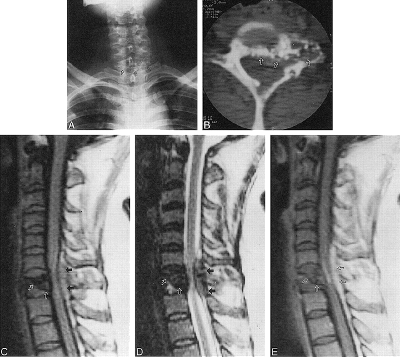
A 45-year-old woman presented with paraparesis, urinary incontinence, and a 3-month history of intermittently recurring pain in the posterior portion of the neck. Physical examination revealed tenderness over the lower cervical spine at the C7 level. Sensation was decreased to pinprick from the T8 dermatome down. Motor strength was 3/5 in the left lower extremity and 2/5 to 3/5 in the right lower extremity. Results of routine blood examination and urinalysis were normal. Bence-Jones protein was not detected in repeated urinalysis. The serum immunoelectrophoresis detected no abnormal band. A bone marrow biopsy specimen showed normal cellularity with no evidence of multiple myeloma or systemic amyloidosis. Results of a rectal biopsy were normal. Plain radiography, CT, MR imaging, and a bone scan with 99mTc-methylene diphosphonate (MDP) were performed and showed an abnormal mass involving the C7 vertebra (Fig 1A-H).

fig 1.
  • Download figure
  • Open in new tab
  • Download powerpoint
fig 1.

45-year-old woman with paraparesis, urinary incontinence, and posterior neck pain.

A, Anteroposterior radiograph of cervical spine shows osteolytic lesion with scattered calcifications (arrows) in the C7 vertebra.

B, Axial CT scan reveals osteolytic lesion with scattered calcifications involving the left lateral mass, pedicle, and anterior portion of the lamina of C7 (arrows).

C and D, Sagittal T1- (C) and T2-weighted (D) MR images show an inhomogeneous low-signal mass and a partially collapsed C7 body (white arrows), with extension to the posterior extradural space (black arrows).

E, Subtle enhancement of amyloid deposits within the partially collapsed vertebral body (white arrows) and extradural mass (black arrows) are noted on sagittal contrast-enhanced T1-weighted MR image.

fig 1.
  • Download figure
  • Open in new tab
  • Download powerpoint
fig 1.

Continued.

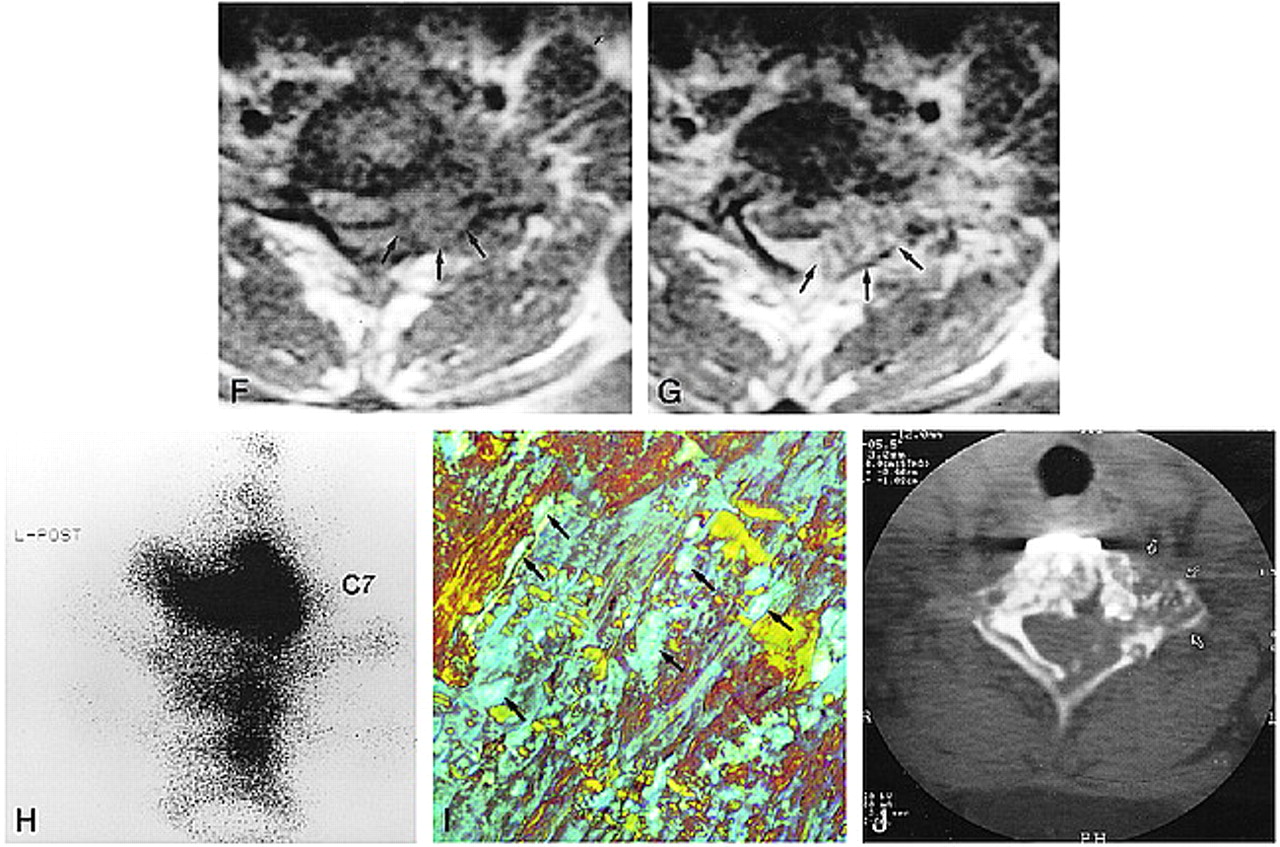
F and G, Axial T1-weighted MR images show the extradural mass encircling the anterior and lateral aspect of the dural sac (arrows, F). After contrast administration (G), the enhancing mass partially compresses the spinal cord (arrows).

H, Posterior pinhole bone scintigram of cervicothoracic junction obtained with 99mTc-MDP shows increased radioactivity in C7. Minimally increased radioactivity is also noted in T1 and T2 vertebral bodies.

I, Biopsy specimen viewed with polarized light is positive for Congo red stain, showing a characteristic green or apple-green birefringence (arrows) (original magnification ×80).

J, At 3-year follow-up, CT scan shows no growth of the residual mass in C7 vertebra (arrows). The metallic instrument for fixation is noted in the anterior aspect of C7.

A decompressive corpectomy with anterior instrumentation and fusion from C6 to T1 was performed via the anterior approach. At surgery, a friable, reddish, extradural mass was found to be compressing the dura. After the operation, the patient's paraparesis and urinary incontinence resolved completely. The biopsy specimen was positive to Congo red stain at histologic examination, with green birefringence to polarized light (Fig 1I), which is characteristic of amyloidoma. During 3 years of follow-up, the patient has reported no specific symptoms despite the presence of a residual mass at C7. A follow-up CT study after 3 years showed no growth of the residual mass (Fig 1J).

Discussion

Amyloidosis may be a systemic or localized process. It may occur spontaneously or in response to chronic disease processes. Primary amyloidosis is characterized by no detectable plasma cell dyscrasia or abnormal serum proteins; secondary amyloidosis involves systemic disease, in which the breakdown of cells is protracted (2).

Primary localized amyloid deposits in bone are unusual and any bone may be involved. Skeletal amyloid deposits often have an associated soft-tissue mass that may contain variable amounts of calcification (4). The lesions grow slowly and can produce significant local destruction of bone and soft tissue. It is this tumor-like appearance and behavior that make the diagnosis difficult to establish on imaging studies (5). Primary amyloidoma involving the spine is extremely rare, and only two cases of involvement of the cervical spine have been reported. Previous reports have described various radiologic findings of spinal amyloid lesions, such as vertebral body fractures and collapse, osteopenia, and lytic lesions. The MR signal characteristics are similar to those of primary amyloid lesions in the nasopharynx and sternum (2, 3, 6, 7) and include low-to-intermediate signal on T1-weighted images, intermediate-to-high signal on T2-weighted images, and variable enhancement on contrast-enhanced T1-weighted images.

In our case, the amyloid tumor arose in the cervical spine. The spinal amyloid deposits appeared as low signal on both T1- and T2-weighted MR images. The exact mechanism for signal hypointensity on both sequences is not entirely clear, although areas of punctate calcification within the amyloid deposit may cause signal loss on both T1- and T2-weighted images. The extradural extension of the amyloid deposits showed prominent enhancement on contrast-enhanced T1-weighted studies, possibly related to reactive inflammatory changes (3). On the basis of the plain radiographic, CT, and MR findings, the differential diagnosis included chondrosarcoma, osteogenic sarcoma, and a calcified metastatic tumor, such as mucinous adenocarcinoma of the colon. Despite its rarity and nonspecific radiologic findings, primary spinal amyloidoma should be included in the differential diagnosis of an osteolytic and calcified mass of the spine.

Footnotes

  • ↵1 Address reprint requests to Young Ha Park, MD, Department of Radiology, St. Vincent's Hospital, The Catholic University of Korea, 93 Chi-dong, Paldal-ku, Suwon, Kyunggi-do, 442–723, Korea.

References

  1. ↵
    Bauer WH, Kuzma JF. Solitary “tumors” of atypical amyloid (paramyloid). Am J Clin Pathol 1949;19:1097-1112
    PubMed
  2. ↵
    Dickmam CA, Sonntag VK, Johnson P, Medina M. Amyloidoma of the cervical spine: a case report. Neurosurgery 1988;22:419-422
    CrossRefPubMed
  3. ↵
    Meyers SP, Mullins KJ, Kazee AM. Unifocal primary amyloidoma of the spine causing compression of the cervical spinal cord: MR findings. J Comput Assist Tomogr 1996;20:592-593
    PubMed
  4. ↵
    Schindel S. Amyloid tumor of the larynx (case report with E.M.). Ann Otolarngol 1972;81:438-444
  5. ↵
    Pawar S, Kay C, Anderson H. Primary amyloidoma of the spine. J Comput Assist Tomogr 1982;6:1175-1177
    PubMed
  6. Hegarty JL, Rao VM. Amyloidoma of the nasopharynx: CT and MR findings. AJNR Am J Neuroradiol 1993;14:215-218
    Abstract/FREE Full Text
  7. Urban BA, Fishman EK, Humphrey RL. Primary amyloidosis of the sternum: imaging findings. Clin Imaging 1994;18:75-78
    PubMed
  • Received August 9, 1999.
  • Copyright © American Society of Neuroradiology
PreviousNext
Back to top

In this issue

American Journal of Neuroradiology
Vol. 21, Issue 3
1 Mar 2000
  • Table of Contents
  • Index by author
Advertisement
Print
Download PDF
Email Article

Thank you for your interest in spreading the word on American Journal of Neuroradiology.

NOTE: We only request your email address so that the person you are recommending the page to knows that you wanted them to see it, and that it is not junk mail. We do not capture any email address.

Enter multiple addresses on separate lines or separate them with commas.
Primary Amyloidoma of the Cervical Spine
(Your Name) has sent you a message from American Journal of Neuroradiology
(Your Name) thought you would like to see the American Journal of Neuroradiology web site.
CAPTCHA
This question is for testing whether or not you are a human visitor and to prevent automated spam submissions.
Cite this article
Sung Su Hwang, Young Ha Park, Jee Young Kim, So Lyung Jung, Myeong Im Ahn, Choon Keun Park, Eun Deok Chang
Primary Amyloidoma of the Cervical Spine
American Journal of Neuroradiology Mar 2000, 21 (3) 601-603;

Citation Manager Formats

  • BibTeX
  • Bookends
  • EasyBib
  • EndNote (tagged)
  • EndNote 8 (xml)
  • Medlars
  • Mendeley
  • Papers
  • RefWorks Tagged
  • Ref Manager
  • RIS
  • Zotero
0 Responses
Respond to this article
Share
Bookmark this article
Primary Amyloidoma of the Cervical Spine
Sung Su Hwang, Young Ha Park, Jee Young Kim, So Lyung Jung, Myeong Im Ahn, Choon Keun Park, Eun Deok Chang
American Journal of Neuroradiology Mar 2000, 21 (3) 601-603;
del.icio.us logo Twitter logo Facebook logo Mendeley logo
  • Tweet Widget
  • Facebook Like
  • Google Plus One

Jump to section

  • Article
    • Abstract
    • Case Report
    • Discussion
    • Footnotes
    • References
  • Figures & Data
  • Info & Metrics
  • Responses
  • References
  • PDF

Related Articles

  • No related articles found.
  • PubMed
  • Google Scholar

Cited By...

  • Imaging of Focal Amyloid Depositions in the Head, Neck, and Spine: Amyloidoma
  • Crossref
  • Google Scholar

This article has not yet been cited by articles in journals that are participating in Crossref Cited-by Linking.

More in this TOC Section

  • Management Outcomes For VO Spine Biopsy
  • Cone Beam CT Myelography
  • Post-Procedural Brachial Neuritis Features
Show more SPINE IMAGING AND SPINE IMAGE-GUIDED INTERVENTIONS

Similar Articles

Advertisement

Indexed Content

  • Current Issue
  • Accepted Manuscripts
  • Article Preview
  • Past Issues
  • Editorials
  • Editor's Choice
  • Fellows' Journal Club
  • Letters to the Editor
  • Video Articles

Cases

  • Case Collection
  • Archive - Case of the Week
  • Archive - Case of the Month
  • Archive - Classic Case

More from AJNR

  • Trainee Corner
  • Imaging Protocols
  • MRI Safety Corner
  • Book Reviews

Multimedia

  • AJNR Podcasts
  • AJNR Scantastics

Resources

  • Turnaround Time
  • Submit a Manuscript
  • Submit a Video Article
  • Submit an eLetter to the Editor/Response
  • Manuscript Submission Guidelines
  • Statistical Tips
  • Fast Publishing of Accepted Manuscripts
  • Graphical Abstract Preparation
  • Imaging Protocol Submission
  • Evidence-Based Medicine Level Guide
  • Publishing Checklists
  • Author Policies
  • Become a Reviewer/Academy of Reviewers
  • News and Updates

About Us

  • About AJNR
  • Editorial Board
  • Editorial Board Alumni
  • Alerts
  • Permissions
  • Not an AJNR Subscriber? Join Now
  • Advertise with Us
  • Librarian Resources
  • Feedback
  • Terms and Conditions
  • AJNR Editorial Board Alumni

American Society of Neuroradiology

  • Not an ASNR Member? Join Now

© 2025 by the American Society of Neuroradiology All rights, including for text and data mining, AI training, and similar technologies, are reserved.
Print ISSN: 0195-6108 Online ISSN: 1936-959X

Powered by HighWire